

나노물질

산업 제조
붕소 기반 나노 물질은 태양 수소 연료 생산 및 환경 개선을 위한 태양 에너지 변환에서 무독성, 지구에 풍부한 (광)전극 촉매 물질로 부상하고 있습니다. 붕소 산질화물(BCNO)은 붕소, 질소, 탄소 및 산소의 조성을 변화시켜 조정할 수 있는 전자, 광학, 물리화학적 특성을 가진 4차 반도체입니다. 그러나 BCNO의 구조와 광촉매 활성 관계 사이의 관계는 아직 탐구되지 않았습니다. 우리는 BCNO 준비에서 두 가지 다른 질소 전구체를 사용하는 효과와 어닐링 온도의 영향을 설명하기 위해 심층 분광 분석을 수행했습니다. BCNO 나노디스크(D =6.7 ± 1.1 nm) 800°C에서 열 어닐링 시 질소 소스 전구체로 구아니딘 염산염을 사용하여 난층 질화붕소 회절 패턴을 사용하여 준비했습니다. BCNO 나노디스크의 X선 광전자 분광법(XPS) 표면 원소 분석 결과 B, C, N, O 조성이 각각 40.6%, 7.95%, 37.7%, 13.8%로 나타났다. 솔리드 스테이트 11 에 따르면 B NMR 분석, 구아니딘 염산염 유래 BCNO 나노디스크는 다양한 삼좌표 BNx의 형성을 보여주었습니다. (OH)3−x 광촉매 활성 부위 중 하나이기도 한 종. XRD 및 심층 분광 분석은 BCNO 도핑된 육방정계 질화붕소 나노디스크의 준비를 확증했습니다. 대조적으로, 질소 전구체로 멜라민을 사용하여 600°C에서 어닐링된 BCNO는 XRD, XPS 및 고체 상태 NMR에 의한 증거로 벌집 격자에서 공유 결합된 B, C, N 및 O 원자로 구성된 층상 나노시트로 구성되었습니다. 분석( 11 B 및 13 다) 분석. 멜라민 유래 BCNO 층 구조의 XPS 표면 원소 조성은 고탄소 조성(75.1%)과 상대적으로 낮은 붕소 조성(5.24%)과 질소(7.27%) 조성으로 구성되어 BCNO가 도핑된 산화 그래핀의 형성을 나타냈다. 적층 시트 구조. 이 일련의 멜라민 유래 BCNO 도핑 산화 그래핀 층 구조는 흑연질화탄소의 광촉매 활성을 능가하는 가장 높은 광촉매 활성을 나타내는 것으로 밝혀졌습니다. 이 계층 구조에서 사좌표 BNx의 형성 (OH)3−x (CO) 종과 풍부한 흑연 도메인은 BCNO가 도핑된 산화 그래핀 층 구조의 광촉매 활성에 중요한 역할을 하는 것으로 제안되었습니다. 광학 밴드 갭 에너지는 BCNO 도핑된 육방정계 질화붕소 나노디스크 및 BCNO 도핑된 산화 그래핀 층 구조에 대해 각각 5.7 eV 및 4.2 eV로 측정되었습니다. 마지막으로 BCNO는 BGH01, BGH03, BMH01, BMH03에 대해 각각 평균 붕괴 수명이 1.58, 2.10, 5.18, 8.14μs인 초장기 광발광을 나타냈습니다. 이 연구는 새로운 무금속 광촉매 시스템을 제공하고 BCNO 기반 광촉매의 기원에 대한 최초의 구조 분석을 제공합니다.
금속이 없는 나노물질은 태양열 연료 생산, 환경 개선, CO2를 포함한 다양한 응용 분야에서 높은 구조적 및 화학적 안정성을 지닌 비용 효율적이고 지구 친화적인 (광) 촉매로 부상하고 있습니다. 유해미생물의 저감, 살균, 유기화합물의 선택적 화학합성을 가능하게 한다[1,2,3,4,5,6,7]. 금속이 없는 촉매와 비교하여 금속이 없는 촉매는 중독 가능성이 적고 사이클 수명이 더 길어집니다. 따라서 안정적이고 효율적이며 비용 효율적인 광촉매 유적인 새로운 재료의 검색 및 개발은 중요하고 도전적인 연구 노력입니다. 흑연질화탄소(CN)[4, 8, 9], 탄소점(C-dot)[2, 3, 10], 그래핀계 재료[7, 11]와 같은 탄소계 재료가 널리 연구되고 있다. 우수한 물리화학적 특성, 구조 및 화학적 안정성, 지구에 풍부한 원소로부터 합성이 용이하기 때문입니다. 최근에 붕소 기반(광)촉매가 놀라운 성능을 가진 금속이 없는 광촉매 시스템으로 개발되었습니다. 특히 경도가 높은 것으로 알려진 탄화붕소는 금속이 없는 가시광선 광촉매 수소 발생을 보여 최첨단 탄소 기반 CN 광촉매를 능가한다[12, 13]. H2에 대한 가시광선 광촉매 활성을 나타내는 고표면적 탄소 도핑된 육방정계 질화붕소(BCN) 나노시트 그리고O2 CO2뿐만 아니라 세대 축소 및 캡처는 사진 시스템에서 새로운 가능성을 제시했습니다[14, 15]. 산질화붕소(BNO)[16, 17], 인화붕소(BP)[18, 19], 붕소 도핑된 그래핀[20], 붕소 탄소 질화물(BCN)[14]과 같은 기타 붕소 함유 (광)전극 촉매, 붕소 도핑된 탄소 질화물(B-도핑된CN ) [21] 및 원소 붕소[22, 23]는 상당한(광) 전기촉매 활성을 입증했습니다[13].
붕소 산질화물(BCNO)은 붕소 기반 나노물질로, 다른 물질보다 덜 연구되었습니다. BCN 이전 제품인 약 2eV의 밴드 갭을 가진 반도체로 산질화물 및 질화물 화합물을 기반으로 하는 독성 형광체를 대체하기 위해 처음 개발되었습니다[24, 25]. B, C, O 및 N 원자를 그래핀 또는 육방정계 질화붕소(hBN) 네트워크로 대체하면 조정 가능한 광발광 특성과 밴드갭이 0eV(그래핀)에서 5.9eV(hBN) 범위인 BCNO 화합물이 생성되었습니다[26] . 이러한 바람직한 반도체 및 광발광 특성은 최근에 연구자들이 더 높은 결정도[27], 제어된 모양[28] 및 원자적으로 얇은 2D 구조[29]를 갖는 저차원 BCNO 나노구조를 합성하기 위한 새로운 합성 방법론을 개발하도록 매료시켰습니다. 이전 연구에서는 심층적인 구조적 특성을 제공하지 않고 BCNO의 광발광 특성을 조절하는 데 어닐링 온도와 시간의 영향을 조사했습니다[30, 31]. 이 논문에서 우리는 BCNO 나노구조의 구조-광촉매 활성에 대한 다양한 질소 소스 전구체, 소성 온도(800°C 대 600C) 및 소성 시간(0.5시간 대 12시간)을 사용하는 효과를 조사했습니다.
붕산 99.99%(H3 BO3 ), 멜라민 99%(C3 H6 N6 ) 및 헥사메틸렌테트라민 ≥ 99%(C6 H12 N4 ) Alfa Aesar에서 구입하여 추가 정제 없이 사용했습니다. 구아니딘 염산염 99.5%(CH5 N3 HCl)은 Arcos Organics에서 구입했습니다. BCNO는 문헌에 따라 저온 어닐링 방법을 통해 합성되었습니다[25, 32]. UPS 분석은 He I 21.22eV를 5V 바이어스의 광자 소스로 사용하여 ULVAC-PHI PHI 5000 Versaprobe II에서 수행되었습니다. BCNO 시료의 형태는 투과전자현미경(JEOL, JEM-ARM200FTH)을 통해 분석하였다. Bruker D2 분광계를 사용하여 XRD 회절을 얻었다. 용액 내 광발광 방출 스펙트럼은 광발광 분광기(PerkinElmer, LS55)를 사용하여 얻었고 용액 내 BCNO의 광 흡수 스펙트럼은 UV-Vis 분광기(HITACHI, U-3900)로 측정했습니다. X-선 광전자 분광법은 Al Ka x-선을 여기 소스로 사용하는 고해상도 X-선 광전자 분광계(ULVAC-PHI, PHI Quantera II)를 통해 분석되었습니다. BCNO 용액은 XPS 특성화를 위해 실리콘 기판에 드롭 캐스팅되었습니다. 결합 에너지는 284.8eV에서 탄소로 보정되었습니다. XPS 피크 디콘볼루션 및 피팅은 CACS XPS 소프트웨어를 사용하여 수행되었습니다. Absolute PLQY는 문헌[33]에 따라 수행되었으며 CCD 카메라(PIXIS 256BR, Princeton Instruments)로 감지되었습니다. 절대적 PLQY의 측정은 보정된 적분구 시스템(Labsphere) 및 전하 결합 소자(CCD) 카메라(PIXIS 256BR, Princeton Instruments)를 포함하는 섬유 기반 분광기를 사용하여 수행되었습니다. 다이오드 레이저(λ =375 nm, Becker &Hickl GmbH)가 펌핑 소스로 사용되었습니다. 시간 분해 광발광은 펄스 질소 레이저(λ =337.1nm, LTB Lasertechnik Berlin GmbH)가 여기 소스로 사용되었으며, 이는 디지털 지연 발생기(DG645, Stanford Research Systems)에 의해 트리거되었습니다. 신호는 광자 계수 광전자 증배관(PMC-100–1, Becker &Hickl GmbH)에 의해 감지되었고 광자 계수는 멀티스케일러 모듈(MSA-300, Becker &Hickl GmbH)로 누적되었습니다. 적외선 스펙트럼은 ATR(Attenuated Total Reflectance)을 사용하여 푸리에 변환 적외선 분광기(Bruker, Vertex 80v)에 의해 기록되었습니다. 4mm MAS(Magic Angle Spinning) 프로브를 사용하여 9.4T 자석이 장착된 Bruker Avance III 400 NMR 분광기를 사용하여 핵 자기 공명 스펙트럼을 얻었습니다. 11 B MAS NMR은 10kHz의 회전 속도로 스핀 에코 방법을 사용하여 기록되었습니다. 4초의 재활용 지연으로 8,000개의 스캔이 수집되었습니다. 화학적 이동은 1M H3를 참조했습니다. BO3 19.6ppm의 수용액. 13 C CP/MAS NMR 스펙트럼은 12.5kHz의 회전 속도로 교차 편광(CP) 시퀀스를 사용하여 기록되었습니다. 4초의 재활용 지연으로 30,000개의 스캔이 수집되었습니다. 모든 13 C 화학적 이동은 아다만탄 CH2의 2차 참조를 사용하여 순수한 트리메틸실란에 대해 참조되었습니다. 38.48ppm에서 피크
이 연구에서는 붕소와 탄소 전구체 소스, 전구체 비율, 어닐링 온도 및 시간을 고정하면서 두 가지 다른 질소 전구체 소스를 사용하여 두 가지 시리즈의 BCNO를 제조했습니다. 이 연구에서 준비한 BCNO 시리즈의 다른 모든 반응 변수를 고정하여 열처리 온도와 시간의 영향을 조사했습니다. 간단히 말해서, 미리 결정된 몰비의 세 가지 전구체 성분 모두를 증류수에 첨가하고 용액이 균질해 보일 때까지 90°C로 가열했습니다(표 1). 찹쌀 혼합물을 오븐에서 밤새 건조시켜 건조된 백색 고체를 얻었다. 백색 고체를 막자사발과 막자를 사용하여 고운 분말로 분쇄하였다. 고체 전구체는 표 1에 나타난 바와 같이 대기압에서 5°C/min의 램핑 속도로 미리 결정된 온도 및 시간에서 노에서 하소되었습니다. 노를 상온으로 자연 냉각시킨 후 황색을 띤 분말 샘플을 미세한 분말로 분쇄하였다.
준비된 BCNO를 물과 에탄올(10mg/mL 농도에서 1:10 v/v)에서 10분 동안 6000rpm으로 원심분리하여 정제했습니다. 원심분리 후, 생성물을 증류수에 재용해시키고, 물:에탄올의 1:10 v/v 비율로 에탄올로 희석하였다. 정제된 BCNO는 TEM 분석을 위해 탄소 코팅된 구리 그리드에 증착되었습니다. SEM 및 XPS 시편 준비를 위해 정제된 BCNO 샘플을 실리콘 웨이퍼에 드롭 캐스팅했습니다. 샘플 증착 전에 실리콘 웨이퍼를 각 용매에서 10분 동안 물, 프로판올, 아세톤으로 초음파 처리하여 세척했습니다. UPS 시편은 샘플이 ITO(인듐 주석 산화물) 코팅 유리에 증착되었다는 점을 제외하고 SEM 및 XPS 샘플에 대해 설명한 절차와 유사하게 준비되었습니다.
벌크 CN은 문헌[34]에 보고된 손쉬운 방법으로 합성되었습니다. 간단히 말해서, 멜라민 분말을 도가니에 넣고 주위 대기압에서 5℃/min의 램핑 속도로 550℃에서 4시간 동안 어닐링했습니다.
다양한 BCNO 샘플의 광촉매 활성은 모델 반응으로 메틸렌 블루(MB) 광분해를 통해 평가되었습니다. 일반적인 염료 분해 실험에서 10mg의 BCNO 샘플을 15mL의 MB 용액(10ppm)이 들어 있는 샘플 바이알에 추가했습니다. 암실에서 10분 동안 교반한 후, 샘플 바이알에 100W 크세논 램프(250nm ~ 1100nm)를 조사했습니다. 총 광촉매 분해 시간이 80분에 도달할 때까지 20분 간격으로 용액에서 피펫을 사용하여 운동 샘플(2mL)을 추출했습니다. UV-vis 분광계를 통해 다른 시간 간격의 운동 샘플을 분석했습니다. MB의 농도 변화는 Beer의 법칙을 사용하여 도출되었습니다.
전하 수송을 용이하게 하기 위해 결정도가 높은 저차원 BCNO 나노구조의 제조를 위한 연구에서, 우리는 뚜렷하게 다른 화학 구조와 광촉매 활성을 갖는 BCNO를 발견했습니다. 문헌[25, 32]에 보고된 BCNO의 합성을 기반으로, 우리는 두 가지 다른 질소 전구체 소스를 사용하는 효과와 열 어닐링 온도 및 BCNO의 구조 속성 변화에 대한 시간의 영향을 조사했습니다. 이 연구에서는 붕소와 탄소원으로 각각 붕산과 헥사메틸렌테트라민을 사용하여 두 가지 시리즈의 BCNO를 준비했습니다(표 1). BCNO는 멜라민 및 구아니딘 염산염을 질소 공급원으로 사용하여 합성되었으며 각각 BMH 및 BGH로 표시됩니다. 각 반응 조건을 체계적으로 조사한 결과 BMH 계열은 600°C의 낮은 온도에서 12시간 동안 열처리했을 때만 광촉매 활성을 보였습니다. BGH 시리즈는 800°C에서 12시간 동안 고온 어닐링으로 광촉매 활성만 나타냈습니다. 두 시리즈 모두에서 붕산, 멜라민 및 구아니딘 염산염의 몰비는 3:1로 고정된 반면, 헥사메틸렌테트라민의 몰비는 0.1에서 0.3으로 다양했습니다. 이러한 헥사메틸렌테트라민 전구체의 비율은 샘플 이름에 각각 BMHH01/BGH01 및 BMH03/BGH03으로 표시됩니다(표 1).
그림 1은 800°C에서 12시간 동안 소성된 BGH의 대표적인 TEM 이미지로, 준구형 나노입자 형태의 결정질 코어와 D =6.7 ± 1nm. 준비된 BGH는 물에 쉽게 분산되었으며(추가 파일 1:그림 S1), TEM 구리 그리드에 개별 나노입자가 증착된 것으로 입증되었습니다. BGH 및 BMH의 수성 분산성은 BGH 및 BMH의 음으로 하전된 표면 전위를 기반으로 하는 정전기 안정화에서 비롯된 가능성이 가장 큽니다. 무접점 11 B NMR 분석은 BMH와 BGH 나노구조 모두에 있는 하이드록실 그룹의 풍부함이 수성 매질에서의 분산성을 확증하는 것으로 나타났습니다. 고배율에서 각 준구형 나노입자는 0.338 nm로 측정된 (002) 평면의 독특한 격자 간격을 나타내었으며 이는 문헌 보고[24, 35](그림 1c)와 일치합니다. 공정염 환경에서 열처리를 통해 얻은 5nm BCNO 나노입자와 비교하여, 본 연구에서 제조된 BGH 나노입자는 높은 결정성을 가졌다[25](그림 1). 다음으로 붕산, 멜라민, 헥사메틸렌테트라민을 사용하여 문헌 보고서를 기반으로 준비하고 600°C에서 12시간 동안 어닐링한 BMH를 특성화했습니다. BGH의 형태와 달리 BMH는 모양이 불분명한 다층 시트로 구성됩니다(그림 1). 더 높은 배율에서 다층 시트 가장자리의 TEM 이미지(그림 1d)는 그림 1e와 같이 구조적 왜곡[36]의 특징을 가진 나노시트를 나타냅니다. 그림 1f는 특징이 없는 마이크론 크기의 응집체를 포함하는 BMH의 대표적인 SEM 이미지를 보여줍니다. 그러나 BGH 시리즈와 달리 멜라민을 질소 소스 전구체로 사용하여 800°C에서 더 높은 온도로 소성하면 나노디스크 형태가 생성되지 않습니다. 다른 반응 조건에서 합성된 다른 BGH 및 BMH 계열 화합물의 TEM, XRD, UV 흡광도 및 광발광을 ESI에서 확인할 수 있습니다.
<그림>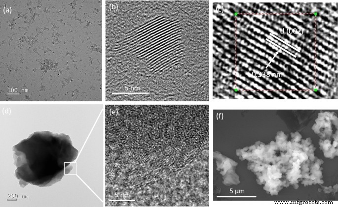
희석된 에탄올 용액에서 증착된 준비된 BGH 및 BMH의 대표적인 투과 전자 현미경 사진(TEM) 및 주사 전자 현미경 사진(SEM)입니다. 아 , b BGH의 고배율 및 저배율 TEM, c 0.338nm의 뚜렷한 (002) 격자 간격을 갖는 BGH의 고해상도 TEM, d 낮은 배율에서 대표적인 BMH의 TEM 이미지, e 그림 1d의 박스 영역을 확대한 TEM 이미지 층상 산화 그래핀 내 구조적 왜곡의 특징을 보여주고, f BMH 대표 SEM 이미지
그림 2는 표 1에 나열된 규정된 반응 조건에서 실험실에서 제조된 BMH03(녹색 추적), BMH01(적색 추적) 및 BGH01(파란색 추적)의 XRD 패턴을 보여줍니다. 43.1°(2θ), 이는 터보층 질화붕소(t-BN)의 대표적인 회절 패턴입니다. 약 26.6°를 중심으로 하는 넓은 피크는 (002) 반사면에서 시작되었으며 43.1° 넓은 피크는 육각형 질화붕소(h-BN)에 의해 유도된 (10) 반사면에 해당합니다[37]. BMH의 XRD 패턴은 약 25.4° 및 42.4°의 2θ 에서 두 개의 넓은 회절 패턴이 지배적이며, 이는 각각 흑연의 육각형 결정 구조의 (002) 및 (10) 밴드에 대한 시그니처 패턴입니다[38]. 일반적으로 표시되는 (10) 밴드는 또한 난층 탄소의 2D 반사와 관련이 있습니다[39]. 또한, 2θ 약 10.9°에서 피크가 없고 약 25.4°에서 광대역의 출현은 그래핀 또는 흑연 산화물의 구조 내에 도펀트 또는 불순물이 혼입되었기 때문입니다[40,41,42,43, 44,45]. 따라서 BMH의 지배적인 구조는 도핑된 산화 그래핀이라고 제안하는 것이 합리적입니다.
<사진>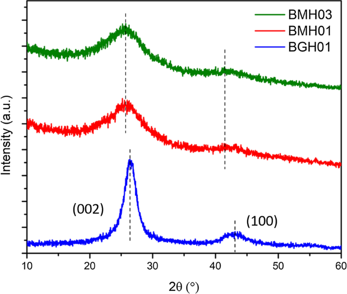
다결정 BMH03(녹색 트레이스), BMH01(빨간 트레이스), BGH01(파란 트레이스), BGH01의 XRD 패턴. ca.에서 BGH01 시리즈의 광범위한 정점. 26.6°는 h-BN의 (002) 평면 반사와 ca에서 또 다른 넓은 피크를 나타냅니다. 43.1°는 h-BN의 미해결 반사면을 나타냅니다. BMH의 회절 패턴은 약 ~ 25.4°와 ~ 42.4°를 중심으로 두 개의 넓은 반사를 보였다. 다른 BCNO 시리즈의 XRD 패턴은 추가 파일 1에 나와 있습니다. 그림 S3)
XPS, FTIR, 고체 NMR 분광법을 수행하여 BGH의 분자 구조에 대한 더 깊은 통찰력을 얻었습니다. XPS는 B, C, N 및 O 원소의 코어 레벨 전자의 존재와 BGH 화합물에서 각각의 화학적 결합을 확인하는 데 사용되었습니다. 그림 3a–e는 BGH 나노디스크의 일반적인 XPS 스펙트럼을 보여줍니다. XPS 표면 원소 조성 분석에 따르면, BGH는 높은 B 및 N 함량(각각 약 40%)을 포함하고 C 및 O 조성은 각각 약 8% 및 13%로 낮습니다(추가 파일 1:표 S3). B 및 N 조성의 '거의' 1:1 화학량론은 XRD 분석에 상응하여 실험실에서 준비된 BGH01이 난류 질화붕소 구조로 구성되었음을 확인했습니다. 모든 XPS 스펙트럼에는 R이 있는 가우스 함수가 장착되었습니다. 2 > 0.99, 이는 각 요소에 대한 XPS 스펙트럼의 빨간색 및 녹색 곡선으로 표시됩니다. BGH 시리즈의 경우 B1s 스펙트럼은 B-O 결합(189.7 eV 결합 에너지에서) 및 B-N 결합(190.7 eV 결합 에너지에서)에 해당하는 두 개의 적합 곡선으로 분해되었습니다. N1 스펙트럼은 397.3eV 결합 에너지를 가진 N-B 결합과 398eV 결합 에너지를 가진 B-N-O 결합으로 구성된 두 개의 가우시안 곡선에 맞춰졌습니다[46]. N-B 및 B-N-O 결합은 모두 O-도핑된 hBN의 존재 또는 BNO 화합물의 형성을 나타냅니다[47]. BGH 합성은 대기 조건에서 수행되었기 때문에 문헌[47]에 보고된 바와 같이 B-O 결합을 위해 산소 원자도 h-BN 도메인에 통합되었습니다. 이 가설은 O1s를 분석하여 확인되었습니다. 스펙트럼은 B-O 결합(532 eV에서 결합 에너지)에 해당하는 단일 피크를 보여주었습니다. 이 결과는 BGH 계열이 O-도핑된 hBN으로 구성되고 대부분의 산소 원자가 붕소 원자에 결합되어 있다는 초기 가설을 뒷받침합니다. C1 BGH 스펙트럼의 3개의 C 종, 즉 C-O, C-B 및 C-O-C로 분해되었으며, 각각 283.7 eV, 285 eV 및 287 eV에서 결합되었습니다. BGH 시리즈에서 C의 낮은 조성과 B와 N의 1:1 화학량론으로 인해 우리는 BGH01의 구조가 C와 O가 도핑된 h-BN이라고 더 추측했습니다. 제안된 구조는 XPS 스펙트럼에서 입증된 바와 같이 hBN 도메인 내에서 C-B, B-O 및 B-N-O 결합의 형성에 의해 지원되었습니다(그림 3). 따라서 800°C에서 제조된 BGH 나노디스크의 구조를 C 및 O가 도핑된 hBN으로 추론하는 것이 합리적입니다. 또한, 분말 XRD 및 고해상도 TEM은 난층 질화붕소의 격자 간격과 유사한 격자 간격을 갖는 벌집형 격자에서 공유 결합된 B, C, N 및 O의 형성을 지원했습니다. 이 원고 전체에서 BGH01의 구조는 BCNO가 도핑된 hBN이라고 합니다.
<그림>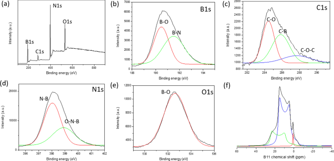
아 BGH01의 XPS 조사 스펙트럼. b의 핵심 레벨 스펙트럼 B1s , ㄷ C1s , d N1s , 및 e O1s . 각 디콘볼루션 피크에는 가우스 함수가 장착됩니다. 에 11 BGH01의 B 고체 MAS NMR 스펙트럼. ( 11 BGH01-LT의 B 고체 MAS NMR은 추가 파일 1에 나와 있습니다. 그림 S10)
우리는 또한 다른 합성 매개변수를 일정하게 유지하면서 BCNO 나노구조의 구조-광촉매 활성에 대한 하소 온도 및 시간의 영향을 조사했습니다. 반응 온도를 600°C에서 800°C로 높이고 반응 시간을 30분에서 12시간으로 늘리면 B-N 및 B-O 결합이 전반적으로 증가했습니다(추가 파일 1:그림 S6). 대조적으로, B-C 결합은 반응 온도 및 시간이 증가함에 따라 감소하는데, 이는 준안정 B-C 결합을 희생시키면서 에너지적으로 안정한 육각형 B-N 및 B-O 결합의 형성을 의미한다[48]. 예상대로 B-C 결합 조성은 다른 매개변수를 일정하게 유지하면서 헥사메틸렌테트라민 전구체(C 공급원)의 비율을 증가시키면 증가합니다[49]. 다양한 소성 온도, 시간 및 전구체 비율에서 준비된 BGH의 화학적 결합 진화에 대한 보다 자세한 경향은 ESI에 제공됩니다(추가 파일 1:그림 S6 및 S7).
1 11 B 고체 MAS NMR은 BGH 및 BMH의 각 B 관련 결합 조성을 정량적으로 정량적으로 분석하는 데 활용되었습니다. 여기에서는 고체 MAS를 활용하여 분자 수준에서 BCNO의 첫 번째 상세한 구조적 특성을 제시합니다 13 C 및 11 B NMR은 특정 C 및 B 관련 결합을 수집합니다. hBN 나노물질과 결함 유발 전자 특성이 상당한 관심을 끌었지만 hBN 가장자리의 분자 구조와 결함 구조는 거의 알려져 있지 않다[50]. 붕소 관련 나노물질의 구조적 특성 규명이 부족한 것은 고체 상태의 분석이 어렵기 때문입니다. 11 B NMR 스펙트럼 때문에 11 B는 반정수 사중극자 핵(I =3/2) [51, 52]. 무접점 11 B NMR은 또한 MAS NMR을 통해 부분적으로만 평균화할 수 있는 신호 왜곡을 초래하는 2차 사중극자 결합으로 인해 해석하기 어렵습니다. 또한 11 의 화학적 이동 범위는 B NMR은 상대적으로 좁기 때문에 다양한 붕소 종의 광범위하고 중첩되며 왜곡된 피크를 지정하는 것이 매우 어렵습니다[54]. 이 연구에서는 11 을 수행했습니다. B 고체 상태 NMR은 9.4T에서 기록되었으며, 스펙트럼은 탑스핀 실선 모양 분석(SOLA)을 사용하여 디콘볼루션되었습니다. CP-MAS 11 준수 문헌에 보고된 B NMR 실험에서 우리는 사중극자 결합 상수(C 질문 ) 및 전기장 기울기(EFG) 텐서 비대칭(η 질문 ) [51, 52, 55]. 그림 3f는 솔리드 스테이트 11 을 보여줍니다. 세 개의 주요 피크와 δ가 있는 BGH의 B 스핀-에코 NMR 스펙트럼 이소 각각 28.3ppm, 20ppm, 1.2ppm을 중심으로 합니다. CP-MAS 11 에 대한 문헌 연구 기반 질화붕소의 B NMR 및 관련 구조, δ 피크 이소 28.3ppm(녹색 추적 적합) 및 C 질문 2.85MHz의 삼각 평면 BN2에 해당 (OH) 단일 수산기를 가진 종 [53,54,55]. 히드록실기 또는 산소 가교 원자가 삼각 평면 붕소 부위 주변의 질소 원자를 대체할 때, δ를 갖는 새로운 B 종 이소 20ppm(파란색 추적 적합)이 나타났습니다. 이 더 낮은 화학적 이동 신호는 2개의 히드록실기 또는 1개의 히드록실기 및 1개의 가교 산소 원자(BN(OH)2 또는 BNO(OH) 사이트). δ에서의 급격한 피크 이소 1.2ppm(적색 추적 맞춤)의 4배위 사면체 B 사이트는 질소 및 다중 하이드록실 그룹 또는 브리징 산소 원자(52–54) 또는 탄소 관련 C-B 결합에 의해 배위될 가능성이 있는 4배위 사면체 B 사이트에 해당합니다[46, 52]. CP-MAS 11 BGH01의 B NMR 분석은 XPS 및 XRD 분석에 상응하는 다양한 B-N, O-B 및 B-C 결합을 보여주었습니다. 또한 솔리드 스테이트 11 B NMR은 또한 대부분의 붕소가 질소 및 BNx로서 하나 이상의 하이드록실 그룹과 결합되어 있음을 보여주었습니다. (OH)3−x 종. 이러한 하이드록실화 종은 수소 결합 및 정전기 안정화를 통해 수용액에서 콜로이드 안정성을 제공하는 것으로 추측됩니다. 우리는 또한 BNx와 같은 사좌표 B-종의 감소를 관찰했습니다. (OH)4−x 또는 BNx (O)(OH)3−x (여기서 O는 가교 산소임) 반응 온도가 증가함에 따라 보록솔 고리[54, 56]가 나타납니다. 동시에 다양한 삼좌표 BNx (OH)3−x 어닐링 온도가 600°C에서 800°C로 증가했을 때 종들이 나타났습니다(추가 파일 1:그림 S8 및 S10). 이 결과는 고온에서 보록솔 고리가 암모니아와 반응하여 다양한 하이드록실화된 BNx을 형성함을 의미합니다. (OH)3−x 또는 BNx (O)(OH)2−x 종 [55] (추가 파일 1:그림 S10).
XPS 및 FTIR은 물론 11 을 통한 강력한 구조 분석을 기반으로 합니다. B 및 13 C 고체 상태 MAS NMR에서 BMH의 구조는 BCNO가 도핑된 산화 그래핀인 것으로 제안되었습니다. XPS 표면 원소 분석에 따르면 BMH는 탄소 종의 75%, 붕소 관련 종의 5%로 구성되었습니다(추가 파일 1:표 S3). 분리된 B1 스펙트럼은 각각 191.7 및 192.4 eV 결합 에너지에서 BCN 및 BN 결합을 보여주었습니다(그림 4). C1 종은 C–C 결합(sp 2 그리고 sp 3 CC 결합) 약 285.0 eV에서 결합 에너지를 가지며 285.6 eV에서 나타나는 B-C-N 결합에서 더 약한 성분이 발생했습니다[57]. 288.0eV 및 288.7eV에서 다른 비교적 작은 신호는 C-N3으로 인한 것입니다. 및 C =O 결합 [46, 57, 58]. 산화된 C 원자(C =O)는 그래핀 옥사이드 도메인의 가장자리에 형성되고 C-N3 결합은 CN의 특징적인 피크였습니다. N1 399.0 eV 결합 에너지를 중심으로 하는 XPS 스펙트럼은 399.2 eV 결합에서 C-N-B 및 400.0 eV에서 C =N 결합으로 구성된 두 개의 가우스 곡선에 맞출 수 있습니다[46, 57, 59, 60]. XPS 분석에 따르면 BMH 시리즈는 BGH 시리즈에서 O 관련 결합의 약 약 35%인 것과 비교하여 O 관련 결합의 약 10–25%만 구성했습니다. O1 531.2 eV 결합 에너지를 중심으로 하는 스펙트럼은 C =O 및 CO 결합으로 분해될 수 있으며, 이는 산화 그래핀 도메인에 기인할 수 있습니다. 13 C 고체 상태 CP-MAS NMR은 40.2ppm에서 흑연 C =C 결합의 존재를 추가로 보여주었으며, 이는 XRD 및 XPS 결과와 확증됩니다[38](그림 5). 이 논문에서 보고된 각 BMH 시리즈에서 13 C NMR은 C에 해당하는 160 및 154ppm에서 탄소 종의 두 가지 등몰비를 보여주었습니다. α 및 C β , 각각 흑연질화탄소 구조에서 일반적으로 발견됩니다[61](그림 5 및 추가 파일 1:표 S4). Compared to the bulk CN synthesized according to the literature procedure [34], the chemical shift of the signature resonance for C α and C β peak appeared at 164 and 156 ppm, respectively (Fig. S13). Boron doping into the CN heptazine structure could have contributed to these slight chemical shift differences between the BMH series and the bulk CN (Additional file 1:Fig. S13).

a Survey XPS spectra of BMH01 and 11 B NMR spectra. Core level spectra of b B1s , c C1s , d N1s , and e O1s . Each core spectra were fitted with a black trace, while the red and green traces under the peak were deconvoluted using a Gaussian function. f 11 B solid-state MAS NMR were deconvoluted using SOLA analysis to tricoordinate and tetracoordinate B-sites. The XPS and 11 B solid-state MAS NMR of other BMH series compounds can be found in Additional file 1:Figure S7, S11, and S12)

Solid-state 13 C MAS NMR of a BMH01 and b BMH03
Due to the abundant evidence of the presence of carbon nitride (CN) in the BMH series based on solid-state NMR spectroscopy (Fig. 5), we considered three possibilities of interactions between BCNO-doped graphene oxide and CN in BMH, namely:(i) bulk phase separation, (ii) disordered 2D network, and (iii) layered intercalation [62]. To eliminate the possibility of bulk phase separation, we examined the XRD pattern of CN nitride andBMH as well as a physical mixture of both in a 1:1 mass ratio. We found that the XRD patterns of the mixtures showed only the diffraction pattern of the bulk CN with a slight reduction in the crystallinity compared to the pristine CN diffraction [63] (Additional file 1:Fig S4). Since the XRD pattern of BMH did not possess any diffraction peaks that corresponded to CN, this experimental result confirmed that CN did not form as a bulk-separated domain during the synthesis of BMH (Additional file 1:Fig. S4). We also considered the formation of a disordered 2D network, in which CN and the doped-graphene oxides are bonded on the same 2D plane [62]. Based on a literature report on a CN/graphene oxide 2D matrix, the XRD pattern of a disordered 2D network showed characteristic peaks for both species with a slight peak broadening and a slight peak shifting [64]. However, the XRD pattern of BMH (Fig. 2) did not contain any signature diffractions of CN. A previous study also showed that characteristic peaks of graphene oxide disappeared in a graphitic CN/amorphous CN/graphene oxide composite due to the layer-by-layer interactions [65]. Therefore, it is reasonable to propose that CN is intercalated between the doped-graphene oxide layers. The FTIR spectrum of the selected BGH and BMH series is shown in Additional file 1:Fig. S5.
Boron-related bonding within the BMH series was investigated using 11 B solid-state MAS NMR at 9.4T, and the broad NMR spectrum was deconvoluted using the SOLA analysis showing the presence of both tricoordinate and tetracoordinate boron site. The SOLA analysis yielded four line fittings under the broad 11 B NMR spectrum, which could be assigned as trigonal planar BN2 (OH) or BN2 O at a δiso of 19.8 ppm and a CQ of 2.85 MHz (green fitting). The bay and corner B sites as in B-doped CN appeared at a δ iso of 5 ppm and δ iso of 11 ppm, respectively [61]. These assignments are also commensurate with the formation of CN based on the 13 C NMR and XPS analyses. Compared to the BGH series, the relative composition of the tetracoordinate B(IV) site of BMH was much higher (ca. 55% in the BMH series vs. 3% in the BGH series). However, the tetracoordinate B(IV) species in BMH appeared at a lower chemical shift than those found in BGH (Fig. 3f) and was therefore presumed to be the BN2 (OC)2−x (OH)x species [46]. Notably, the h-BN domain was absent from the BMH series prepared via thermal annealing at a lower temperature (600 °C). However, upon increasing the thermal annealing temperature from 600 °C to 800 °C, the structures of BMH01HT-30 min and BMH01HT-12 h showed a high composition of tetracoordinate BN2 (OH)2 species and the tricoordinate BN3 bonding (Additional file 1:Fig. S11 and S12). The presence of a high composition of tetracoordinate BN2 (OH)2 and BN3 bonding was shared among all the inactive BCNO investigated in this study. Moreover, although BMH01HT-12 h possessed an identical surface elemental composition to that of BGH01, the solid-state 11 B NMR revealed that both compounds possessed significant structural differences, which explained for their differences in photocatalytic activity (Additional file 1:Table S3 and Fig. S12).
In light of the moderate photocatalytic activity of BMH01 and BGH01 (Fig. 7), further synthesis optimization was performed to expand their light absorption spectrum into the visible light region. Previous literature showed that increasing the composition of the hexamethylenetetramine precursor (as a carbon source) could modulate the bandgap and photoluminescence properties of BCNO. Based on these reports, BMH and BGH compounds with a higher ratio of hexamethylenetetramine were prepared accordingly while keeping the other parameters constant. The optimized BCNO with a higher carbon content is denoted as BMH03 and BGH03, in which the molar ratio of carbon source was increased from 0.1 to 0.3. The higher ratio of hexamethylenetetramine precursor yielded BMH03 with a higher composition of the graphitic domain as in sp 2 C = C, and a small peak emerged which corresponded to BCN bonding at 191 eV binding energy (Additional file 1:Fig. S7). The increase in the graphitic sp 2 C = C domain upon increasing the concentration of hexamethylenetetramine is consistent with the role of hexamethylenetetramine as both a C and N source in the synthesis of N-doped graphite [66]. The increased sp 2 C = C graphite bonding in BMH03 was further confirmed via 13 C CP-MAS NMR with the emergence of a more prominent peak centered at 40.2 ppm, which is the signature of graphitic C = C(H) bonding (Additional file 1:Table S4).
The optical properties of BMH and BGH were investigated using UV–visible absorption and photoluminescence spectroscopy, as shown in Fig. 6. Since BGH and BMH series possessed distinctively different structures, and the optical properties of nanomaterials are highly correlated with their structures, the origin of absorption and luminescence for both series were also found to be different. In this study, BGH01 quasi-spherical nanoparticles showed a featureless UV–vis spectrum. (Fig. 6a, red trace). As the ratio of hexamethylenetetramine increased, the intensity of absorbance peaked at 237 nm for BGH03 and increased with the emergence of an additional broad absorption peak centered around 330 nm (Fig. 6a, blue trace). The origin of the optical properties of BCNO is controversial due to the lack of structural analysis of BCNO nanomaterials. The most widely cited origin of photoluminescence of BCNO is attributed to the formation of B, C, N, and O self-interstitial sites, substitutional impurities, and native point defects within hBN or the graphene matrix [50, 67, 68]. Other possible photoluminescence mechanisms in the BCNO system include the electronic transition from the nitrogen-vacancy (VN ) levels to the carbon impurity levels, the electronic transition between the closed-shell BO − and BO 2− anions, and intrinsic state emission and defect state emission (surface energy traps). The featureless UV absorbance of BGH01 can be attributed to the electronic transition between the valence band (VB) and conduction band (CB) of boron nitride with a bandgap energy of ca. 5.9 eV. The lower energy absorption in BGH03 was a result of the mid-gap absorption from the valence band to the nitrogen-vacancy (VN ) level located approximately 0.7–1.0 eV below the conduction band of hBN.(62) The absorption peak at ca. 330 nm could be attributed to the presence of C-related impurities level located ca. 2–4 eV below the conduction band of hBN [69,70,71]. Under 365 nm excitation, BGH01 produced a broad emission with three bumps located at 412 nm, 445 nm, and 489 nm, respectively. Based on the BGH structural analysis, in which the dominant structure was composed of BN and BO-related bonding, the photoluminescence of the BGH series could be most likely originated from the B-O luminescence centers [24, 47, 72]. The yellow-green emission at 445 and 489 nm could be induced by the transition from the VN level to the carbon-related and oxygen defect levels (2–4 eV) below the conduction band of h-BN [69,70,71]. As the C composition increased in BGH03, the emission wavelength was further red-shifted to 506 nm, consistent with literature reports [73, 74]. The stacked photoluminescence (PL), UV absorbance, and Tauc plot for different BGH series prepared in this study are presented in Additional file 1:Fig. S14.

a Overlay UV absorbance spectra of BMH01, BMH03, BGH01, and BGH03. b Stacked photoluminescence spectra of BMH01, BMH03, BGH01, and BGH03 upon excitation at 365 nm c stacked photoluminescence lifetime decay spectrum of BMH01, BMH03, BGH01, and BGH03 monitored at a predetermined λ max for each sample. Samples were excited with 337 nm laser pulses at 298 K, d Overlay Tauc plot (αhv) 1/2 vs. hv, for BMH01 (black trace) and BMH03 (green trace). The optical band gap is represented by thesolid line, while the interband state for BMH03 is represented by the red dashed line
Based on XRD and various spectroscopic analyses, the structure of BMH was deduced to be dominated by BCNO-doped graphene oxides (Fig. 4, Additional file 1:Fig. S7, and Fig. S12, and Table S4). Thus, the optical properties of the BMH series are hypothesized to be more closely related to the carbon-quantum dot (CD) [75, 76] and doped-graphene oxides systems [44, 77,78,79]. Based on the origin of photoluminescence of the CD and graphene oxides, the optical properties of BMH prepared in this study can be attributed to the intrinsic state emission [76, 80, 81], electron–hole recombination [82, 83], and defective state emission [84]. Intrinsic emission of BMH is speculated to have originated from isolated sp 2 luminescence centers embedded within the sp 3 matrix of the carbonaceous film. The sp 3 matrix of graphene oxides is composed of C–OH, C–O–C, and C = O edge sites, whose energy levels lie between the energy levels of π–π* states of the sp 2 C = C domain, thus giving rise to multiple absorption bands. Both BMH01 and BMH03 possessed a strong UV absorbance band at ca. 240 nm, corresponding to the π to π* transition of C = C within the graphene oxides domain. With the increasing ratio of hexamethylenetetramine in the BMH03 sample, an additional bump at 288 nm emerged, which can be ascribed to the n-π* transition of the C = O and C = N bonds of the oxidized graphitic region [77, 85, 86]. The latter absorbance band at ca. 288 nm was induced by oxygen, nitrogen, and boron defect sites, creating new radiative recombination sites [82, 87,88,89]. Upon photoexcitation at 365 nm, BMH01 and BMH03 exhibited a maximum emission wavelengths at 429 and 447 nm, respectively. The BMH03 sample showed a slight red-shift emission, unlike BMH01 due to the greater extent of graphitization [77, 84] (Additional file 1:Figs. 5 and 6). Both BMH samples revealed a broad and much lower energy emission wavelengths than BGH samples due to lower energy emissive centers arising from O, N, and B defects and surface states. According to the electron–hole recombination mechanism, these photoexcited electrons from each defect state recombine with their corresponding holes in the HOMO, thus yielding a broad photoluminescence emission [90] (Fig. 6b). Interestingly, only BMH01 exhibited a pronounced excitation-dependent photoluminescence as observed in other BCNO [72] and carbon quantum dot systems [76]. The presence of O, and N impurities embedded within the graphene oxides matrix was shown to create a large number of surface emissive traps that corresponded to a diverse energy levels within the bandgap, thus yielding an excitation dependent fluorescence spectra in BMH01 (Additional file 1:Fig. S15). In contrast, the lack of an excitation dependent emission in BMH03 could be explained by the formation of a greater extent of graphitization (C = C) with a concurrent reduction in the surface states population (eg.:C = O) [44].
The bandgap values of BMH and BGH prepared in this study were estimated from Tauc's formulation:(αhν) 2 − hν, where α is the absorbance (Fig. 6d). The bandgap was estimated by extrapolating the photon energy intercept at (αhν) 2 = 0. For the BMH series, the presence of multiple energy levels within the optical bandgap may have originated from the electronic transition from various π-π* (C = C bonds) and n-π* of C = O or other surface groups [76, 82, 87]. As for the BGH series, carbon substituted on boron sites (CB ), nitrogen-vacancy sites (VN ), and interstitial carbon defect levels gave rise to the emergence of interband states between the bandgap of hBN. The presence of multiple energy levels was supported by the photoluminescence spectra of lower energy radiative recombinations [24, 47, 66] (Fig. 6b and Table 2). The BGH03 sample exhibited two large bandgaps at 5.7 eV and 3.8 eV, corresponding to the bandgap of hBN and the transition from the valence band to VN levels, respectively [50, 74] (Additional file 1:Fig. S14).
Long-lived charge carriers that can persist into the microseconds and milliseconds timescales in semiconductor photoelectrodes such as CN photocatalysts, have been proposed as an important parameter in enhancing photocatalytic activity by reducing charge recombinations [91,92,93,94,95]. Time-resolved photoluminescence (TRPL) experiments were conducted to gain insight into the recombination processes of the photogenerated charge carriers of BCNO (Fig. 6c). The µs-PL decay kinetics could be fitted with three exponential decays according to the following equation.
$$I\left( t \right) =I_{1} \exp ( - t/\tau_{1} ) + I_{2} \exp ( - t/\tau_{2} ) + I_{3} \exp \left( {t/\tau_{3} } \right)$$The multiple exponential decays imply that BCNO undergoes complex recombination from both intrinsic and defect states of BCNO [32, 72]. In the equation, I 1 through I 3 are constants with values of emission intensity measured at t =0, andτ 1 through τ 3 are the lifetimes of three channels responsible for the decay, respectively. Through multiexponential fitting of the entire decay curves for the BMH and BGH series, the average lifetimes were calculated to be 1.58, 2.10, 5.18, and 8.14 µs for BGH01, BGH03, BMH01, and BMH03, respectively. The values of I and τ in Eq. 1 for the BMH and BGH series are reported in Additional file 1:Table S5. The persistent lifetime of the charge carrier in the BCNO system has been attributed to the presence of shallow traps composed of nitrogen-vacancy (VN ) stabilized by carbon impurities, which were located ca. 0.7 eV-1.0 eV below the conduction band of h-BN [72, 74]. Shallow traps in CN photocatalysts have been attributed to charge separation states with long life-times due to chemical defects [95]. According to works related to prolonged photoluminescence in CN and other nanostructured photoelectrodes [91, 92], the microseconds lifetimes of BCNO-doped graphene oxides and BCNO-doped hBN are associated with the enhanced charged separation within the BCNO domain. The ultralong lifetimes are speculated to be a critical factor in facilitating heterogeneous photocatalysis [91].
As a proof of concept demonstration, the photocatalytic performance of BMH and BGH was evaluated by the photodegradation of methylene blue (MB) under UV–visible light irradiation. Details of the experimental procedure and analysis of the photocatalytic dye degradation are available in the ESI. Figure 7 shows the percent degradation (C /C 0 × 100%) for BMH03 (red line), BMH01 (blue line), BGH01 (green line), BGH03 (orange line), and CN (purple line), where C is the concentration of MB at a time, t and C 0 is the initial concentration MB after the dark equilibrium. According to the Langmuir–Hinshelwood model, ln(C /C 0 ) = kt , where k is the rate constant, the dye degradation rate constant values were calculated to be 2.31 × 10 −3 min −1 for BMH03, 1.52 × 10 −3 min −1 for BGH01, 1.48 × 10 −3 min −1 for BMH01 and 9.38 × 10 −4 min −1 for BGH03. Compared to the state-of-the-art metal-free photocatalyst, BMH03 exhibited a 25% improvement in the photocatalytic dye degradation rates (Fig. 7). This proof-of-concept demonstration warrants a more in-depth investigation of the structure–property relationship of this new metal-free, boron-based photocatalyst.

UV–visible light-induced photocatalytic MB degradation using BGH and BMH. a Photodegradation of MB under UV–visible light (plot of C /C 0 ) and b pseudo-first-order rate reaction kinetics for MB dye using BGH, BMH, and CN as a photocatalyst. c The proposed structures for BGH01, BGH03, BMH01, and BMH03 and their corresponding activities toward photodegradation of MB dye. Each domain is color-coded, i.e., BCNO (orange), h-BN and graphene oxides (blue), tricoordinate boron BN2 (OH) and BO2 (OH) (green), boroxol ring (purple), tetracoordinate B sites (aqua blue), and functional groups or dangling bonds (black).
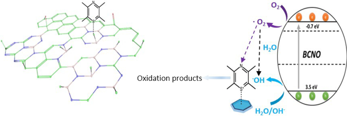
Based on the detailed structural analysis and the proposed structure in Fig. 7c, the highest photocatalytic activity of BMH03 consisted of BCNO-doped graphene oxides [1, 65]. This ternary metal-free photocatalyst is reported to enhance the photocatalytic performances by increasing the charge separation and migration to the reaction site [1]. Additionally, the incorporation of boron into graphene-based materials [11, 20], CN [21, 96], and carbon nanotubes [97] has also exhibited enhanced performance compared to their pristine material without a dopant due to multiple synergistic effects. The large differences in electronegativity between boron, carbon, and nitrogen (2.04 vs. 2.55 vs. 3.04, respectively) yielded a strongly polarized bonding towards C and N atoms. As a result, a local positive charge was formed on boron that turned boron into a strong acidic defect site for preferential adsorption sites of pollutants [98], O2 [97], HOO
−
and OH
−
[99, 100]. Therefore, electron-rich pollutants such as MB (used as a model reaction) were speculated to preferentially adsorbed onto the electropositive B sites of the BMH photocatalyst. The photocatalytic degradation of MB on BCNO was proposed to undergo an indirect dye degradation mechanism [101]. In the indirect photodegradation mechanism, the photogenerated holes on the surface of BCNO produced highly oxidative hydroxyl radicals and attack the  bond of MB. Meanwhile, photogenerated electrons from the conduction band of BCNO formed highly reducing superoxide radical anion O2
−
species that could directly attack MB. Figure 8 illustrates the proposed mechanism of BCNO in photocatalyzing the degradation of MB
bond of MB. Meanwhile, photogenerated electrons from the conduction band of BCNO formed highly reducing superoxide radical anion O2
−
species that could directly attack MB. Figure 8 illustrates the proposed mechanism of BCNO in photocatalyzing the degradation of MB

Schematic illustration of the proposed mechanism of BCNO in catalyzing the degradation of MB upon light irradiation. The positively charged MB was selectively absorbed on the N2 B-OH sites. The band structures of BMH03 were determined via UPS and Tauc plot (Fig. 6 and Additional file 1:Fig. S17). The redox potentials of O2 /O2 = − 0.13 eV vs ENHE and H2 O/OH = 2.72 eV vs. ENHE are given in black dashed lines as reference
According to the XPS and solid-state NMR analyses, BMH03 possessed a higher composition of graphitic sp 2 C = C with a simultaneous reduction in tetracoordinate B site compared to BMH01 (Additional file 1:Fig. S9 and Table S4) The high composition of tetracoordinate B-sites in BMH01 (33%) also translated to a reduction in the number of boron Lewis acid sites serving as the active catalytic center, which explains the trend of photocatalytic activities among the BMH series [13]. However, the BGH series comprised the C, and O-doped h-BN domain with various tri- and tetracoordinate boron sites. Interestingly, BGH01 annealed at a lower temperature (Table 1, BGH01LT) was found to be inactive but exhibited a high absorbing ability in removing dye from the solution (Additional file 1:Fig. S1 for TEM morphology; Fig. S16 for photodegradation MB UV–vis absorbance).
The previous report also concluded that BCNO was photocatalytically inactive towards dye degradation but exhibited an excellent absorbing ability in removing dye from the solution [102]. The inactive BGH01LT was comprised primarily of tetracoordinate B sites and boron oxides-related bonding. Based on the SOLA analysis and the literature , the tricoordinate boron site with a δ iso of 16.7 ppm was related to boroxol rings (B2 O3 ) [54, 56]. Our results are also supported by other works on the catalytic activation of peroxymonosulfate using amorphous boron [22]. However, the photocatalytic active BGH01, BMH01, and BMH03 possessed a high composition of BN(OH)2 and BN2 OH bonding sites. Our investigation suggested that the photocatalytic activity of BCNO is highly dependent on the local structure of the boron site. While the exact catalytic site and mechanism are yet to be explored, we proposed that BNx (OH)3−x served as one of the BGH and BMH series catalytic sites. For the BMH series, the formation of tetracoordinate B-O sites was detrimental in catalyzing dye degradation due to the reduction in the Lewis acid site. At the same time, the increasing composition of the sp 2 C = C graphitic domain enhanced the photocatalytic activities of the BGH and BMH series.
In summary, BCNO structures and their photocatalytic activities have been presented here and found to be highly dependent on the choice of precursor, precursor ratios, annealing temperatures, and times. In this study, two types of distinctly different BCNO nanostructures were prepared via low-temperature annealing (600 °C–800 °C):(1) BCNO-doped boron nitride and (2) BCNO-doped graphene oxides. Through systematic investigation of using two different nitrogen precursors, crystalline BCNO with a quasi-spherical shape was prepared at 800 °C for 12 hr using guanidine hydrochloride as the nitrogen source. This series of BCNO exhibited moderate photocatalytic activity through the emergence of BN2 (OH) or BN(OH)2 tricoordinate boron serving as the Lewis acidic site. However, BCNO prepared using melamine as the nitrogen source at 600 °C yielded multi-layered sheets with ill-defined shapes. These BCNO-doped graphene oxides layered structures exhibited the highest photocatalytic activity, surpassing the state-of-art metal-free photocatalyst, CN. For the melamine-derived BCNO layered structures, the presence of tricoordinate boron species as BN2 (OH) or BN(OH)2 and a higher composition of graphitic sp 2 C = C were speculated to play an important role in promoting their photocatalytic activity. This study demonstrates the potential of BCNO as a photocatalyst for energy conversion and environmental remediation applications. Further structural optimization on this new B-C-N–O photocatalyst system is expected to facilitate the development of a sustainable catalyst for applications including solar hydrogen fuel production, environmental remediation, electrocatalytic oxygen reduction reaction, and catalytic oxidative dehydrogenation reaction.
All data are fully available without restriction.
Boron carbon oxynitride
Carbon nitride
Hexagonal boron nitride
Time-resolved photoluminescence
Magic angle spin
Cross polarization
Solid line shape analysis
Intensity
Attenuated total reflectance
나노물질
철강의 탈산 제강 공정은 산화 분위기에서 수행되는 뜨거운 금속을 강으로 정련하는 것으로 구성됩니다. 정제 과정에서 산소는 강철에 용해됩니다. 다음은 강철의 주요 산소 공급원입니다. 산소 분사 제강 공정 중 산화 슬래그 및 철광석 사용 티밍 작업 중 액강에 의한 대기 중 산소 채취 안감의 산화 내화물 녹슬고 젖은 스크랩. 탈산은 제강의 마지막 단계입니다. 제강 중 태핑 시의 강욕은 400~800ppm의 활성산소를 함유하고 있다. 태핑하는 동안 적절한 양의 철 합금 또는 기타 특수 탈산제를 가득 찬 국자에 첨가하여 탈산을
다양한 화학 성분이 강철에 미치는 영향 알아보기 철강은 항공우주에서 주방용품에 이르기까지 모든 종류의 제품에서 발견됩니다. 이러한 다양한 응용 분야에는 다용도 재료가 필요하며 강철도 적합합니다. 강철은 실제로 수백 가지 용도별 등급이 있는 전체 금속 합금 제품군을 설명하지만 대부분의 사람들은 강철을 탄소강과 스테인리스강의 두 가지 광범위한 범주로 이해합니다. 탄소강과 스테인리스강은 철과 탄소의 기본 성분이 동일합니다. 주요 차이점은 합금 함량입니다. 탄소강은 10.5% 미만의 합금 함량을 갖는 반면 스테인리스강은 10.5%