

나노물질

산업 제조
우리는 최근 몇 년 동안 그래핀 계열 재료의 바이오 응용에 대한 연구에서 풍부한 돌파구를 목격했습니다. 나노 크기, 큰 비표면적, 광발광 특성 및 항균 활성으로 인해 그래핀 계열 물질은 뼈 조직 공학, 약물/유전자 전달 및 생물학적 감지/영상 응용 분야에서 엄청난 잠재력을 가지고 있습니다. 이 리뷰에서 우리는 그래핀 연구의 최근 진행 상황과 성과를 회고하고 뼈 조직 재생을 위한 그래핀 계열 물질의 다양한 생물 의학적 응용 가능성과 생물학적 안전성을 비판적으로 분석하고 논의합니다.
심각한 악안면 감염, 외상, 종양 및 턱뼈 결함을 앓고 있는 선천적 기형의 희생자는 일반적으로 장기간의 회복이 필요합니다. 다른 많은 조직과 달리 뼈는 손상되었을 때 재생하는 뛰어난 능력을 가지고 있습니다[1, 2]. 그러나 인간 골격의 제한된 자가 재생 능력은 충분히 크거나 임계 크기의 골 결손의 재건을 임상 치료에 있어 중요한 도전 과제로 만듭니다[3]. 어떤 경우에는 심한 환자는 광범위한 뼈 확대 수술이 필요합니다. 현재 골 재생을 위한 치료법은 자가이식, 동종이식, 이종이식으로 구성된다[4]. 자가골은 면역원성 없이 골전도, 골유도 및 골형성 기능을 가진 "골드 스탠다드" 골 이식 재료로 간주됩니다. 그러나 자가이식을 아직 임상에서 사용하기 어려운 이유는 기증자 감염의 위험성과 회복기간이 길기 때문이다[5]. 다른 사람에게서 얻은 동종이식편은 종종 차선책으로 간주됩니다. 그러나 동종이식편의 사용은 감염 및 면역 거부반응의 위험이 급격히 증가하는 등 잠재적인 위험이 있습니다[4, 6]. 산-소화된 탈회골 기질 및 소 콜라겐과 같은 이종이식 재료는 쉽게 입수 및 제조됩니다. 이제 이종이식은 임상 실습의 주요 접근 방식입니다. 그러나 골유도 능력이 낮습니다[7]. 현재 뼈와 비교하여 생물학적 또는 기계적 특성이 우수하거나 심지어 동일한 이종 또는 합성 뼈 대체물이 없습니다[5]. 이러한 치료법은 유용한 것으로 입증되었지만 고유한 문제를 안고 있습니다. 따라서 적절한 뼈 재생 요법에 대한 연구와 개발이 여전히 필요합니다. 분명히, 뼈 조직 공학 및 재생 의학 연구는 결과를 개선하고 뼈 결함이 있는 환자의 빠른 회복을 위한 길을 열어줍니다[8]. 조직 공학 뼈 구조는 뼈 치유를 증가시키기 위한 적절한 자가 이식 및 동종이식 재료의 부족으로 인해 발생하는 수요를 완화할 가능성이 있습니다[9]. 골 결손 부위의 골 부피를 증가시키기 위해 스캐폴드[1, 6], 코팅[10], 유도 골 재생(GBR)을 위한 차단막[11, 12]을 포함한 다양한 골 재생 방법이 개발되었습니다.
현재, 그래핀 계열 물질의 잠재력은 다양한 유형의 줄기 세포를 신경성[13,14,15], 연골성[16, 17], 근성[18]으로 분화하기 위한 2D 평면 코팅 또는 3D 다공성 지지체로서 엄청난 주목을 받고 있습니다. , 지방 생성 [19] 및 골 생성 계통 [20, 21]. 따라서 그래핀 계열 물질은 다음 뼈 재생 물질로 선택될 가능성이 더 높습니다. 단일 또는 소수의 sp 2 레이어로 정의되는 그래핀 -혼성화된 탄소 원자는 2004년 Novoselov와 Geim에 의해 흑연에서 처음으로 분리되었습니다[22]. 연구 관심이 증가함에 따라 그래핀 산화물(GO), 카르복실 그래핀(CXYG), 환원 그래핀 산화물(rGO), 그래핀 양자점(GQD)을 포함한 그래핀 계열의 재료에 대한 연구가 광범위하게 이루어지고 있습니다. 그래핀은 기계적, 전도성, 열적, 광학적 특성이 매우 뛰어나 [23,24,25] 전자, 생명 공학 및 고분자 과학 [26]에 널리 적용되었습니다. 전도도가 유망한 전도성 물질은 세포 활동을 강화하고 뼈 조직 복구를 자극하여[27, 28] 항균 활성도 잘 나타내는 것으로 알려져 있습니다[29]. 산화 그래핀(GO)과 카르복실 그래핀(CXYG)은 모두 그래핀의 유도체입니다. 산소화된 작용기(에폭사이드, 카복실 및 하이드록실 그룹)의 존재로 인해 GO 및 CXYG는 친수성 용매에서 더 잘 분산되며 이는 생물의학 응용에 필수적입니다[30, 31]. 환원그래핀옥사이드(rGO)는 특정 조건에서 특정 환원제로 GO를 환원시켜 합성할 수 있습니다. 일부 특수한 π-π 화학적 상호작용의 감소 덕분에 rGO는 그래핀 및 GO보다 특정 물리적 및 화학적 특성이 더 우수합니다[32, 33]. 그래핀 양자점(GQD)의 원료는 GO이다. GQD는 강한 양자 구속과 광발광 특성을 가지고 있습니다[34]. GQD의 강한 형광성으로 인해 세포 이미징에 유용합니다. 그래핀 계열 물질의 우수한 특성으로 인해 약물/유전자 전달, 생물학적 감지/영상 응용 및 조직 공학에 대한 엄청난 잠재력을 가지고 있습니다[35,36,37,38,39]. 그러나 그래핀 계열 물질의 세포 골 형성 분화를 유도하는 장기적인 생물학적 안전성과 능력에 대한 도전은 여전히 존재합니다. 여기에서 우리는 그래핀 및 그 파생물의 최근 진행 상황과 성과를 종합적으로 검토합니다. 동시에, 우리는 시험관 내 및 생체 내 생체 안전성을 비판적으로 분석하고 뼈 조직 재생을 위한 그래핀 계열 물질의 다양한 생물 의학적 응용 가능성에 대해 논의합니다.
그래핀 계열 물질이 임상 시험을 위해 고려되기 전에 세포 독성 및 생체 적합성에 의해 엄격하게 평가되어야 합니다[38]. “그래핀은 생체적합성 물질인가?” 대답은 여전히 논란의 여지가 있습니다. 기능화가 없는 원시 그래핀은 소수성이며 수성 매질에서 쉽게 응집됩니다[34, 40]. 소수성 표면에서 비특이적 단백질의 조밀한 층이 표면에서 물을 대체하고 물질에 즉시 축적되어 나노입자의 면역학적 인식을 초래할 수 있습니다[41]. 따라서 산화, 환원 및 작용기의 도입을 포함한 화학적 기능화는 그래핀의 친수성을 증가시키는 생물 의학 응용 분야에 사용되는 그래핀의 전제 조건입니다. 서로 다른 화학적 성질을 가진 서로 다른 기능을 가진 그래핀 계열 물질은 서로 다른 독성을 나타냅니다[13]. Soumen et al. rGO가 GO보다 독성이 덜하다는 것을 발견했습니다. rGO 표면의 산소 작용기 밀도가 증가함에 따라 산화 스트레스가 증가한다는 사실을 확인하는 것은 흥미로웠습니다. 그들은 GO 시트의 작용기 밀도가 세포 세포 독성을 매개하는 핵심 요소 중 하나라고 결론지었습니다[31]. 표면 기능화 외에도 그래핀 계열 물질의 세포 독성은 농도, 크기 및 모양을 포함한 수많은 요인의 영향을 받았습니다[42].
첫째, 일부 연구에서는 그래핀 계열 물질이 시간 의존적 세포독성과 함께 또는 없이 용량 의존적 세포독성을 갖는다는 것이 입증되었습니다. 예를 들어 Chang et al. 고농도의 GO(≥ 50μg/mL)에서 세포 생존력의 약간의 손실이 관찰되었으며 GO는 세포 내 축적을 유도하고 폐암 상피 세포주에서 용량 의존적 산화 스트레스를 유발할 수 있다고 보고했습니다(A549) [43]. Wei et al. 원시 GO는 10μg/mL의 높은 농도에서 골 중간엽 줄기 세포(BMSC)의 증식을 억제하는 반면 0.1μg/mL의 낮은 농도에서 BMSC의 증식을 향상시키는 것으로 나타났습니다[44]. 유사하게, 감소된 세포 수는 200μg/mL의 GO에서 분명히 관찰되었으며 더 큰 세포 독성 효과는 300μg/mL의 GO에서 보고되었습니다[45]. 게다가 Kim et al. 골아세포(MC3T3-E1) 생존력이 <62.5μg/mL 농도에서 rGO에 의해 약간 영향을 받았지만 유의하게(p <0.05) 더 높은 농도(≥ 100μg/mL)에서 감소했습니다[23]. 또한, CXYG, GQD 모두 낮은 농도에서 적용될 때 세포독성 가능성이 거의 나타나지 않았습니다[34, 46]. 간단히 말해서, 그래핀 계열 물질은 세포 형태, 생존율 및 증식에 거의 부정적인 영향을 미치지 않고 낮은 농도에서 세포 적합성이지만 농도가 단일 관련 요소는 아닙니다.
둘째, 층, 나노시트 및 플레이크, 리본 및 점과 같은 다양한 모양이 그래핀 계열의 세포독성 복잡성에 기여하는 것으로 나타났습니다[40]. Talukdar et al. 그래핀 나노-양파(GNO), GO 나노리본(GONR) 및 GO 나노혈소판(GONP)의 세포 독성을 평가했습니다. CD50 값은 GNOs> GONRs> GONPs 경향을 따랐으며, 이는 GONR이 GONP에 비해 더 세포독성이 있음을 나타냅니다[47]. 따라서 그래핀 계열 나노물질의 모양은 세포독성을 매개하는 핵심 요소이기도 하다. 예를 들어, 그래핀과 다중벽 탄소나노튜브(MWNT)는 모양이 다르지만(그래핀의 경우 평평한 원자 시트 및 나노튜브의 경우 관형) 화학적 조성과 결정 구조는 유사합니다. GO는 50μg/mL가 될 때까지 SK-N-SH 세포의 세포 성장 억제 활성을 나타내지 않았습니다. 이에 비해 MWCNT는 낮은 농도(6.7μg/mL)에서 세포의 증식을 억제하여 급성 세포독성을 나타냅니다. HeLa 세포의 경우 GO는 최대 50μg/mL 농도에서도 약간의 성장 억제 활성을 나타내는 반면 MWCNT는 HeLa 세포에서 중간 정도의 세포 독성을 나타냅니다[48]. 그들은 이 현상을 다른 모양과 다양한 물리적/화학적 방식에 의존했습니다. 그래핀 계열 물질은 평평한 모양으로 인해 세포막과 약간의 상호 작용이 있을 것으로 예상되었습니다. MWCNT의 관 모양은 막의 침투를 촉진하여 세포 독성을 유발합니다[48,49,50]. 또 다른 중요한 정보는 나노구조 그래핀 유도체의 세포독성이 기능화, 농도, 크기 및 모양의 의존성 외에도 세포 유형에 의존적이라는 것입니다. 신경 세포주로서 SK-N-SH 세포는 나노구조 그래핀 유도체의 역효과에 대해 HeLa 세포보다 더 민감함을 나타냈다[48].
셋째, 크기도 그래핀 계열 물질의 생체 안전성에 중요한 역할을 한다. Yunet al. 세포 기반 전기 화학적 임피던스 바이오 센서를 통해 그래 핀 나노 플레이크의 크기 의존적 세포 독성 효과를 평가했습니다. 그들은 더 작은 그래핀 나노플레이크(30.9 ± 5.4 nm)가 세포에 의한 더 높은 흡수로 인해 세포자멸사를 유도하는 반면, 세포막에 대부분 응집된 더 큰 그래핀 나노플레이크(80.9 ± 5.5 nm)는 독성을 덜 유발한다는 것을 발견했습니다[51]. 나노 물질의 세포 흡수 특성이 세포 증식, 분화 및 나노 입자 배설에 영향을 줄 수 있다는 것은 잘 알려져 있습니다[52]. Mu et al. 단백질로 코팅된 GO 나노시트의 가능한 크기 의존적 흡수 메커니즘을 자세히 설명하고 더 큰 나노시트(860 ± 370 nm)가 먼저 세포 표면에 부착된 다음 막 함입, 가성족(pseudopodia)의 확장 및 마지막으로 주로 식균 작용을 통해 세포에 들어가는 반면 더 작은 나노시트( 420 ± 260 nm)는 주로 클라트린 매개 세포내이입을 통해 세포에 들어갔다[33]. Das et al. 10μg/mL의 GO 및 rGO에 인간 제대 정맥 내피 세포(HUVEC)를 다양한 크기의 시트(800nm 및 400nm)로 시드합니다. 결과는 MTT 분석에서 더 작은 크기의 시트가 더 큰 시트보다 더 독성이 있음을 보여주었습니다. 그런 다음 더 큰 크기의 GO와 rGO(800nm)를 초음파 처리하여 더 작은 크기(70nm)로 나누었습니다. 초음파 처리 후 증가된 세포 독성이 관찰되었으며, 이는 더 작은 크기의 GO 및 rGO가 더 많은 독성을 나타냄을 나타냅니다[31]. 유사하게, MCF7 세포는 4가지 크기의 GO 샘플(744 ± 178nm, 323 ± 50nm, 201 ± 28nm, 100 ± 10nm)에 노출되었습니다. 처리되지 않은 세포와 비교하여 더 큰 크기의 GO 분산액(744 ± 178nm)에 72시간 노출된 후에도 시험관 내에서 세포 독성이 관찰되지 않은 반면, 100 ± 10nm 크기의 GO 분산액으로 처리하면 세포 증식이 감소했습니다. 처리되지 않은 세포의 약 50%까지 [53]. 위의 결과로부터 30nm에서 860nm까지 다양한 크기의 그래핀 계열 물질이 연구되었습니다. 그리고 우리는 더 작은 크기의 그래핀 계열 물질이 더 큰 크기보다 더 독성이 있다는 결론을 얻은 것 같습니다. 그러나 다른 팀은 그래핀과 그 파생물의 크기 규모를 정의하는 기준이 다릅니다. 따라서 이 결론은 논쟁의 여지가 있을 수 있습니다. 한편, 나노 크기의 그래핀 계열 물질은 생물의학 응용에 훨씬 더 안전한 것으로 보고되었다[54]. 그래핀 계열 물질의 크기 조절 합성은 후속 연구에서 신중하게 고려되어야 합니다.
그래핀의 세포독성은 그래핀 계열의 다양성, 화학적 기능화, 농도, 모양 및 크기와 결정적으로 관련이 있다고 결론지었습니다. 앞으로 다양한 유형의 작용기로 그래핀 패밀리를 변형하여 농도와 크기를 더 잘 제어하여 세포, 조직 또는 유기체와 더 잘 상호 작용하는 생체 적합성 장치를 제작하는 것을 목표로 합니다.
그래핀 계열 물질이 생체 적합성 물질인지 여부를 추가로 감지하고 광범위한 응용 분야에서 제안된 사용을 향상시키기 위해서는 생체 내 실험이 필수 방법입니다. 생체 내 그래핀 계열 물질의 생체 적합성 및 생체 분포에 대한 많은 연구는 세포 연구와 거의 일치합니다. Chowdhury et al. zebrafish 배아를 더 큰 크기의 GO 분산액에 적용한 결과 대조군에 비해 배아 사망률이 증가하지 않았으며, 작은 크기의 GO 분산액에서는 배아 생존율이 감소하는 것으로 관찰되었습니다[53]. GO는 배아에서 세포자멸사를 크게 증가시키지 않았지만 MWCNT는 25mg/L의 상대적으로 낮은 농도에서도 발달 중인 배아에서 심각한 형태학적 결함을 초래했습니다[48]. 이 연구는 또한 생체 내 독성이 그래핀과 그 유도체의 크기, 농도 및 모양에 크게 좌우된다는 것을 보여주었습니다. 또한 그래핀 계열 물질은 일반적으로 정맥 주사, 흡입 또는 피하 이식을 통해 동물 모델에 노출됩니다. 따라서 독성, 일반 조직학 및 생체 분포의 변화가 다양합니다. Li et al. 는 정맥 주사를 통해 마우스에서 나노 규모 GO의 독성을 평가하고 GO가 대부분 간, 폐 및 비장에 유지되고 손상, 만성 간염 및 폐 섬유증을 유발한다는 것을 발견했습니다. GO(GO-PEG)의 폴리에틸렌 글리콜(PEG) 코팅은 간, 폐 및 비장에서 GO의 체류를 감소시키고 급성 조직 손상을 완화할 수 있습니다[55]. Duch et al. 그들은 GO가 쥐의 폐에 직접 투여될 때 응집된 그래핀 및 Pluronic-분산 그래핀보다 더 높은 독성을 발견하여 심각하고 지속적인 폐 손상을 유발한다는 것을 발견했기 때문에 폐에서 그래핀 나노물질의 독성 효과를 줄이는 전략을 탐구했습니다. 독성은 액상 박리를 통한 깨끗한 그래핀의 제조에 의해 상당히 완화되었으며 블록 공중합체 Pluronic과 함께 분산될 때 더욱 최소화되었습니다[56]. Zhaet al. 피하 이식 쥐 모델에서 3D 그래핀 폼(GF) 또는 그래핀 산화물 폼(GOF)의 단기(이식 후 첫 2주) 및 장기(7개월) 생체 내 독성과 성능을 확인했습니다. 혈액 분석에 따르면 GF 및 GOF는 이식 후 눈에 띄는 혈액학적, 간 또는 신장 독성을 유발하지 않았으며 이식 후 최소 7개월 후에는 유의한 분해가 관찰되지 않았습니다. 이식 부위에 오랫동안 존재하던 육아종만이 관찰되었다. H 염색 이미지는 생체 내 생체 적합성이 더 우수함을 보여주었습니다(그림 1)[40]. Zha et al. 앞서 언급한 바와 같이 다른 연구보다 더 긍정적인 결과를 얻은 것은 아마도 투여 경로가 다르기 때문일 것입니다. 피하 실험은 이식된 물질의 생체 내 생체 적합성을 평가하는 매우 직접적이고 효과적인 방법이며[57], 생체 내에서 그래핀 계열 나노 물질의 접촉 패턴, 증착 위치, 분해 경로에 영향을 미칠 수 있습니다[58]. 복합 재료의 분해를 제어하는 것은 조직 공학에서 매우 중요합니다.
<그림>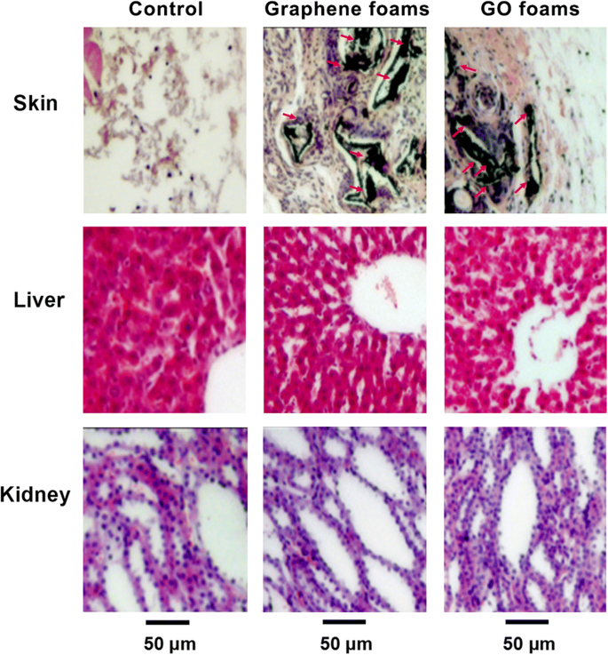
이식 후 14일째에 그래핀 발포체, GO 발포체 또는 아무것도 이식되지 않은 주요 장기(쥐에서 수집한 이식 부위, 간 및 신장)의 대표적인 HE 염색 이미지. 명백한 장기 손상이나 병변은 관찰되지 않았습니다. ref에서 재생산. [40] Journal of Nanoparticle Research의 허가를 받아
일반적으로 세포 연구는 예비 세포 독성 분석, 세포와의 상호 작용 메커니즘 이해에 탁월합니다. 그러나 생체 내에서는 훨씬 더 복잡한 미세 환경입니다. 습한 부식성 미세 환경에서 그래핀 계열 물질이 어떻게 작용하는지 이해하는 것도 중요합니다. 그래핀 계열 물질의 생체 적합성은 농도, 작용기의 종류, 그래핀 계열의 유형, 크기 및 모양과 밀접한 관련이 있습니다. 그러나 메커니즘은 여전히 자세히 자세히 조사해야합니다. 그러나 생체 내 생체 안전성에 대한 평가는 상대적으로 많지 않으며 특히 장기간 생체 적합성 및 생체 분포에 더주의를 기울여야합니다. 일부 논문에서 바이오 안전성에 대한 우려가 제기되고 있지만, 그래핀 제품군이 고유하게 제공하는 잠재적인 다용성으로 인해 바이오메디컬 애플리케이션을 위한 경쟁력 있는 후보가 되었습니다.
뼈 재형성과 새로운 뼈 형성은 뼈 결손의 살균된 미세 환경 없이는 완전히 성공할 수 없습니다. 사실 감염성 골결손의 치료는 여전히 주요 과제입니다[59]. 큰 골 결손과 감염 문제로 인해 치료가 어렵고 환자는 장기간의 회복 기간이 필요합니다. 따라서 그래핀 계열 물질의 박테리아 억제 능력은 많은 도움이 됩니다. 그래핀 계열 물질은 항균력이 있는 것으로 여겨집니다(표 1). Liu et al. (1) 그래핀 기반 물질의 초기 세포 침착, (2) 날카로운 나노시트와의 직접 접촉으로 인한 막 응력, (3) 이어지는 슈퍼옥사이드 음이온 비의존 산화를 포함하는 3단계 항균 메커니즘을 제안했습니다[60]. 그러나 Mangadlao et al. 그래핀의 표면은 가장자리가 아니라 주로 항균 활성을 담당한다고 생각했습니다. 박테리아와 접촉할 때 그래핀은 독립적인 산화 스트레스를 생성하는 박테리아 막에서 전자를 펌핑하는 전자 수용체 역할을 합니다[61]. 한편, Li et al. 그래핀 필름의 항균 작용에 대한 더 나은 이해를 위한 새로운 통찰력을 제공했습니다. 그들은 그래핀 계열 물질의 항균 활성이 활성산소(ROS) 매개 손상에 기인한 것이 아니라 미생물막에서 그래핀으로의 전자 전달 상호작용에 의한 것이라는 의견을 견지하고[62], Panda et al. 비산화성 전자 전달 메커니즘의 시너지 효과와 그에 따른 박테리아에 대한 ROS 매개 산화 스트레스가 천연 유래 GO 금속 필름의 향상된 항균 활성을 유도함을 입증했습니다[63].
그래핀 기반 시트의 물리화학적 특성이 항균 활성에 어떻게 영향을 미치는지는 아직 불확실하지만 그래핀 계열 물질의 항균 능력은 우리가 연구하고 더 활용할 가치가 있습니다.
많은 학자들은 그래핀이 세포(예:치수 줄기 세포[64, 65], 골수 줄기 세포[8, 20, 66, 67], 치주 인대 줄기 세포[68])의 부착 및 증식을 허용할 뿐만 아니라 ], 인간 조골세포[69], 섬유아세포 세포[70], 종양 세포[43]) 명백한 세포독성의 징후가 없지만 초기 세포 조골 분화를 유도하고 높은 정도의 광물화를 생성할 수 있습니다[20, 64,65,66,67, 68]. 현재 수많은 팀에서 그래핀 계열 나노물질을 지지체 또는 지지체에 첨가제로, 기질 물질 표면에 코팅으로, 가이드 뼈 재생막 및 약물 전달 수단으로 적용하는 새로운 전략을 설계하기 위해 많은 연구를 했습니다. (그림 2). 그들은 그래핀 계열 물질을 사용하여 기질 물질의 특정 특성을 더욱 개선하고 기질 기반 복합 재료에 생리 활성 특성을 부여하려고 했습니다.
<그림>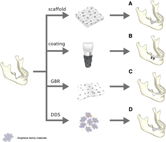
A. 뼈 재생을 위한 지지체 또는 지지체의 보강재로서의 그래핀 계열 재료. B. 골 재생을 위해 기판에 전사된 코팅으로서의 그래핀 계열 물질. C. 유도 골막의 첨가제로서의 그래핀 계열. D. 뼈 재생을 촉진하는 약물 전달 시스템으로서의 그래핀 계열 물질
뼈 조직 공학을 위한 가장 일반적인 전략은 뼈 재형성 및 재생의 자연적 과정을 시뮬레이션하는 것입니다. 이 전략은 3차원(3D) 생체 적합성, 생분해성, 골전도성 또는 골유도성 지지체에 의해 충족될 수 있습니다[3]. 이러한 종류의 스캐폴드는 골형성 세포 부착, 이동, 증식 및 분화뿐만 아니라 성장 인자의 운반체를 위한 세포외 기질(ECM)을 모방하는 이상적인 미세 환경을 제공할 수 있습니다[6]. 유망한 생체 적합성 지지체로서 그래핀은 세포 확산 및 기질에서 골 형성 분화에 사용할 수 있는 큰 표면적을 만들 수 있습니다[20]. 예를 들어, 인간 중간엽 줄기 세포(hMSC)의 배양 기질로 사용된 3D 그래핀 폼은 줄기 세포 생존력을 유지하고 골형성 분화를 촉진할 수 있다는 증거를 제공했습니다[66]. 또한, 3D 그래핀(3DGp) 지지체와 2D 그래핀(2DGp) 코팅은 더 높은 수준의 광물화 및 상향 조절된 뼈 관련 유전자 및 화학 인덕터를 사용하거나 사용하지 않고 그래핀의 단백질 [68].
요즘은 발판 역할을 하는 다양한 생체재료들이 버섯처럼 솟아나고 있다. 뼈 재생에 사용하기에 잠재적으로 적합한 합성 지지체에는 수산화인회석(HA)[71]과 같은 인산칼슘; β-인산삼칼슘(β-TCP)[72]; 폴리락트산(PLA)[73], 폴리글리콜산(PLGA)[74], 폴리카프로락톤(PCL)[75], 키토산(CS)[1], 콜라겐[76]과 같은 합성 또는 바이오 폴리머 ]; 및 위에서 언급한 재료의 합성물 [77, 78]. 그러나 이제 가장 중요한 관심사 중 하나는 비계의 기계적 특성입니다. 천연 뼈는 7-27GPa 범위의 영률 값을 갖는 초탄성 생체 역학적 특성을 나타내기 때문에 이상적인 지지체는 자연 뼈의 강도, 강성 및 기계적 거동을 모방해야 합니다. 그래핀 계열 재료는 기계적 특성을 강화하고 물리화학적 특성화를 개선하기 위해 스캐폴드에 강화 재료로 추가할 수 있습니다. 예를 들어 순수 PCL 스캐폴드의 인장 강도는 1.61MPa, 연신율은 122%, 영률은 7.01MPa입니다. GO(2%)를 추가하면 인장 강도가 3.50MPa로, 연신율이 131%로, 영률이 15.15MPa로 크게 증가했습니다[80].
그래핀 계열 재료를 강화 재료로 사용한 성공에 자극을 받아 많은 팀이 합성 또는 생체 고분자가 제공하는 생체 적합성과 그래핀 계열 재료의 놀라운 물리적 특성을 결합했습니다. 그들은 새로운 뼈 형성을 지원하고 유도하기 위해 개선된 기계적 특성, 적절한 다공성, 구조적 디자인 및 우수한 생체 적합성을 갖춘 이상적인 복합 지지체를 얻을 것으로 기대했습니다.
인간의 뼈는 30%의 유기물(대부분 콜라겐)과 70%의 무기물(대부분 하이드록시아파타이트(HA; Ca10) (PO4 )6 (OH)2 ) [81, 82]. HA, β-TCP(β-tricalcium phosphate), CPC(calcium phosphate cements)와 같은 합성 인산칼슘계 재료는 뼈의 천연 광물상과 유사한 조성 및 구조와 우수한 골 형성성으로 인해 널리 사용되는 비계 재료입니다. 능력 [83,84,85]. 특히, HA의 우수한 골전도 및 골유도 능력 때문에[86], 골 결손 부위를 수복하기 위한 정형외과 또는 악안면 수술에서 인공 골 이식재로 오랫동안 널리 사용되어 왔다[11, 71]. 그러나 성형 어려움, 독특한 취성 및 낮은 파괴 인성과 같은 HA 재료의 고유한 단점을 개선해야 합니다[87, 88]. 그래핀 계열 재료 강화 HA 복합 재료가 개발되어 파괴 인성과 생물학적 성능이 크게 향상되었다고 보고되었습니다. 예를 들어, HA/그래핀 합성물은 HA 허용 강도를 부여하는 스파크 플라즈마 소결(SPS)에 의해 준비되었습니다[89]. Raucci et al. In situ sol-gel approach와 biomimetic approach의 두 가지 접근 방식으로 HA와 GO를 결합했습니다. in situ sol-gel 접근법으로 얻은 HA-GO는 골 형성 인자를 사용하지 않고 hMSC의 세포 생존 능력을 향상시키고 조골 세포 분화를 유도했습니다. 생체모방 접근법을 통해 형성된 HA-GO는 세포 생존과 증식을 지속시켰다[90]. 또한 환원그래핀옥사이드(rGO)는 HA의 보강재로도 사용될 수 있다. HA-rGO 복합 재료의 파괴 인성은 3.94MPa m 1/2 에 도달했습니다. , 순수 HA에 비해 203% 증가. HA-rGO는 인간 조골 세포의 알칼리성 인산 가수분해 효소(alkaline phosphatase, ALP) 활성에 의해 평가된 세포 증식 및 조골 분화를 향상시켰다[91]. 또한 Nie et al. 자가 조립을 통해 rGO 및 나노 하이드록시아파타이트(nHA) 3D 다공성 복합 지지체(nHA@rGO)를 성공적으로 합성했습니다. 자가 조립 과정을 유도하기 위해 가열된 nHA 물 현탁액과 혼합된 GO 용액. 마지막으로 반응 생성물을 동결 건조하여 3차원 다공성 지지체를 얻었다. nHA@rGO 스캐폴드는 쥐 뼈 중간엽 줄기 세포(rBMSC)의 세포 증식, ALP 활성 및 골형성 유전자 발현을 상당히 촉진할 수 있습니다. 그리고 생체 내 실험에서 20% nHA가 포함된 rGO(nHA@rGO) 다공성 지지체가 토끼의 원형 두개골 결손 치유를 가속화할 수 있음이 밝혀졌습니다[92]. 또한, 이중 성분뿐만 아니라 삼성분도 우수한 세포 적합성 및 향상된 친수성 및 기계적 특성으로 우수한 성능을 보였다[93,94,95].
인산삼칼슘의 유사체인 인산삼칼슘은 뼈회[Ca3라고도 알려진 3차 인산칼슘입니다. (PO4 )2 ]. 그것은 쉽게 흡수될 수 있는 칼슘과 인의 풍부한 출처 역할을 합니다. Beta-tricalcium phosphate(β-TCP)는 생체 적합성이 높으며 결손 부위 내에 재흡수 가능한 연동 네트워크를 생성하여 치유를 촉진합니다[96]. Wu et al. 2D β-TCP-GO 디스크와 3D β-TCP-GO 스캐폴드를 성공적으로 합성했습니다. β-TCP 및 블랭크 대조군과 비교하여, 2D β-TCP-GO 디스크는 Wnt 관련 신호 전달 경로를 활성화하여 hBMSC의 증식, ALP 활성 및 골형성 유전자 발현을 유의하게 향상시켰으며, 이는 GO-의 우수한 시험관내 골자극 특성을 나타냅니다. 변형된 β-TCP[85]. Wnt 표준 신호 전달 경로는 세포 증식, 분화 및 형태 형성과 같은 세포 활동을 조절하는 데 중요한 역할을 하는 것으로 알려져 있습니다[97, 98]. 생체 내 연구는 3D β-TCP-GO 스캐폴드가 순수한 TCP 스캐폴드보다 두개골 결손에서 더 큰 새로운 뼈 형성을 갖는 것으로 나타났습니다(그림 3)[85]. GO-Cu 나노복합체 지지체(CPC/GO-Cu)가 통합된 새로운 스캐폴드, 인산칼슘 시멘트는 rBMSC의 접착 및 골형성 분화를 촉진했으며, 이는 Erk1/2를 활성화하여 rBMSC에서 Hif-1α의 발현을 상향조절할 수 있음을 확인했습니다. 신호전달 경로와 혈관내피성장인자(VEGF)와 BMP-2 단백질의 분비를 유도한다. 또한, CPC/GO-Cu 스캐폴드를 임계 크기의 두개골 결손이 있는 쥐에 이식한 결과, 스캐폴드(CPC/GO-Cu)가 결손 부위의 혈관신생 및 골형성을 유의하게 촉진하는 것으로 나타났습니다[99].
<그림>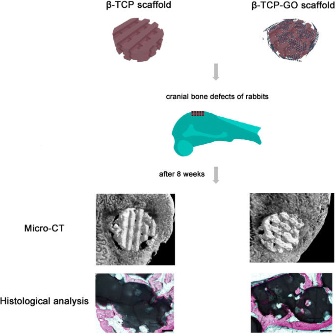
β-TCP 및 β-TCP-GO 스캐폴드에 대한 계획 설명은 생체 내 골형성을 자극했습니다. 8주 동안 토끼의 두개골 결함에 이식한 후 β-TCP 및 β-TCP-GO 지지체에 대한 생체 내 골 형성 능력의 마이크로 CT 분석 및 조직학적 분석. ref에서 재생산. [85] Journal of Carbon의 허가를 받아
Chitosan (CS), a highly versatile biopolymer, derived from the shells of crustaceans [1, 87], has a hydrophilic surface that promotes cell adhesion and proliferation and its degradation products are nontoxic. Chitosan is biocompatible, osteoconductive, hemostatic, and can be easily converted into the desired shapes [2]. Besides, chitosan can promote bone matrix of mineralization [1] and minimize the inflammatory response after implantation [100]. All properties above make chitosan especially attractive as a bone scaffold material. But the most challenging part is the obtainment of CS-based scaffolds with good mechanical properties and processability [101]. Interestingly, CS/GO scaffolds have high water-retention ability, porosity, and hydrophilic nature [101, 102]. The CS-based 3D materials were enriched with GO in different proportions (0.5 wt% and 3 wt%). The new developed CS/GO 3 wt% scaffold was expected to be ideally designed for bone tissue engineering applications in terms of biocompatibility and properties to promote cell growth and proliferation [103]. Another CHT/GO scaffold with 0, 0.5, and 3 wt.% GO were prepared by freeze-drying method. Similarly, the CS/GO 3 wt% scaffolds significantly enhanced the ALP activity in vitro and the new bone formation in vivo, suggesting a positive contribution of 3 wt% GO to the efficiency of osteogenic differentiation process (Fig. 4) [3]. All results proved that CS/GO scaffolds could be a feasible tool for the regeneration of bone defects, and the addition of a 3 wt% of GO to material composition could have a better impact on cell osteogenic differentiation.

아 ALP activity in mice calvaria defects implanted with CHT/GO and b histomophometric analysis of Masson Goldner trichrome-stained sections. ###p < 0.001 vs CHT; **p < 0.01 vs control; ***p < 0.001 vs control. Reproduced from ref. [3] with permission from the Journal of Scientific Reports
Moreover, some tricomponent composites, such as CS, GO, and HA can release more Ca and P ions compared to the pure HA nanoparticles, displaying a high bioactivity of the composite scaffold [87]. Ravichandran et al. fabricated a unique composite scaffold, GO–CS–HA scaffold, and the incorporation of GO enhanced the tensile strength of CS up to 8.2 MPa and CS–HA to 10 MPa. And the results demonstrated that GO–CS–HA scaffolds facilitated cell adhesion and proliferation, meanwhile showed improved osteogenesis in in vitro tests [2]. Another tricomponent composite scaffold, containing CS, gelatin (Gn), and different concentrates of graphene oxide (0.1%, 0.25%, 0.5%, and 1% (w /v ) GO) showed better physic-chemical properties than CS/Gn scaffolds. The addition of GO at the concentration of 0.25% to CS/Gn scaffolds exhibited enhanced absorption of proteins, extensive apatite deposition. The 0.25% GO/CS/Gn scaffolds were cyto-friendly to rat osteoprogenitor cells, and they enhanced differentiation of mouse mesenchymal stem cells into osteoblasts in vitro (Fig. 5). The tibial bone defect filled with 0.25% GO/CS/Gn scaffolds showed the growth of new bone and bridging the defect area, indicating their biocompatible and osteogenic nature [104]. Thus, no matter bicomponent or tricomponent composites scaffolds, the addition of graphene family materials to chitosan can favorably improve the mechanical properties and regulate the biological response of osteoblasts, promoting osteogenic differentiation.

아 MTT assay after incubation of CS/Gn scaffolds and 0.25% GO/CS/Gn scaffolds with media for 48 h. The asterisk indicates a significant increase versus control, and the pound sign indicates a significant decrease versus control (p <0.05). ㄴ , ㄷ Expression of osteogenic-related genes (RUNX2, ALP, COL-1, and OC) in mMSCs cultured on CS/Gn scaffolds and 0.25% GO/CS/Gn scaffolds for 7 and 14 days measured by quantitative RT-PCR. Reproduced from ref. [104] with permission from the Journal of International Journal of Biological Macromolecules
Sponge scaffolds of type I collagen, the major organic component of bone [81], have been clinically applied as scaffolds to regenerate bone tissue [105, 106]. Because collagen scaffolds (elastic moduli:14.6 ± 2.8 kPa) are relatively soft, the combination with GO is expected to enhance the elastic modulus of collagen scaffolds and to improve the osteogenic differentiation of MSCs for bone regeneration. The covalent conjugation of GO flakes to 3D collagen scaffolds (elastic moduli:38.7 ± 2.8 kPa) increased the scaffold stiffness by threefold and did not negatively affect the viability of BMSCs. The enhanced osteogenic differentiation observed on the stiffer scaffolds were likely mediated by BMSCs mechanosensing because the molecules involved in cell adhesion to stiff substrates were either upregulated or activated [107]. Moreover, the development of new biomaterials utilizing graphene family materials with high osteogenic capacity is urgently pursued (Table 2).
Up to now, these improved tricomponent systems for bone tissue engineering scaffolds possess good biocompatibility, which can promote cell attachment, proliferation, and have been reported mechanical properties matchable to those of natural bone. But the response to specific biological signals expressing, as well as the capabilities of enhancing cell differentiation and finally bone tissue regeneration, still needs to be explored further. Moreover, it has been reported that the pore structure (pore size, pore morphology, and pore orientation) and the elasticity of scaffolds were manipulated to regulate osteogenesis [108,109,110]. However, due to the complicated structure of porous and different elasticity accurately controlled of the scaffolds, it remains a major challenge to individually design specific pore architectures and elasticity 3D porous scaffolds that can stimulate bone regeneration. With the rapidly development of the science and technology, the emerging of the 3D-printing method may overcome this problem and open an avenue for bone tissue regeneration [85]. The in vitro bioactivity and excellent in vivo bone-forming ability of graphene family nanomaterials present a new prospect of developing a broad new type of multifunctional scaffolds for biomedical applications. Thus, we believe that the unraveled the molecular mechanisms behind will be revealed soon and graphene family materials still have attractive potential of applications in bone regeneration waiting us to explore.
Graphene family materials have been widely applied in diverse forms of medical applications for bone regeneration. As a coating, graphene family materials can be transferred on two dimensional (2D) flat non-metal or metal substrates to induce spontaneous osteogenic differentiation of several types of mesenchymal stem cells (MSCs) [64]. Nayak et al. transferred graphene to four 2D non-metal substrates (polydimethylsiloxane (PDMS), polyethylene terephthalate (PET), glass slide, and silicon wafer with 300 nm SiO2 (Si/SiO2 ).) and investigated the influence of graphene on BMSCs differentiation. They summarized that the graphene coating was cytocompatible and contributed to enhance the osteogenic differentiation of BMSCs at a rate comparable to differentiation under the influence of BMP-2 in the osteogenic medium [20]. Similarly, Elkhenany et al. found that goat BMSCs, seeded on 2D graphene-coated plates underwent osteoblastic differentiation in culture medium without the addition of any specific growth factors [8]. Simultaneously, Lee et al. tried to explain the origin of how graphene coating could accelerate stem cell renewal and differentiation. They deemed that the strong noncovalent binding abilities of graphene allowed it to serve as a preconcentration platform for osteoblastic inducers, which facilitated BMSCs osteogenic differentiation [67]. The capability of graphene in modulating osteogenic differentiation is evident. How about its derivatives? GO coatings and rGO coatings all showed favorable cytocompatibility and enhanced spontaneous osteogenic differentiation by upregulating levels of ALP activity [111, 112].
Since titanium (Ti) and medical-grade Ti alloy have been extendedly applied in the orthopedic and dental fields [113,114,115], satisfactory osseointegration for titanium and its alloys is still a major challenge and need to be explored deeply in order to help the clinicians to promote the success or survive rate of implants and diminish the likely complications encountered after their placement [114, 116, 117]. Graphene family materials coated titanium and its alloys, serving as a new method to improve their capabilities of osseointegration at the tissue-implant interface, attracted widespread attention. For example, GO-coated titanium enhanced cell proliferation, upregulated levels of ALP activity and gene expression level of osteogenesis-related markers, and promoted the protein expression of BSP, Runx2, and OCN [117]. Qiu et al. made different thickness GO coatings on the pure titanium surfaces respectively by cathodal electrophoretic deposition. Interestingly, with the increasing thickness of GO, the ALP-positive areas improved, ECM mineralization increased [118]. Moreover, Zeng et al. firstly fabricated GO/HA composite coatings by electrochemical deposition technique on Ti substrate. The addition of GO facilitated both the crystallinity of deposited apatite particles and the bonding strength of the as-synthesized composite coatings [119]. It is well known that hydrophilic surface is biocompatible compared to hydrophobic surface. In the case of rGO coating, the rapid adsorption of serum protein improves hydrophilia of graphene surface and enhances cell adhesion. Jia et al. used evaporation-assisted electrostatic assembly and one-pot assembly to fabricate 2D GO-coated Ti and rGO-coated Ti, with tailored sheet size and surface properties. Compared to the contact angle of titanium (60.4°), the contact angle of GO-coated Ti and rGO-coated Ti were 20° and 14.2°, respectively, indicating the successful interfacial assembly of graphene and excellent wettability properties. The rGO-coated Ti elicited better cell adhesion and growth than bulk GO, while the latter evoked higher activity of osteogenic differentiation [120].
Osseointegration is a complicated biological process determined by the surface properties of implants [114]. The graphene-based coatings above all lack 3D morphology. The 3D porous surface structure of coating can mimic the special macrostructures of the nature bone tissues [115]. Qiu et al. first synthesized 3D porous graphene-based coating on the pure titanium plates (GO@Ti and rGO@Ti). Water contact angles showed super hydrophilic surfaces of GO@Ti and rGO@Ti. Surface wettability exerts great effect on the biocompatibility of materials, which is strongly related to biomolecules adsorption [121]. GO@Ti and rGO@Ti both showed the excellent cytocompability and the optimal capability of osteoinduction [39]. Morin and his co-workers even transferred single or double chemical vapor deposition (CVD) grown graphene coatings onto 3D objects with differences in 3D geometries and surface roughness, such us dental implant, locking compression plate and mandible plate (Fig. 6) [64]. CVD is a very stable coating fabrication method, with substrate-independent properties and versatile surface functionalization. Besides, surface active CVD coatings are good platforms for immobilizing biomolecules, which is very important to bone regeneration [122].

아 The calvarial defects of rats were enclosed with a GO-Ti membrane. ㄴ New bone formation of the rat calvarial defects after the implantation of Ti or GO-Ti membrane at postoperative week 8. *p < 0.05 vs control; #p < 0.05 vs Ti. ㄷ Images of HE staining of the rat calvarial defects after the implantation of Ti or GO-Ti membrane at postoperative week 8. Reproduced from ref. [128] with permission from the Journal of Applied Spectroscopy Reviews
Overall, the strategy of applying graphene family materials as coating onto a surface is charming. Through currently available techniques or methods, such as CVD [123], electrochemical deposition [119], with diverse substrates (e.g. polymers, metals), graphene, and its derivatives can be obtained efficiently, with dimensions ranging from nanometer to macroscopic scales [120]. Then, graphene family nanomaterials can be transferred onto the substrate, either as 2D coatings/films/sheets or 3D porous structures of coating, to enable the binding of biomolecules, absorb the serum protein, and facilitate osteogenic differentiation of stem cells. But the different physical and chemical properties of the substrates and the type or frequent use of chemical inducers for osteogenic differentiation (e.g., dexamethasone, bone morphogenetic protein-2) that may cover up the effects exerted by graphene family materials alone [65]. Therefore, these methods still require to be well-directly improved and further studied.
Barrier membranes are standardly used in oral surgical procedures, applying in guided tissue regeneration (GTR) and guided bone regeneration (GBR), for the treatment of periodontal bone defects and peri-implant defects, as well as for bone augmentation [124, 125]. GBR is considered to be one of the most promising methods for bone tissue regeneration. The concept of GBR is using a non-resorbable or absorbable membrane serving as a barrier to prevent the ingrowth of soft connective tissue into the bone defect and offer a space to “guide” the bone reconstruction [126, 127]. An ideal GBR membrane should have excellent biocompatibility and mechanical property to promote the regeneration of bone tissues and prevent soft-tissue ingrowth. Ti membrane is a non-resorbable membrane with excellent mechanical properties for the stabilization of bone grafts. Park et al. fabricated GO-coated Ti (GO-Ti) membranes, with increased roughness and higher hydrophilicity. GO endowed the pure Ti membranes better biocompatibility and enhanced the attachment, proliferation, and osteogenesis of MC3T3-E1 in vitro. Moreover, GO-Ti membranes were implanted into rat calvarial defects (Fig. 6) and new bone formation significantly in full-thickness calvarial defects without inflammatory responses was observed [128].
However, non-resorbable membranes need to be removed by a second operation. Thus, a resorbable membrane is recommended owing to avoid a second intervention during operation, which can diminish the risk of infection and the loss of the regenerated bone. But the resorbable membranes made of collagen or chitosan usually has poor mechanical property. The addition of graphene family materials improves the weaknesses of resorbable membrane. For instance, De et al. attempted to prepare absorbable collagen membranes enriched with different concentrations of GO. The presence of GO on the membrane altered the mechanical features of the membrane, by conferring lower deformability, improving stiffness, and increasing roughness [129]. Tian et al. made 3D rGO (3D-rGO) porous films, which can accelerate cell viability and proliferation, as well as significantly enhanced ALP activity and osteogenic-related gene expressions [130].
Although pristine graphene is basically incompatible with organic polymer to form homogeneous composite, and even decrease the cell viability in some cases if the amount of graphene is excessive [131]. The incorporation of graphene family materials can enhance the bioactivity and mechanical properties of composite membranes. Because of the potent effects on altering mechanical drawbacks, stimulating osteogenic differentiation, and exhibiting superior bioactivity, graphene family material-modified membranes can be applied effectively to GBR.
Due to their small size, intrinsic optical properties, large specific surface area, low cost, and useful noncovalent interactions with aromatic drug molecules, graphene family materials exhibit excellent efficacy as delivery vehicles of genes and biomolecules. Moreover, simple physisorption via π-π stacking, hydrogen bonding, and electrostatic interaction is able to assist in high drug loading of hydrophobic drugs without compromising potency or efficiency [38]. The therapeutic efficacy of drugs is always related to the drug delivery carrier, which should enable the loading of large doses, controlled release, and retention of the bioactivity of the therapeutic proteins [132]. At present, anticancer drugs, including doxorubicin [133,134,135,136,137], paclitaxel [138, 139], cisplatin [140], and methotrexate [141, 142] loaded by graphene family nanomaterials showed amazing cancerous effect for the selective killing of cancer cells.
For better bone regeneration, we sometimes need the help of osteogenic drug or macromolecular osteogenic protein. It was reported that the adsorbed drugs or loaded growth factors on graphene or its derivatives could enhance the osteogenic differentiation of cells due to the increased local concentration [143]. For example, simvastatin (SIM) chosen as a model drug was loaded on the 3D porous scaffolds, which were made of silk fibroin (SF) and GO. SIM is an inhibitor of the competitive 3-hydroxy-3-methyl coenzyme A (HMG-CoA) reductase [144]. The effects of SIM on bone formation are associated with an increase in the expression of bone morphogenetic protein-2 (BMP-2) mRNA and enhanced the vascular endothelial growth factor (VEGF) expression [145, 146]. SIM can release sustainedly (30 days), and the release rate was relevant to the GO content within the scaffolds. In vitro, compared with the blank scaffolds, the SF/GO/SIM showed better biocompatibility, and the cells cultured on them exhibited faster proliferation rate [147]. Dexamethasone (DEX) is an osteogenic drug for which can facilitate osseointegration. Jung et al. firstly loaded DEX on rGO-coated Ti by π-π stacking. The loading efficiency of DEX on rGO-Ti was 31% after drug loading for 24 h and only 10% of total loaded DEX was released for 7 days, indicating that the drug delivery system can induce a long-term stimulation of stem cells for osteogenic differentiation. The DEX/rGO-Ti significantly facilitated MC3T3-E1 cells growth and differentiation into osteoblasts [143]. Similarly, Ren et al. also employed the GO-Ti and rGO-Ti as drug vehicles to absorb DEX. The presence of DEX-GO and DEX-rGO helped to promote the cell proliferation and largely enhanced osteogenic differentiation [115]. The graphene family materials coating on Ti alloys with controlled drug delivery can stimulate and enhance cellular response around implant surface to reduce the osseointegration time, expected to be applied for various dental and biomedical applications [143].
Not only small molecular osteogenic drug, but also macromolecular proteins can be loaded by graphene family materials for bone regeneration. Bone morphogenetic proteins (BMPs) are the most potent osteoinductive protein for bone regeneration. Thus, BMP-2 was loaded on the surface of Ti/GO through π-π stacking and the interaction between negatively charged carboxylic groups at the edges of GO and positively charged amino acid residues of BMP-2 [132]. Ti/GO/BMP-2 exhibited the high loading and the sustained release of BMP-2 with preservation of its 3D conformational stability and bioactivity. In vitro, the capability of Ti/GO/BMP-2 is to enhance osteogenic differentiation of hBMSCs. In a mouse calvarial defect model, compared to Ti/BMP-2 implants, Ti/GO/BMP-2 implants around had much more extensive bone formation [132]. Xie et al. used GO-modified hydroxyapatite (HA) and GO-modified tricalcium phosphate (TCP) as an anchor for adsorbing BMP-encapsulated BSA- nanoparticles (NPs) respectively. The charge balance and BMP-2 sustained release capability of the new scaffolds synergistically improved BMSCs proliferation, differentiation, and bone regeneration in vivo [148]. Poor osteointegration and infection are the most serious complications leading to failures of Ti implantation [10]. Han et al. incorporated GO onto polydopamine (PDA)-modified Ti scaffolds. Then, BMP-2 and vancomycin (Van) were separately encapsulated into gelatin microspheres (GelMS). After that, drug-containing GelMS were loaded on GO/Ti scaffolds and anchored by the functional groups of GO (Fig. 7). The new scaffolds were endowed with dual functions of inducing bone regeneration and preventing bacterial infection [149]. Substance P (SP) is a highly conserved 11 amino acid neuropeptide [150], involved in many processes, such as the regulation of inflammation, wound healing, and angiogenesis, and it is expected to promote MSC recruitment to the implants [151]. Therefore, apart from BMP-2, La et al. added this peptide, SP, on the surface of GO-coated Ti. The dual delivery system via GO-coated Ti showed sustained release of BMP-2 and SP and the potential of SP for inducing migration of MSCs. In vivo, Ti/GO/SP/BMP-2 group showed the greater new bone formation in the mouse calvaria than Ti/GO/BMP-2 group may be due to the MSCs recruitment by SP to the implants [152].

Schematics and scanning electron micrographs of the preparation the new GO/Ti scaffold:BMP2- and Van-loaded CGelMS were immobilized on the GO/Ti scaffold through electrostatic interactions between the functional groups of GO and CGelMS. Reproduced from ref. [149] with permission from the Journal of Biomaterials Science
Currently, more and more teams get down to designing new drug delivery system to improve the practical applications. The loading of large doses, controlled release, and retention of the bioactivity of the therapeutic proteins are still difficulty in research on drug delivery system.
Studies on the graphene family materials on biological applications is emerging rapidly, especially their potential applications for bacteria inhibition and inducing stem cell osteogenic differentiation. Before their biological applications are considered for clinical trial, the biocompatibility of graphene family materials is of vital importance. However, the challenges exist and must be overcome. These challenges include a thorough understanding of the graphene-cell (or tissue, organ) interaction and cellular uptake mechanism as well as mechanism(s) of potential toxicity. We summarize and analyze several articles and conclude that the cytotoxicity and in vivo biocompatibility of graphene family materials are influenced by numerous factors, including surface functionalization, concentration, size, and shape. At low concentration, graphene family materials are cytocompatible, with little negative influence on cell morphology, viability, and proliferation. Furthermore, it was reported that graphene family materials with flat shapes having better biocompatibility, because the flat shape materials were expected to have minor interaction with the cellular membranes [47]. Although the different criteria were used to define the size scale and shape of graphene and its derivatives, it was true that nano-sized graphene family materials were much safer for biomedical applications [54]. Size-control synthesis of graphene family materials needs to be considered prudently in subsequent researches. Moreover, the major challenge for researchers lies in understanding how graphene family materials behave in complicated microenvironment and establishing the long-term biocompatibility of graphene and its derivatives. Thus, researchers should spare no efforts to keeping studying the bio-safety of graphene family materials in vivo, as well as in vitro, to further understand the intricate interaction between cells and the materials. Although some papers raise concerns about bio-safety, after better control of the modifying of graphene family materials during synthesis, the potential versatility that graphene family uniquely offers has made it a competitive candidate of option for biomedical applications.
On the one hand, a lot of researches have pointed out that graphene family materials possess the capability of bacteria inhibition, due to their functional chemical groups, sharp edges, and synergistic effect with other drugs. Besides, bone remolding and regenerating successfully in an infective bone defect area is challenging. Peri-implant infection and poor osseointegration are also major challenges we confront. The use of graphene family materials in the design and development of antimicrobial bone regeneration application will capture tremendous attention in the future.
On the other hand, lots of teams painstakingly did researches to design and fabricate the new strategies of applying graphene family materials in bone tissue engineering. 3D graphene-based scaffold is a promising biocompatible scaffold, which can enhance pre-osteoblasts or stem cells osteoblastic differentiation. Graphene family materials also can be added as a reinforced material aiming to strengthen the composite scaffold mechanical properties and improve physicochemical characterization. In addition, the strategy of applying graphene or its derivatives as coating onto a surface is charming, which is expected to possess the antibacterial activity and better osseointegration, especially the 3D coating. It has been generally hypothesized that the surface characteristics of graphene family materials including nanostructures, surface roughness, protein absorption ability, electrostatic interactions, and surface hydrophilicity, exert an enormous effect on the molecular pathways which control the fate of stem cells [39, 115]. The 3D structure of scaffold or coating allows nutrients to be freely delivered, which influences the biocompatibility of the graphene family. But the manufacturing method of 3D scaffold or coating is relatively difficult and complicated. However, with the rapidly development of the science and technology, the emerging of the 3D-printing method may overcome this problem and open an avenue for bone tissue regeneration.
Moreover, graphene family materials show great potential in GBR and DDS as well. Graphene family materials improve poor mechanical property of the resorbable membranes made of collagen or chitosan without compromising their intrinsic property. Osteogenic drug or macromolecular osteogenic protein can be adsorbed on graphene or its derivatives via π-π stacking, hydrogen bonding, and electrostatic interaction with high loading and good efficiency. Taking the varied merits into consideration, graphene family materials hold great potential to bone tissue regeneration.
Considering that many supreme properties graphene and its derivatives have, especially in vitro osteogenesis enhancing ability and excellent in vivo bone-forming ability, although they still have drawbacks, graphene family materials still are promising candidates used for bone regeneration applications.
Alkaline phosphatase
Bone morphogenetic protein-2
Bone mesenchymal stem cells
Calcium phosphate cements
화학 기상 증착
Carboxyl graphene
Dexamethasone
세포외 기질
Guided bone regeneration
Gelatin microspheres
Graphene nano-onions
산화 그래핀
Graphene oxide nanoplatelets
Graphene oxide nanoribbons
Graphene quantum dots
Hydroxyapatite
Human umbilical vein endothelial cells
A murine pre-osteoblastic cell line
Multi-walled carbon nanotubes
Oxygen plasma etching
Polycaprolactone
Polydopamine
Poly (d, l-lactic acid)
Periodontal ligament stem cells
Polydimethylsiloxane
폴리에틸렌 글리콜
폴리에틸렌 테레프탈레이트
Poly-lactic acid
Poly-glycolic acid
폴리피롤
환원그래핀옥사이드
활성 산소 종
Simvastatin
Substance P
Spark plasma sintering
Titanium
Vancomycin
Vascular endothelial growth factor
β-Tricalcium phosphate
나노물질
과학자들은 항상 일반인이 결코 이해할 수 없는 물질로부터 극미량의 에너지를 얻으려고 노력하고 있습니다. 그러나 우리의 순수한 경이의 작은 세계에서 정확히 무슨 일이 일어나고 있는지 아는 것은 항상 좋은 일입니다. 그래서 저는 여러분에게 바로 눈앞에서 세상을 바꿀 수 있는 몇 가지 정보를 알려 드리겠습니다. 그래핀. 간단히 말해서 그래핀은 단일 탄소 시트 층입니다. 연필의 흑연을 상상해보십시오. 흑연의 가장 작고 단일한 층은 그라펜입니다. 원자 1개 두께의 순수한 탄소 시트입니다. 그러한 단순한 물질이 실제로 이렇게 인상적인 특성을
하이브리드 클라우드는 높은 유연성, 안정성 및 성능을 제공하는 비용 효율적인 클라우드 솔루션입니다. 그러나 하이브리드 클라우드의 채택에는 몇 가지 문제가 있습니다. 대부분의 문제는 초기 클라우드 설정 중에 발생하므로 이러한 문제를 조기에 해결하는 것이 우선입니다. 이 문서에서는 하이브리드 클라우드 채택의 가장 일반적인 11가지 과제를 검토합니다. . 또한 기업이 어떻게 문제를 극복하고 하이브리드 클라우드 환경을 개선해야 하는지 설명합니다. 기업이 하이브리드 클라우드 솔루션을 선택하는 이유 하이브리드 클라우드는 2개 이상의 클라우