암은 복잡한 병태생리와 함께 사망 및 이환율의 주요 원인 중 하나입니다. 전통적인 암 치료법에는 화학 요법, 방사선 요법, 표적 요법 및 면역 요법이 있습니다. 그러나 특이성의 결여, 세포독성, 다제내성 등의 한계는 유리한 암 치료에 상당한 도전이 되고 있다. 나노기술의 출현은 암 진단 및 치료 분야에 혁명을 일으켰습니다. 나노입자(1~100nm)는 생체 적합성, 독성 감소, 안정성 향상, 투과성 및 체류 효과 향상, 정확한 표적화와 같은 특정 이점으로 인해 암 치료에 사용할 수 있습니다. 나노 입자는 몇 가지 주요 범주로 분류됩니다. 나노입자 약물 전달 시스템은 특히 종양 및 종양 환경 특성을 활용합니다. 나노입자는 기존 암치료의 한계를 해결할 뿐만 아니라 다제내성을 극복할 수 있다. 또한, 새로운 다제내성 기전이 밝혀지고 연구됨에 따라 나노입자에 대한 연구가 더욱 활발해지고 있습니다. 나노제형의 다양한 치료적 의미는 암 치료에 대한 새로운 관점을 창출했습니다. 그러나 대부분의 연구는 생체 내 및 시험관 내 연구에 국한되어 있으며 승인된 나노 약물의 수는 수년 동안 크게 증가하지 않았습니다. 이 검토에서는 암 치료에서 종양학적 의미에 대해 다양한 유형의 나노입자, 표적화 메커니즘 및 승인된 나노치료제에 대해 논의합니다. 또한 임상 번역의 현재 관점, 장점 및 과제도 요약합니다.
소개
암은 통제되지 않는 무작위 세포 분열 및 침습성을 특징으로 하는 일련의 질병에 대한 총칭입니다. 수년에 걸친 광범위한 노력은 암의 다양한 위험 요소를 탐지하는 데 집중되었습니다. 일부 암의 경우, 병인학은 방사선 및 오염과 같은 특정 환경(후천적 요인)과 영향을 미칩니다. 그러나 균형이 맞지 않는 식단, 담배 소비, 흡연, 스트레스, 신체 활동 부족과 같은 건강하지 못한 생활 방식은 암 위험 결정에 큰 영향을 미칩니다[1, 2]. 이러한 외부 요인이 암의 주요 원인으로 인식되고 있지만 원종양유전자의 돌연변이, 종양 억제 유전자 발현 패턴 및 DNA 복구에 관여하는 유전자의 관련성은 추정하기 어렵습니다. 암 사례의 5-10%만이 유전적 유전과 관련이 있습니다[3]. 고령화는 암과 많은 개별 암 유형에 대한 또 다른 중요한 위험 요소입니다.
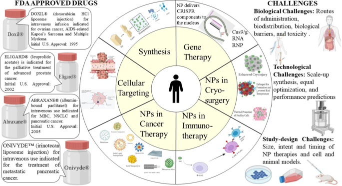
암은 전 세계적으로 중요한 공중 보건 문제 중 하나이며 두 번째 주요 사망 원인입니다. 미국암학회(American Cancer Society)에 따르면 2021년 말까지 신규 환자 수는 190만 명에 이를 것으로 예상된다[4]. 암 치료에 사용되는 기존의 치료법에는 수술, 화학 요법, 방사선 요법, 표적 요법, 면역 요법 및 호르몬 요법이 있습니다[5, 6]. 화학요법 및 방사선 요법은 세포 정지 및 세포 독성 능력을 가지고 있지만[7], 이러한 접근법은 종종 급성 부작용 및 높은 재발 위험과 연결됩니다. 에 의해 유발되는 가장 흔한 부작용은 신경병증, 골수 억제, 위장 및 피부 장애, 탈모 및 피로를 포함합니다. 이 외에도 안트라사이클린, 블레오마이신에 의한 심장독성, 폐독성 등의 약물 특이적 부작용이 있다[8](그림 1).
<그림>
암 치료용 나노 입자
표적 치료의 출현으로 정밀 치료가 성장했습니다[9]. 그러나 여전히 다제내성 등의 불가피한 부작용이 많아 치료효과를 제한하고 있다[8]. 면역항암제는 원발성 암의 치료뿐만 아니라 원격전이를 예방하고 재발률을 낮춤으로써 유망한 결과를 보여주고 있다[10]. 그럼에도 불구하고 자가면역질환은 면역요법의 주요 부작용이다. 또한 연구와 증거에 따르면 면역 요법은 림프종보다 고형 종양에 덜 효과적입니다[11]. 이러한 암은 면역 세포가 침투하기 어려운 특이한 세포외 기질(ECM)을 생성합니다[12]. 이러한 새롭게 진화된 표적 요법 및 면역 요법은 악성 행동과 표피 및 진피의 정상적인 항상성 기능에 필수적인 신호 전달 경로를 방해하고 피부과적 부작용(dAE)을 유발합니다[13].
이러한 모든 세부 사항을 고려할 때 최근 몇 년 동안 암의 정확한 치료를 위한 새로운 전략의 발전에 대한 요구가 탄력을 받고 있습니다. 최근 나노입자를 이용한 기존 치료적 접근의 한계를 해결하기 위한 노력이 이루어지고 있다. 나노입자 기반 약물 전달 시스템은 우수한 약동학, 정확한 표적화, 감소된 부작용 및 약물 내성을 입증함으로써 암 치료 및 관리에 이점을 반영했습니다[14, 15].
나노기술의 발전에 따라 다수의 나노치료 약물이 상업화되어 널리 판매되고 있으며 2010년 이후 더 많은 약물이 임상 단계에 진입했습니다. 약물 병용 요법의 기회를 제공하고 약물 내성 기전의 억제를 통해 내성(MDR)을 감소시킵니다[16]. 1960년대 취리히 ETH에서 나노기술을 의학에 적용하기 위한 선구적인 노력이 있었습니다[17]. 이 조합은 다양한 진단 장치와 더 나은 치료법을 개발하는 데 더 나은 융합으로 입증되었습니다. 이 검토는 주로 나노치료제의 응용에 대한 기본 원칙, 현재의 도전 과제 전망에 초점을 맞추고 미래 연구의 경로를 설명합니다.
나노입자
나노입자(NP)는 일반적으로 동일한 재료의 대량 샘플에서 발견되지 않는 고유한 특성을 가진 100nm 미만의 1차원 입자로 기술적으로 정의됩니다[18]. 나노 입자의 전체 모양에 따라 0D, 1D, 2D 또는 3D로 분류할 수 있습니다[19]. 나노입자의 기본 구성은 표면층, 쉘층, 그리고 기본적으로 나노입자의 중심 부분이며 일반적으로 나노입자 자체라고 불리는 코어로 구성되어 상당히 복잡하다[20]. 높은 표면:부피 비율, 비유사성, 서브마이크론 크기 및 향상된 타겟팅 시스템과 같은 탁월한 기능으로 인해 이러한 재료는 다학문 분야에서 많은 중요성을 갖게 되었습니다.
NP는 EPR(Enhanced Permeability and Retention) 효과를 증가시키기 위해 깊은 조직 침투를 갖는 것으로 밝혀졌습니다. 또한 표면 특성은 상피 천공을 효과적으로 가로질러 생체이용률과 반감기에 영향을 미칩니다[21]. 예를 들어, 친수성 고분자인 폴리에틸렌 글리콜(PEG)로 코팅된 나노입자는 옵소닌화를 감소시키고 면역 체계 제거를 우회합니다[22]. 또한, 입자 폴리머 특성을 조작하여 약물 또는 활성 부분의 방출 속도를 최적화할 수 있습니다. 전체적으로 NP의 고유한 특성은 암 관리 및 치료에서 치료 효과를 조절합니다.
NP 합성
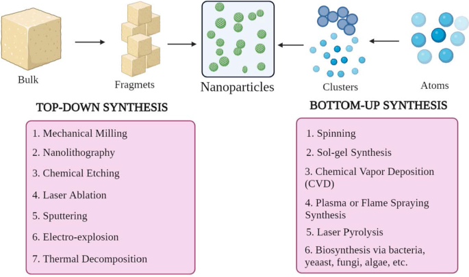
NP는 모양, 크기 및 구조가 다릅니다. 이를 달성하기 위해 수많은 합성 방법이 채택됩니다. 이러한 방법은 크게 1) 상향식 접근 방식과 2) 하향식 접근 방식의 두 가지 주요 그룹으로 분류할 수 있습니다. 이러한 접근 방식은 반응 조건 및 작동에 따라 다른 하위 클래스로 더 분류할 수 있습니다(그림 2).
<그림>
NP 합성의 분류 a 하향식 및 b 상향식 접근 방식
상향식 접근 방식
이 방법은 원자에서 클러스터, NP에 이르기까지 재료를 구성하는 것입니다. 즉, 구성적 방법으로 알려진 더 단순한 물질로 구성합니다. 일반적으로 사용되는 방법에는 회전, 졸겔 합성, 화학 기상 증착(CVD), 플라즈마 또는 화염 분무 합성, 레이저 열분해 및 생합성이 있습니다.
하향식 접근 방식
그것은 또한 NP를 합성하기 위해 벌크 물질 또는 물질을 감소시키는 파괴적 방법으로 알려져 있다. 더 큰 분자는 분해되거나 더 작은 단위로 분해되어 NP로 변환됩니다[24]. 여기에는 기계적 밀링, 나노리소그래피, 화학적 에칭, 레이저 제거, 스퍼터링, 전기 폭발 및 열 분해와 같은 기술이 포함됩니다.
놀랍게도, 나노입자의 크기, 모양 및 전하와 같은 형태학적 매개변수는 반응 조건 및 기타 합성 매개변수를 변경하여 수정할 수 있습니다[25]. 게다가, 성장 메커니즘은 또한 NP의 화학적 특성을 결정합니다. 따라서 성장 메커니즘을 이해하는 것은 필요한 NP를 합성하는 데 필수적입니다.
셀룰러 타겟팅 메커니즘
효과적인 암 치료를 위해서는 정상의 건강한 세포를 제외하고 종양 세포를 표적으로 삼는 능력이 뛰어난 약물 또는 유전자 전달 시스템을 개발하거나 조작하는 것이 필수적입니다. 그것은 치료 효능을 향상시켜 세포 독성의 영향으로부터 정상 세포를 보호합니다. 암 세포를 간접적으로 표적으로 하는 종양 미세 환경(TME)으로 NP를 잘 조직화하여 전달함으로써 달성할 수 있습니다. 이러한 나노제형은 수많은 생리학적 및 생물학적 장벽을 통과해야 합니다. 이러한 장벽은 여러 층(상피, 내피 및 세포막)과 구성요소(기계적 및 물리화학적 장벽 및 효소적 장벽)의 복잡한 시스템입니다. 이러한 사실은 비특이적 표적화를 방지하기 위해 NP의 크기, 생체 적합성 및 표면 화학에 대한 사양을 부과합니다. 그러나, NP 약물 분자의 단순한 세포질 내재화는 그것이 세포내 표적에 도달한다는 것을 의미하지 않습니다. 세포 또는 핵 표적을 활성화하려면 특정 엔지니어링 및 최적화가 필수입니다.
지금까지 여러 연구가 수행되었으며 NP 기반 약물 표적화 설계를 발견하기 위해 더 많은 연구가 진행 중입니다. 이러한 나노운반체는 일반적으로 1) 표적 TME에 도달할 때까지 혈관계(혈액)에서 안정적으로 유지되는 능력, 2) 세망내피계(RES) 제거를 탈출하는 능력, 3) 단핵성 식세포계를 탈출하는 능력( MPS), 4) 종양 혈관계를 통해 TME에 축적, 5) 종양 유체로의 고압 침투, 6) 표적에 도달하고 종양 세포와만 상호작용한다[26]. 표면 기능화, 물리화학적 특성 및 병태생리학적 특성과 같은 중요한 측면은 NP 약물 표적화 과정을 조절합니다.
일반적으로 암 치료에 적합한 것으로 간주되는 나노입자의 직경 범위는 10~100nm입니다. NP 운반체와 암세포 및 종양 생물학 간의 상호 작용 및 누화 과정을 이해하려면 표적화 메커니즘을 해결하는 것이 중요합니다. 타겟팅 메커니즘은 크게 수동 타겟팅과 능동 타겟팅의 두 그룹으로 분류할 수 있습니다.
수동 타겟팅
암세포에서 소수의 거대분자가 우선적으로 축적된다는 관찰은 1980년대 후반에 발견되었습니다. 종양에 축적되는 것으로 보고된 최초의 거대분자는 Matsuura와 Maeda에 의한 폴리(스티렌-코-말레산)-네오카르지노스타틴(SMANCS)이었다[27]. 추가 연구에서 이러한 우선적 분포는 손상된 종양 혈관에서 발견되는 구멍의 발생과 불량한 림프 배액에 기인하며, 이러한 결합을 "향상된 침투 및 유지 효과"라고 합니다.
저산소증이나 염증과 같은 특정 조건에서 혈관의 내피층이 더 투과성이 됩니다[28]. 저산소증 상황에서 빠르게 성장하는 종양 세포는 더 많은 혈관을 작동시키거나 대처하기 위해 기존 혈관을 삼키는 경향이 있습니다. 이 과정을 신생혈관 형성이라고 합니다. 이 새로운 혈관은 구멍이 커서 새는 혈관으로, 정상 혈관에 비해 종양 혈관의 투과 선택성이 나쁘다[29, 30]. 이러한 큰 구멍 또는 구멍은 암 유형, TME 및 국소화에 따라 200~2000nm 범위입니다[31]. 이 빠르고 결함이 있는 혈관신생은 혈관외유출에 대한 저항이 거의 없으며 NP가 이러한 혈관에서 확산되어 궁극적으로 암세포 내에 모이도록 합니다.
정상 조직에서 림프관으로 ECF(세포외액)의 배수는 평균 유속 0.1–2 µm/s로 자주 발생하여 일정한 배수와 재생을 유지합니다[32]. 종양이 형성되면 림프 기능이 탈선하여 간질액 흡수가 최소화됩니다[33]. 이 기능은 NP가 제거되지 않고 종양 간질에 축적되기 때문에 NP 보유에 기여합니다. 이 과정은 EPR 효과의 강화된 유지 부분을 나타냅니다. 이는 순환시간이 짧은 분자에는 적용되지 않고 암세포에서 빠르게 씻겨나간 특성이다. 따라서 이러한 상황을 개선하기 위해 이러한 작은 분자를 나노 크기의 약물 담체에 캡슐화하여 약동학을 향상시키고 종양 선택성을 제공하며 부작용을 줄이기 위해 일상적으로 수행됩니다[34].
EPR 효과에 비해 TME는 수동 타겟팅에서 중요한 기능입니다. 빠르게 증식하는 종양 세포의 중요한 대사 기능 중 하나는 해당 작용입니다. 세포 분열의 주요 에너지원이며[35] 주변 환경을 산성화합니다. 이렇게 낮아진 TME의 pH는 낮은 pH에서 약물을 방출하는 pH에 민감한 NP를 사용하는 데 활용될 수 있습니다[36].
이러한 유형의 종양 표적화를 "수동적"이라고 합니다. 수동적 표적화는 주로 다른 종양 생물학(혈관성, 누출) 및 운반체 특성(크기 및 순환 시간)에 의존합니다. 이러한 유형의 종양 표적화는 특정 유형의 종양 세포에 대한 특정 리간드를 보유하지 않습니다. EPR 효과는 1) 혈관신생 및 림프관신생의 정도 또는 정도, 2) 혈관주위 종양 침습의 정도 또는 정도, 3) 종양내압과 같은 근본적인 종양 생물학에 크게 의존합니다. 이러한 요인들은 나노입자의 물리화학적 특성과 결합하여 나노입자 약물 전달 시스템의 효율성을 결정합니다(그림 3).
<그림>
수동적 셀룰러 타겟팅
수동 타겟팅의 예
탁산은 암 치료에 사용되는 가장 성공적인 약물 그룹 중 하나입니다. 파클리탁셀은 광범위한 암에 대해 강력한 효능을 보여주었습니다. 유방암, 폐암(소세포 및 비소세포) 및 난소암은 탁산으로 가장 많이 치료되는 조직학입니다. 2005년 US-FDA는 진행성 또는 전이성 유방암(MBC)에 사용되는 Abraxane®(알부민 결합 파클리탁셀, Abraxis Bio-Sciences)을 승인했습니다.
Abraxane®은 해중합을 방지하여 미세소관을 안정화시키는 항미세소관 약물입니다. 약물이 튜불린 이량체로부터 미세소관 조립을 촉진할 때 발생합니다. 이렇게 얻은 안정성은 간기 및 유사분열 세포 기능 동안 매우 중요한 미세소관 재구성을 방해합니다. 세포 주기와 유사 분열 동안 잘 사용되는 탁산인 파클리탁셀은 각각 여러 개의 과꽃과 함께 특이한 미세소관 배열을 유발합니다. Abraxane® 단독 또는 젬시타빈과 같은 다른 세포독성제와 조합은 췌장암 이종이식 마우스 모델에서 췌장 기질을 감소시킵니다[37].
Genexol PM®은 파클리탁셀과 CrEL이 없는 멸균 동결건조 고분자 미셀 제형의 혁신적인 나노 제형입니다. 실험에 따르면 Genexol PM®은 누드 마우스에서 최대 허용 용량(MTD)이 3배 더 높은 것으로 밝혀졌습니다. 게다가, 생체 분포는 간, 비장, 신장, 폐와 같은 다른 조직에서 2-3배 더 높은 수준을 나타내었고 암세포에서 더 두드러지게 나타났습니다. 한국에서 MBC 치료용으로 승인되었습니다. 췌장암을 치료하기 위해 미국에서 아직 2상 임상 연구 중입니다[38].
DaunoXome®(리포솜 다우노루비신; Gilead Science/Diatos)은 종양 세포 성장을 감소시키는 항암제입니다. 활성 물질은 다우노루비신입니다. 피부, 폐 및 장에 영향을 미치는 암의 한 형태인 카포시 육종을 치료하는 데 사용되는 다우노루비신(리포솜 형태)의 독특한 제형입니다. US-FDA는 1996년에 이를 승인했습니다[39].
신생혈관 및 혈관신생이 NP 확산에 영향을 미치지만, 이는 NP의 축적을 억제하는 더 큰 간질압으로 이어집니다. 또한, 이질적인 혈액 공급으로 인해 종양 세포의 성장이 불규칙합니다. 즉, 혈관에 가까운 세포가 혈관에서 멀리 떨어져 있거나 코어를 형성하는 저산소 또는 괴사 영역에서 깊은 세포보다 빠르게 분열합니다. 종양. 높은 간질압을 유발하는 이러한 불규칙한 누출은 약물 전달 및 축적을 방해하고 신생혈관 생성 과정을 늦춥니다[34]. 그러나 기계적 또는 화학적으로 EPR 효과를 제어하는 것은 가능합니다. 여기에는 산화질소, 과산화질산염, 브래디키닌, VPF(혈관 투과성 인자), 초음파, 방사선, 온열 요법 등이 포함됩니다. 그러나 특정 제한 사항과 금기 사항이 있습니다.
활성 타겟팅
능동 표적화는 표적 세포(병이 있는 기관, 조직, 세포 또는 세포내 도메인)에서 특이적으로 발현되거나 과발현되는 분자 또는 수용체에 결합하는 트랜스페린 및 엽산과 같은 특정 리간드 또는 분자에 의존합니다[40]. 이러한 유형의 표적화를 리간드 매개 표적화라고 합니다[41]. 여기서, 머무름 및 흡수와 같은 특정 기능을 가진 리간드를 보유하는 NP는 더 큰 친화도가 있도록 표적에 근접해야 합니다. 이 전략은 암세포에 결합하는 NP의 변화를 향상시켜 약물 침투를 향상시킵니다. 1980년에 리포솜 표면에 이식된 항체가 가장 먼저 나타났고[34], 펩티드, 앱타머와 같은 다른 다양한 유형의 리간드가 뒤따랐습니다. 따라서 주요 방법은 전체 생체 분포를 변동시키지 않고 NP와 대상 사이의 누화를 증가시키는 것입니다[42]. 활성 표적화 또는 리간드 매개 표적화의 중요한 메커니즘은 표적 기질 수용체에 의한 리간드 식별입니다. 예시적인 리간드에는 단백질, 펩타이드, 항체, 핵산, 당, 비타민과 같은 소분자 등이 포함될 수 있습니다.[43]. 가장 일반적으로 연구된 수용체는 트랜스페린 수용체, 엽산 수용체, 당단백질 및 표피 성장 인자 수용체(EGFR)입니다. 리간드-표적 상호작용은 수용체-매개 세포내이입을 통해 막의 접힘 및 NP의 내재화를 유발합니다. 적극적인 타겟팅이 발생하는 다양한 메커니즘이 있습니다. 대부분의 종양 표적화는 일반적으로 NP에 의한 종양 세포 표적화에 의해 수행됩니다. 이 과정은 세포 침투를 향상시킵니다. 이전에 언급했듯이 트랜스페린은 널리 연구된 수용체 중 하나입니다. 철분을 세포로 운반하는 데 도움을 주는 일종의 혈청 당단백질입니다. 이 수용체는 대부분의 종양 세포, 특히 고형 종양에서 과발현되는 것으로 밝혀졌으며 건강한 세포에서는 더 낮은 수준으로 발현됩니다. 따라서 우리는 트랜스페린을 특이적으로 표적으로 하는 리간드와 함께 NP를 수정할 수 있습니다[44]. 예를 들어, A2780 난소 암종 세포는 트랜스페린을 과발현합니다. 이 기능은 이러한 세포를 특이적으로 표적으로 하는 트랜스페린 변형 PEG-포스파티딜-에탄올아민(Tf-Mpeg-pe) NP에 의해 사용됩니다[45]. 또 다른 대안적인 방법은 혈관신생 내피 세포와 같은 암세포에 인접한 세포를 표적으로 삼는 것입니다. 이 세포는 또한 종양 혈관과 밀접하게 접촉합니다. 이 전략은 암세포로의 혈액 공급을 줄임으로써 저산소증과 괴사를 유발할 수 있습니다. 종양 조직이 정상 조직보다 더 산성인 것으로 밝혀졌습니다. 이것은 Warburg 효과[46]에 의해 광범위하게 설명되었습니다. 이것은 암세포 대사가 해당과정으로 전환되어 젖산을 형성하는 것을 설명합니다. 젖산이 축적되면 세포가 죽습니다. 이 상황에 대처하기 위해 세포는 과잉 젖산을 세포 외 환경으로 내보내는 양성자 펌프를 과발현하여 더 산성으로 만듭니다. 따라서 리포솜 기반 pH 민감성 약물 전달 시스템이 연구되었습니다.
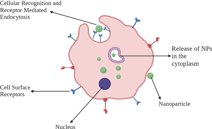
NP의 다가 특성은 리간드 코팅된 NP와 표적 암세포의 누화를 개선합니다. 이러한 NP의 설계는 NP 아키텍처와 리간드 표적 화학이 전체 방법의 효능에 영향을 미치기 때문에 복잡합니다. 투여 경로, 리간드 밀도와 같은 물리화학적 특성[47], 나노입자의 크기[8]와 같은 기타 요인이 시스템의 성공에 기여합니다(그림 4).
<그림>
활성 세포 타겟팅의 그림 표현
활성 타겟팅의 예
티로신 키나제(TK) 수용체의 ErbB 계열의 구성원인 EGFR은 다양한 유형의 암, 특히 편평 세포 조직학에서 과발현됩니다. 항-EGFR-PEG-AuNP 및 항-IgG-PEG-Au 나노입자를 포함하는 금 나노입자는 인간 SCC를 표적화하는 데 사용할 수 있습니다[48].
허셉틴®은 유방암 세포 표면에서 과발현되는 인간 EGF 수용체-2(HER2)를 표적으로 하는 치료제입니다. HER2를 표적으로 하는 PEG화 리포솜 독소루비신은 안트라사이클린의 알려진 부작용인 심장 독성을 줄이기 위해 개발되었습니다[49].
종양 내피의 표면은 혈관 신생 과정에 관여하는 혈관 세포 부착 분자-1(VCAM-1)로 알려진 당단백질을 발현합니다. 한 연구는 유방암 모델에서 VCAM-1을 표적으로 하는 NP를 강조하여 잠재적 역할을 나타냅니다[50].
비타민 B9로도 알려진 엽산은 뉴클레오티드 합성에 필수적입니다. 엽산은 세포에서 발현되는 엽산 수용체에 의해 내재화됩니다. 그러나 종양 세포는 FR-α(엽산 수용체의 알파 isoform)를 과발현하는 반면, FR-β는 액체 암세포에서 과발현됩니다[51]. NP에 의한 엽산 수용체 표적화는 현재 특정 암 치료를 위한 것이었습니다[52, 53].
암 치료의 나노입자
약물 전달 시스템에서 광범위하게 사용되는 NP는 유기 NP, 무기 NP 및 하이브리드 NP를 포함합니다(그림 5).
<그림>
암 치료에 사용되는 다양한 나노물질
유기 나노입자
고분자 나노입자
고분자 나노입자(PNP)는 서로 다른 단량체에 의해 형성된 특정 구조적 구조를 가진 "콜로이드 거대분자"로 잘 정의되어 있습니다[54]. 약물은 NP' 외부에 갇히거나 부착되어 표적에서 조절된 약물 방출을 달성하기 위해 나노구 또는 나노캡슐을 생성합니다. 초기에 PNP는 폴리아크릴아미드, 폴리메틸메타크릴레이트(PMMA) 및 폴리스티렌과 같은 비생분해성 폴리머로 구성되었습니다[56]. 그러나 이들이 축적되면 시스템에서 제거하기가 어려워 독성이 발생했습니다. 폴리락트산, 폴리(아미노산), 키토산, 알지네이트, 알부민과 같은 생분해성 고분자가 현재 사용되고 있으며 독성을 감소시키고 약물 방출 및 생체 적합성을 향상시키는 것으로 알려져 있습니다[57]. 입증된 연구에 따르면 PNP를 폴리소르베이트로 코팅하고 폴리소르베이트를 사용하면 계면활성제 효과가 나타납니다. 외부 코팅은 혈뇌장벽(BBB)의 내피 세포막과 나노입자의 상호작용을 향상시킵니다[58].
한 연구에 따르면 인도메타신이 로딩된 나노캡슐은 쥐의 이종이식 신경교종 모델에서 종양 크기의 실질적인 감소와 생존 개선과 관련이 있음을 보여주었습니다[59]. 항암제를 포함하는 10개 이상의 고분자 나노입자가 임상 개발 중에 있는 성장하는 분야입니다. 몇 가지 예에는 파클리탁셀 폴리글루멕스(Xyotax), PEG-캄프토테신(프로테칸), 수정된 덱스트란-캄프토테신(DE 310), HPMA 공중합체-DACH-백금(AP5346), HPMA 공중합체-백금(AP 5280), HPMA 공중합체-파클리탁셀( PNU166945) 및 HPMA 공중합체-독소루비신 갈락토사민(PK2) [60].
덴드리머
덴드리머는 정의된 하이퍼브랜치 아키텍처를 가진 구형 고분자 고분자입니다. 고도로 분지된 구조는 덴드리머의 특징입니다. 일반적으로 덴드리머의 합성은 암모니아 코어를 아크릴산과 반응시켜 시작됩니다. 이 반응은 에틸렌디아민과 추가로 반응하여 GO 생성물인 "트리아민"을 생성하는 "삼중산" 분자를 형성합니다. 이 생성물은 추가로 아크릴산과 반응하여 6산을 생성하고, 이는 추가로 "헥사아민"(1세대) 생성물 등을 생성한다[61]. 일반적으로 덴드리머의 크기는 1~10nm입니다. 그러나 크기는 최대 15nm에 이를 수 있습니다[62]. 정의된 분자량, 조정 가능한 가지, 생체 이용률 및 전하와 같은 특정 구조를 감안할 때 이들은 핵산을 표적화하는 데 사용됩니다. 널리 사용되는 일부 덴드리머는 폴리아미도아민(PAMAM), PEG(폴리(에틸렌글리콜)), PPI(폴리프로필렌이민) 및 TEA(트리에탄올아민)[63]입니다.
PAMAM 덴드리머는 처음에 MDR 관리를 달성하기 위해 설계되었습니다. DNA 조립 PAMAM 덴드리머는 광범위하게 설명되어 있습니다. 단일제제 화학요법을 받은 동물과 비교하여 합성된 덴드리머는 상피암 이종이식편의 성장을 유의하게 지연시켰다[64].
mAb 나노입자
단일클론항체는 특정한 표적화 능력 때문에 암 치료에 널리 사용된다[65]. 이러한 mAb는 이제 NP와 결합하여 항체-약물 접합체(ADC)를 형성합니다. 이들은 세포독성 약물 또는 mAb 단독보다 매우 특이적이고 설득력 있는 것으로 입증되었습니다. 예를 들어, 파클리탁셀 코어와 트라스투주맙으로 변형된 표면으로 구성된 항체 약물 NP는 HER2 양성 유방 상피 세포 대조군에서 단일 제제 파클리탁셀 또는 트라스투주맙 단독보다 더 나은 항종양 효능과 더 낮은 독성을 나타냈다[66].
세포외 소포
세포외 소포(EV)는 50–1000 nm n 크기 범위의 이중층 인-지질 소포입니다[67]. EV는 다양한 세포 유형에 의해 지속적으로 분비되며 기원, 크기 및 구성이 다양합니다. EV는 1) 엑소좀, 2) 미세소포, 3) 세포사멸체의 세 가지 클래스로 나뉩니다[68]. 엑소좀과 결합된 나노입자는 지질과 분자가 기원 세포와 매우 유사하여 널리 사용됩니다. 게다가, 그들은 면역 감시를 피하고 암세포 내에서 매우 빠르게 내재화합니다. 그들은 표적 부위에 세포독성 약물 및 기타 항종양 약물을 전달함으로써 천연 매개체 역할을 합니다. 독소루비신(exoDOX)이 로딩된 엑소좀이 가장 좋은 예입니다. exoDOX는 유방암 치료에 사용되며 세포독성을 높이고 심장독성을 피함으로써 보존적 독소루비신 치료에 비해 우수한 결과를 보였다[69]. Exosome NP는 합성 NP에 비해 고유한 생체 적합성 기능, 고급 화학적 안정성 및 세포 내 통신을 가지고 있습니다. 그럼에도 불구하고 엑소좀 분리 및 정제를 위한 표준 조건의 결핍과 같은 결점은 매우 중요하며 해결해야 합니다[70, 71].
리포솜
이들은 약물 분자를 캡슐화하기 위해 단일 층 또는 다중 층일 수 있는 인지질을 포함하는 구형 소포입니다[72]. 리포솜은 낮은 고유 독성, 약한 면역원성, 생물학적 불활성과 같은 특성을 갖는 것이 독특합니다[73]. 리포솜은 1965년에 승인된 최초의 나노규모 약물이다[74]. 전형적인 리포솜 구조는 "친수성 코어"와 "소수성 인지질 이중층"으로 구성됩니다. 이 독특한 구조로 인해 친수성 및 소수성 약물을 모두 포획하여 순환 중 환경 저하로부터 포획된 약물을 효과적으로 보호할 수 있습니다[75].
리포솜은 더 높은 항종양 효능과 향상된 생체이용률을 보여줌으로써 독소루비신, 파클리탁셀 및 핵산과 같은 약물 전달을 위한 우수한 플랫폼을 제공합니다[76]. Doxil® 및 Myocet®은 MBC 치료에 사용되는 승인된 리포솜 기반 다우노루비신 제제입니다[77, 78]. 그러나 캡슐화 효능 감소, MP에 의한 빠른 제거, 세포 흡착, 짧은 유통기한 등의 단점으로 인해 리포솜 기반 나노입자의 적용에는 한계가 있다.
고체 지질 나노입자(SLN)
이들은 인지질 단층, 유화제 및 물로 구성된 콜로이드성 나노운반체(1~100nm)입니다[79]. 이들은 0차원 나노물질로 알려져 있습니다. 지질 성분은 트리글리세리드, 지방산, 왁스, 스테로이드 및 PEG화 지질일 수 있습니다[80]. Unlike conventional liposomes, SLNs have a “micelle-like structure” within which the drug is entrapped in a non-aqueous core. Examples include mitoxantrone-loaded SLN, which has shown reduced toxicity and enhanced bioavailability [81]. The incorporation of doxorubicin and idarubicin by SLN in “P388/ADR leukemia cells” and the “murine leukemia mouse model” has shown positive results [82].
Nanoemulsions
Nanoemulsions are colloidal NPs with heterogeneous mixtures of an oil droplet in aqueous media ranging from 10–1000 nm [83]. Three representative types of nanoemulsions can be made in:1) oil-in-water system, 2) water-in-oil system, and 3) bi-continuous nanoemulsions. Membrane-modified nanoemulsions have been extensively studied. For instance, nanoemulsions loaded with spirulina and paclitaxel showed an improved anti-tumor effect by regulating immunity through TLR4/NF-kB signaling pathways [84]. Nanoemulsion consisting of rapamycin, bevacizumab, and temozolomide is known to treat advanced melanoma [85]. Nanoemulsions are different from liposomes and certainly have enhanced characteristics than others, such as optical clarity, stability, and biodegradability [86]. However, there are challenges to clinical applications of these nanoemulsions as these involve high temperature and pressure and instruments such as homogenizers and microfluidizers that are expensive.
Cyclodextrin Nanosponges
Cyclodextrins are usually used as stabilizers to increase the drug loading capacity of NPs [87]. Nanosponges are tiny, mesh-like structures [88]. Β-cyclodextrin nanosponges loaded with paclitaxel have shown sound cytotoxic effects in MCF-7 cell line culture [89]. Similarly, camptothecin has shown improved solubility and stability when formulated with cyclodextrin-based nanosponges [90].
Inorganic Nanoparticles
Carbon Nanoparticles
Carbon NPs as the name suggests are based on the element carbon. They have been widely utilized in medical arenas because of their optical, mechanical, and electronic properties combined with biocompatibility [91]. Due to their inherent hydrophobic nature, carbon NPs can encapsulate drugs through π-π stacking [92]. Carbon NPs are further categorized into graphene, carbon nanotubes, fullerenes, carbon nanohorns, and graphyne. Although all these are carbon-based, they vary in their structure, morphology, and properties.
“Graphene” is 2D crystal with sp2-hybridized carbon sheet that holds extraordinary mechanical, electrochemical, and high drug loading properties. Further, based on composition, properties, and composition, graphene can be divided as follows:1) single-layer graphene, 2) graphene oxide (GO), 3) reduced graphene oxide (rGO), and 4) multi-layer graphene [93]. GO and rGOs are widely used due to their ability to target hypoxia [94] and irregular angiogenesis in TME [95]. Studies have shown that GO-doxorubicin exhibits higher anticancer activities in cellular models of breast cancer [96].
Fullerenes are large carbon-cage molecules composed of carbon allotrope with different conformation types such as sphere, ellipsoid, or tube. They are the most widely studied nanocarriers as they have typical structural, physical, chemical, and electrical properties [97]. These are used in photodynamic therapy as they have triple yield and generate oxygen species due to the presence of extended π-conjugation and the ability to absorb light [98]. PEG-modified fullerenes showed promising photodynamic effects on tumor cells [99].
Carbon nanotubes (CNTs) are cylindrical tubes, most often considered as rolls of graphene, were discovered in the late 1980s. They are classified into two groups:1) single-walled CNTs and 2) multi-walled CNTs. As they are carbon-based, they can bring upon immune response by interacting with immune cells, thereby suppressing the tumor growth. Traditionally, they have been used as DNA delivery vectors and for thermal ablation therapy. For instance, a fluorescent single-walled CNT with mAb encapsulating doxorubicin is used to target colon cancer cells. Such CNTs form a complex which is effectively engulfed by the cancer cells leading to the intracellular release of doxorubicin, whereas the CNTs are retained in the cytoplasm [100].
Quantum Dots
Quantum dots are typically nanometer-scale semiconductors with a broad spectrum of absorption, narrow emission bands, and high photostability, allowing them to be widely used in biological imaging [101]. Based on carbon, these are divided into:1) graphene quantum dots, 2) nanodiamond quantum dots, and 3) carbon quantum dots. Besides biological imaging, quantum dots are being actively investigated in cancer treatment. The most commonly used quantum dots is graphene quantum dots due to their inherent biocompatibility and rapid excretion. For example, quantum dots aptamer—doxorubicin conjugate targets prostate cancer cells [102]. However, the deficiency of optimized process in producing quantum dots is the major obstacle.
Metallic Nanoparticles
Metallic nanoparticles are commonly explored in “biological imaging” and targeted DDS due to their remarkable optical, magnetic, and photothermal properties. Some of the most commonly used metallic NPs are gold NPs, silver NPs, iron-based NPs, and copper NPs. Gold NPs are used as intracellular targeting drug carriers because the size and surface properties are easily controlled [103]. Moreover, their visible light extinction behavior makes it possible to track NP trajectories in the cells. “Anti-HER2 functionalized gold-on-silica nanoshells” have been shown to aim HER2 positive breast cancer cells [104]. Combidex®, an iron oxide NP formulation, is presently in the late-stage clinical testing phase to detect nodal metastases [105]. Feraheme®, a ferumoxytol containing iron oxide NP formulation, is used to treat iron-deficiency anemia. This is also used to treat nodal metastases in prostate and testicular cancer and was approved by FDA in June 2009 [106, 107].
Magnetic Nanoparticles
Magnetic NPs are generally used in MRI imaging, and drug delivery contains metal or metal oxides. These are usually covered with organic substances like polymers and fatty acids to enhance stability and biocompatibility [108]. LHRH-conjugated superparamagnetic iron oxide NPs are effective in targeting and imaging of breast cancer [109]. Moreover, magnetic NPs are used in magnetic hyperthermia for thermal ablation of cancer cells [110, 111]. Some of the magnetic NPs that are in the market or in the clinical trial phase are Feridex® and Resovist® for liver metastasis and colon cancer [112].
Calcium Phosphate Nanoparticles
“Calcium phosphate NPs” is biologically compatible, biodegradable, and do not cause any harsh adverse reactions. Hence, they are used as a delivery agent for insulin, growth factors, antibiotics, and contraceptives [113]. They are also used in the delivery of oligonucleotides and plasmid DNA [114]. Calcium phosphate NPs combined with either viral or non-viral vector has been positively used as delivery vectors in cellular gene transfer. A “liposomal nanolipoplex formulation” of calcium and glycerol has shown decreased toxicity and enhanced transfection features [115, 116].
Silica Nanoparticles
Silica being a significant component of many natural materials was only studied concerning biology recently. Silica NPs are commonly used to deliver genes by functionalizing the NP surface with amino-silicanes [117]. N-(6–aminohexyl)–3–aminopropyl–trimethoxysilane functionalized silica NPs have shown excellent efficiency in the transfection of Cos-1 cells with minimal toxicity and is now commercially available [118]. Mesoporous silica NPs are considered one of the best drug carriers due to their better pharmacokinetic properties. They have been extensively used in immunotherapy. According to a study, colorectal cancer cells have shown successful uptake of camptothecin-loaded mesoporous silica NPs.
Mechanism of NPs in Overcoming Drug Resistance
Drug resistance is one of the chief problems in cancer therapy and management. It prevails across all types of cancer and all possible treatment modalities. Drug resistance is a phenomenon that results when diseases become tolerant to pharmaceutical treatments. Drug resistance can be classified into two types:1) innate and 2) acquired [119]. Innate resistance usually results from pre-existing mutations in the genes that are involved in cell growth or apoptosis. Acquired resistance is defined as the type of resistance that is developed after a particular anti-tumor treatment, which may result from the development of new mutations or from alterations in the TME during treatment. Nanoparticles, due to their extraordinary ability to co-encapsulate multiple therapeutic agents, can also be used to overcome cancer-related drug resistance.
Targeting Efflux Transporters
Efflux transporters are classified under the family of “ATP-binding cassette (ABC) transporters.” These have a significant role in MDR. The primary function of these transporters is to pump out drugs out of the cell and reduce the concentration. “P-glycoprotein (P-gp)” is one such efflux transporter that is overexpressed by drug-resistant cancer cells [120, 121].
Overexpression of P-gp has been linked with inadequate treatment response, especially in breast cancer [122] and ovarian cancer [123]. NPs can be used to tackle efflux pumps. As NPs internalize the cell via “endocytosis” instead of diffusion and release the drug at the “perinuclear site,” which is distant from active efflux pumps, NPs can bypass the efflux pumps [124]. Besides, by modifying the control of drug releases, such as by utilizing low pH levels and redox as triggers, NPs can effectively bypass efflux pumps [125, 126].
Combination therapy is yet another method to overcome MDR. NPs can be loaded with multiple drugs within a single drug carrier [127]. Inhibiting efflux transporter expression instead of just dodging them would be another viable option. This can be achieved by building NPs in such a way that it can entrap both efflux pump inhibitors and chemotherapy agents [128]. A recent study positively reflected upon reversing MDR in breast cancer cells by using NPs that co-deliver COX-2 inhibitors and doxorubicin [129]. Similarly, using silica NP that encapsulates miRNA-495 and doxorubicin has proved effective in overcoming drug resistance in lung cancer cells [130]. Another interesting study found out that using NPs in the tumor neo-vasculature targeting KDR receptors is a more effective anti-tumor function than P-gp inhibitor combination therapy. Yet, another way of overcoming drug resistance is by depleting the source of ATP, which is essential for the functioning of ABC transporters. This can be done by targeting mitochondria which leads to a decrease in ATP production.
Targeting an Apoptotic Pathway
Cancer cells proliferate due to faulty apoptotic machinery and upsurge their survival adding to drug resistance [131]. The faulty apoptotic pathway gets activated by “deregulation of Bcl-2” and “nuclear factor kappa B (NF-κB).” These are the most widely investigated anti-apoptotic proteins and can be potentially used as the target for reversing drug resistance. Using a classic process of co-delivery of “Bcl-2 siRNA and chemotherapeutics” by NPs is a way to overcome MDR [132]. NF-κB inhibitors have been used in combination with “pyrrolidine dithiocarbamate (PDTC)” [133] and curcumin [134]. Besides suppressing anti-apoptotic factors, triggering pro-apoptotic factors is another to fight “apoptotic pathway-mediated drug resistance.” For instance, a combination of ceramide and paclitaxel is a good example [135]. Ceramide restores the expression of a chief tumor suppressor, p53 protein, by regulating alternative pre-mRNA splicing. Delivering ceramide via NPs is an excellent way to correct the p53 missense mutation [136]. Owing to its potential, a combination of ceramide and paclitaxel has shown significant therapeutic efficacy in cancer drug resistance models. Transfecting the p53 gene by cationic SLNs has been reported in lung cancer cases [137]. Similarly, transfecting the p53 gene by PLGA has been carried out in breast cancer cells models that have shown potent induction of apoptosis and inhibition of tumor growth [138].
Some NP-based DDS act by impeding efflux pumps and encouraging apoptosis [139]. A pioneering study conducted to prove both pump- and non-pump-mediated drug resistance used an “amphiphilic cationic NP” entrapping paclitaxel and Bcl-2 converter gene in drug-resistant liver cancer models. NP complex diminished P-gp-induced drug efflux and the apoptosis activation. Similarly, co-delivery of “doxorubicin and resveratrol encapsulated in NPs” has shown noteworthy cellular toxicity on doxorubicin resistance breast cancer cells by downregulating the expression of Bcl-2 and NF-κB, thereby initiating apoptosis as well as through the inhibition of efflux transporter expression [140]. A similar study was done on multi-drug resistant prostate cancer cells by using folic acid-conjugated planetary ball milled NPs encapsulated with resveratrol and docetaxel. This worked by downregulating anti-apoptotic gene expression while inhibiting ABC transporter markers [141].
Targeting Hypoxia
Hypoxia is yet an additional aspect that backs MDR [142]. Due to abnormal blood vessels in the vicinity of the tumor and due to the increasing demand of oxygen by the rapidly growing tumor, some tumor cells are repeatedly in a hypoxic condition. The part of the tumor that is in hypoxic condition often escapes from the chemotherapy drugs. Hypoxia creates an oxygen ramp inside the tumor that intensifies tumor heterogeneity, encouraging a more aggressive phenotype. Moreover, the hypoxia condition has been established to facilitate the overexpression of efflux proteins [143]. The major protein, “hypoxia-inducible factor 1α (HIF-1α)” acts an important role. Hence targeting HIF-1α or silencing HIF-1α gene is a way to overcome drug resistance. NPs containing HIF-1α siRNA can be used to reduce hypoxia-mediated drug resistance [144]. Instead of directly targeting HIF-1α, indirect inhibition of HIF-1α signaling can be used. For example, the “PI3K/Akt/mTOR pathway” is known to control the expression of HIF-1α. Inhibition of this pathway effectively downregulates the expression of HIF-1α, which enhances the sensitivity of MDR cells to cancer treatment [145]. NPs like PLGA-PEG and PEGylated and non-PEGylated liposomes can be used effectively. In addition, “heat shock protein 90 (HSP90)” is needed for transcriptional activity of HIF-1 and inhibition of HSP90, which downregulates the expression of HIF-1α [146]. The HSP90 inhibitor in “17AAG loaded NPs” has dramatically improved MDR in bladder cancer treatment [147].
Nanoparticles and Proteomics
When NPs are subjected to the biological system, they are surrounded by cellular and serum proteins which form a structure known as protein corona (PC) [148]. Based on the degree of interaction of these proteins with the NPs, there are classified into the hard corona and soft corona. “Hard corona” is formed when these proteins have a high binding affinity towards the NPs. “Soft corona” is produced when these proteins are loosely bound to the NPS. It has been established that the most protein forming a PC first will be eventually substituted by proteins with higher affinities. This is known as Vroman effect [149]. Hence developing the technology that can manufacture NPs with desired properties is essential. Several proteomic approaches such as MS, LC–MS, SDS-PAGE, isothermal microcalorimetry (ITC), etc. [150], are being used. PC affects the crosstalk of NP with the biological setting and thereby governs the application and usage of the same in the medical field.
Cancer proteomics studies the number of proteins in cancer cells and serum, which supports hunting proteins and biomarkers that aids in diagnosis, treatment, and prognosis [151]. It also helps in understanding cancer pathogenesis and drug resistance mechanism. Post-translational modifications (PTMs) play an indispensable part in occurrence, recurrence, and metastasis. Besides using chemotherapy and kinase inhibitors, novel agents like siRNA, mRNA, and gene editing are central therapeutics used with NPs.
Nanotechnology for Small Interfering RNA (siRNA) Delivery
siRNAs are small ds RNA molecules (around 21 nucleotides long) that suppress the expression of genes in the target. This process is known as “RNA interference.” A few siRNA-based NPs that are currently under clinical investigations are ALN-TTR01 that is used to target the transthyretin gene to treat transthyretin-mediated amyloidosis, and Atu027, which is a liposomal siRNA that targets protein kinase N3 and TKM-ApoB that knock downs the expression of ApoB [152, 153].
Nanotechnology for Tumor microRNA Profiling and Delivery
MicroRNAs are a class of endogenous “single-stranded non-coding RNA” molecules that control post-transcription gene expression by blocking translation of the target mRNA or repressing protein production by destabilizing mRNA [154]. These are emerging as vital biomarkers that are a significant target for cancer diagnosis, therapy, and treatment. The base priming nature of nucleic acid forms the very foundation for nanotechnology used miRNA profiling techniques. Several profiling techniques use biosensors or surface plasmon resonance imaging techniques in combination with molecular biology enzymatic reactions. Nanotechnology can be used for the delivery of MicroRNAs. For example, biodegradable polycationic prodrugs showed promising results in the regulation of polyamine metabolism [155]. MicroRNA-loaded polycation-hyaluronic acid NPs of single-chain antibody fragments have shown progressive downregulation of “survivin expression” in high metastatic cancer load in the lung of murine B16F10 melanoma.
DNA Nanotechnology for Cancer Therapy
DNA-based nanostructures have been synthesized for DNA sensors to detect nucleic acid, DNA-coated gold NPs for lead sensing by hybridizing Pb-activated DNAzyme to the linking DNA, scaffolds to organize organics, inorganic, and biomolecules into distinct morphology molecular transporters, and drug delivery (Table 1).
Advantages of Nanoparticles in Cancer Therapy
The utilization of nanotechnology in the diagnosis, treatment, and management of cancer has led to a whole new era. NPs, either by active or passive targeting, augment the intracellular concentration of drugs while avoiding toxicity in the healthy tissue. The targeted NPs can be designed and altered as either pH-sensitive or temperature-sensitive to establish and regulate the drug release. The pH-sensitive drug delivery system can deliver drugs within the acidic TME. Similarly, the temperature-sensitive NPs release the drugs in the target site due to changes in temperature brought in by sources like magnetic fields and ultrasound waves. In addition, the “physicochemical characteristics” of NPs, such as shape, size, molecular mass, and surface chemistry, have a significant part in the targeted drug delivery system. Further, NPs can be modified according to the target and used to target a particular moiety.
Conventional chemotherapy and radiation therapy have several disadvantages concerning efficacy and side effects because of uneven dispersal and cytotoxicity. Therefore, cautious dosing is required that effectively kills cancer cells without any significant toxicity. To reach the target site, the drug has to pass several fortifications. Drug metabolism is a very complex process. In physiological conditions, the drug needs to pass TME, RES, BBB, and kidney infiltration. RES or macrophage system is made up of “blood monocytes, macrophages, and other immune cells” [160]. MPS in the liver, spleen, or lungs react with the drugs and activate “macrophages or leukocytes” that rapidly remove the drug. This leads to a short half-life of the drug [161]. To overcome this, NPs with “surface modification,” such as PEG, bypass this mechanism and increase the “drug half-life.” Besides, kidney infiltration is a crucial function in the human body. Proper kidney infiltration thus minimizes the toxicity caused by NPs.
The brain-blood barrier (BBB) is a specialized protection structure offered to protect the CNS from harmful and toxic agents. “Brain capillary endothelial cells” are arranged in the form of a wall that provides essential nutrients to the brain. Since the primary function of BBB is to block toxic agents to reach the brain, currently available chemotherapy agents for brain cancer are highly limited to intraventricular or intracerebral infusions [162]. However, NPs are known to cross BBB. Now, several approaches such as EPR effect, focused ultrasound, peptide-modified endocytosis, and transcytosis are used to deliver NPs. Glutathione PEGylated liposome encapsulated with methotrexate showed improved methotrexate uptake in rats [163]. Au-NPs are often used as they have proven to help transport drugs to induce apoptosis [164].
NPs being carriers also increase the drug stability by preventing the degradation of the encapsulated cargo. Additionally, a large volume of drugs can be encapsulated without any chemical reaction. Dry solid dosage forms are more stable than nanoliquid products [165]. Stabilizers can be used to enhance stability. Yet another way to increase stability is to use porous NPs.
Tumor has unique pathophysiology features such as extensive angiogenesis, flawed vascular architecture and defective lymphatic drainage. The NPs use these features to target tumor tissue. Due to reduced venous return in tumor tissue and meager lymphatic clearance, NPs are effectively retained. This phenomenon is known as EPR. Similarly, by targeting the adjacent tissues, tumor-targeting can be accomplished [166].
NPs can be administered through several routes like oral, nasal, parenteral, intra-ocular etc. NPs have a high surface-to-volume ratio and intracellular uptake. Studies have reported that NPs are more effective than microparticles as drug carriers [167].
Nanoparticles in Immunotherapy
The immune system sets an important part in the establishment and development of cancer cells. The advancement of immunotherapy has revolutionized cancer therapy. It is found that NPs not only help in target delivery of chemotherapy but can also be used in combination with immunotherapy. There are several approaches in immunotherapy aimed at activating the immune system against cancer cells [168] by “immune checkpoint blockade therapy,” “cancer vaccine therapy,” “chimeric antigen receptor (CAR)-T cell therapy,” and “immune system modulator therapy” [169,170,171]. NP-based immunotherapy includes “nanovaccines,” “aAPCs (artificial antigen-presenting cells),” and “immunosuppressed TME targeting.”
Nanovaccines specialize in delivering “tumor-associated antigens” and “adjuvants” to antigen-presenting cells, such as dendritic cells (DCs) [172]. Moreover, these can also be employed as adjuvants to enhance “APC antigen presentation” and promote DC maturation that leads to the stimulation of cytotoxic T cells that have anti-tumor function [173, 174]. Liposomes, PLGA NPs, gold NPs are found to have the ability to deliver TAAs into DCs in the cytoplasm [175]. Mesoporous silica, the most used inorganic NP, has exhibited an adjuvant role, leading to immune response stimulation [176]. Artificial APCs interact with MHC-antigen complexes directly which binds to T cells. They also bind to co-stimulatory molecules that bind to co-stimulatory receptors leading to T cell activation [177]. Targeting the immunosuppressed TME is yet another method of using NPs in immunotherapies. This is done by targeting essential cell types in TME such as “tumor-associated macrophages (TAMs),” regulatory T cells, and “myeloid-derived suppressor cells (MDSCs).”
Besides, the combination of chemoimmunotherapy has been demonstrated to be a capable approach in cancer therapy. For instance, a study has shown that co-loading Nutlin-3a, which is a chemotherapeutic agent and cytokine GM-CSF, in “spermine-modified acetylated dextran (AcDEX) NPs” improved cytotoxic CD8( +) T cells proliferation and activated an immune response [178].
“Programmed cell death protein 1 (PD-1)” and “programmed cell death ligand 1 (PD-L1)” are some of the essential immune checkpoints [179]. Hence immune checkpoint inhibitors are used to target these using NPs. According to a study, conventional immune checkpoint inhibitors of PD-L1/PD-1 displayed inconsistent responses. To enhance the chances and bonding of immune checkpoint inhibitors and immune checkpoints, multivalent poly (amidoamine) dendrimers were used. Usage of these dendrimers not only showed enhanced PD-L1 blockade but also showed improved drug accumulation at the tumor site [180].
Nanoparticles in Cryosurgery
Cryosurgery is an advanced practice of freeze-destroying cancer tissue. Although this is less invasive and causes intraoperative bleeding and postoperative complications, certain drawbacks like inadequate freezing capacity and damage to adjacent cells need to be addressed [181]. The rise of nanotechnology has enabled the use of NPs in cryosurgery.
The primary working of nanocryosurgery is introducing NPs with particular properties into the cancer cells and causing freezing [182]. During this process, ice is formed within the cells, which causes damage to it. This is an important process and can be carried out effectively using NPs. The thermal conductivity property of NPs can be exploited, which significantly freeze the tumor tissue and cause tumor damage [183]. Besides, they cool down rapidly, and it is feasible to regulate the “growth direction” and “direction of the ice ball” (Fig. 6).
Diagrammatic representation of NPs in cryosurgery
When the location of the tumor makes it not feasible for cryosurgery or if other adjacent organs are at risk, there are high chances that the freezing can damage healthy tissue. Recently, phase change materials (PMs) made up of NPs are used to protect the adjacent normal healthy tissue during cryosurgery [184]. For instance, liposome-based microencapsulated phase change NPs have shown incredible results in protecting surrounding healthy tissue [185]. These NPs are deemed to possess large latent heat and low thermal conductivity, making them perfect for cryosurgery.
Significant Challenges in the Clinical Application of Nanoparticles
At present, as nanotechnology has bloomed, the amount of knowledge and research put into nanoparticles has steeply raised. But only a few of them actually make it up to clinical trials. Most of them only halt at in vivo and in vitro stages. Each individual nanoformulation has particular challenges in their clinical translation, but most NPs face similar challenges that can be divided into biological, technological, and study-design related.
Biological challenges include lack of routes of administration, tempering biodistribution, the channel of NPs across the biological barriers, their degradation, and toxicity [186]. NPs are usually injected via intravenous injections directly into the blood, which takes away NPs, making it challenging to stay and interact with the target site. As a result, a high concentration drug is used, which might not provide desired therapeutic effects [187]. However, magnetic NPs can be used to overcome this as many in vivo and in vitro studies have proved the usage of 3D magnetic fields to control the movement of NPs against blood flow. But, the effect of magnetic fields on the human body, crosstalk between magnetic fields, and a large number of NPs has to be researched upon.
Controlling the biological fate of NPs is very hard and needs a lot of focus. Even though NPs are made up of biosafety materials and are modulated accordingly to increase the retention time and half-life, there runs a risk of lung, liver and kidney damage. Some factors that govern toxicity are surface area, particle size and shape, solubility, and agglomeration [188]. NPs have shown greater deposition in the lung with inflammatory, oxidative and cytotoxic effects [189]. Studies reveal that healthy cells often suffer from free radicals generated by NPs [190]. Fabricating NPs with more biocompatible substances like chitosan and materials that disintegrate after near infrared light irradiation may be potential solutions.
Another tricky challenge is avoiding the “mononuclear phagocytic system (MPS).” In biological fluids, NPs adsorb proteins to produce PC, which attacks MPS to uptake NPs. To escape this, NPs have been coated with materials that prevent the formation of the protein corona. However, they have not shown any significant results. Designing NPs that target “macrophages” and using those as new drug vehicles can be pitched to overcome this problem. Currently, preventing macrophage recruitment, depleting and reprograming TAMs, and obstructing “CD47-SIRPα pathways” are commonly used strategies [191].
Technological challenges of NPs include scale-up synthesis, equal optimization, and performance predictions. These are very crucial in safeguarding the clinical success of NPs. Most of the NPs that are used in vivo and in vitro studies are usually produced in minor batches, and scale-up for huge quantities is not constantly feasible given instrumentation and other reasons. The lead clinical candidates that prove to be the best in animal models are not systematically designed optimized. To overcome this, we can use certain methods that can test numerous nanoformulation and by selective iterations selecting a single optimized formulation [192,193,194]. However, such hits shouldn’t be introduced directly in human testing. Predicting nanoparticle efficacy and performance is hard and replicating the in vivo results in human trials is a herculean task. Computational or theoretical modeling along with experimental results can be designed to imitate physiological tissue and surrounding. For instance, organs-on-chips are being actively studied and can improve NP predictions of efficacy and performance.
Study-design challenges like study size, intent, and timing of NP therapies during the therapy impact significantly during clinical studies. Most of the studies revolve around “cell and animal models” that may not provide comprehensible results in human trials. Therefore, the usage of a single model is tough to imitate natural reactions in the human body. In addition, “models of cancer metastasis” should be actively researched as metastasis is one of the significant properties of cancer. Moreover, N = 1 clinical studies will be required if we focus on personalized medicine. This needs to count in many factors such as genetic, environmental, and past medical history. [195, 196]. Another major challenge is that NPs are never used as first-line therapies. Although we have effectively approved nanoformulations, they are usually saved for further treatment if disease progression is found in the clinical trial scenario. Most of the patients have either had progressed on multiple lines of therapies or have gained drug resistance. These situations often skew the clinical trial results and lessen the chance of NP treatment to benefit those who are likely still treatable.
Conclusion and Future Perspective
Nanotechnology has shown a promising new era of cancer treatment by delivering small molecules for cancer detection, diagnosis, and therapy. Cancer therapies based on the exceptional features of NPs are being vastly used in the clinical setting of several cancer types. NP-based DDS is linked with enhanced pharmacokinetics, biocompatibility, tumor targeting, and stability compared to conventional drugs. Moreover, NPs provide an excellent platform for combination therapy which helps in overcoming MDR. With increasing research, several types of NPs, such as polymeric NPs, metallic NPs, and hybrid NPs, have shown improved efficacy of drug delivery. Researchers must be well attentive to the features of the nominated nanoplatforms and the properties of therapeutic agents. However, there are certain limitations like deficiency of in vitro models that precisely replicate in vivo stage, immunotoxicity, the long-term toxicity, and neurotoxicity. Although “nanovaccines” and “artificial APCs” have proved improved efficacy compared to conventional immunotherapy, the clinical efficacy is substandard. The safety and tolerance of these new modalities should to be inspected. Additionally, developing “immunomodulatory factor-loaded NPs” may advance the efficiency of vaccines for immunotherapy.
This is an emerging area, and it is anticipated that with growth in proteomics research on the “mechanism of cancer origin, MDR, occurrence,” more NP-based drugs can be exploited. Compared to the mammoth amount of investigations, only a few NP-based drugs are actually in use, a few others in clinical trials, and most in the exploratory stage. For rational nanotechnology design, more efforts must be reserved in “understanding toxicity, cellular and physiological factors that regulate NP-based drug delivery, EPR, and PC mechanism” in the human body. Based on the evidence cited above, we presuppose that the revolution in clinical translation for NP-based cancer therapy will be attained with nanotechnology and cancer therapy development.