시험관 내 종양 세포 및 생화학적 매개변수, 생체 내 세포 혈액 조성에 대한 탄소 나노튜브 및 그 유도체의 영향
초록
제안된 작업의 목적은 세포 및 유기체 수준에서 독소루비신(CNT-Dox) 및 플루오레세인(CNT-FITC)에 의해 기능화된 산화 탄소 나노튜브(CNTox)의 독성을 분석하는 것이었습니다. CNTox, CNT-Dox 및 CNT-FITC의 세포독성 효과는 시험관내 종양 세포(2-D, 3-D 배양) 및 생체내 Balb2/c 마우스 모델에서 분석되었습니다. 그 결과, CNT 표면에 doxorubicin 고정화 가능성과 CNT 표면에서 doxorubicin(Dox)의 조절 방출 가능성을 입증하였다. 유리 Dox와 비교하여 감소하는 세포독성 효과 CNT-Dox와 일치하는 Dox 고정. CNT 표면과의 펩타이드 결합의 분해는 독소루비신의 방출과 CNT 및 Dox의 세포독성 효과의 용량 의존적 향상으로 이어졌습니다. 종양 세포의 생존에 대한 CNT, Dox 및 트립신의 결합된 세포독성 효과가 나타났습니다. 유기체 수준에서 얻어진 나노구조가 실험동물의 간 효소계 상태, 단백질 대사 및 세포혈액 조성에 미치는 영향을 조사하였다. CNTox 영향 in vivo 모델은 통계적으로 대조군과 동일했습니다. CNT-Dox는 순수한 독소루비신과 비교하여 더 낮은 총 유기체 독성 효과를 입증했습니다. 세포 혈액 조성의 편차는 CNT-Dox의 일반적인 독성 효과를 나타내었지만 순수한 doxorubicin에 비해 더 온건했습니다. 얻은 데이터에서 우리는 독소루비신과 CNT를 결합하면 혈액의 일반적인 생화학적 지표에 대한 독소루비신의 독성과 생체 내 혈액 세포 구성의 위반을 줄일 수 있다고 결론지었습니다. 동시에 약물 방출 후 CNT와 독소루비신의 복합 효과로 시험관 내 종양 성장 억제에 더 큰 효능을 얻을 수 있었습니다.
<섹션 데이터-제목="배경">
배경
탄소 나노튜브의 가장 눈에 띄고 유망한 응용 분야 중 하나는 의학입니다. 탄소 나노튜브(CNT)는 독특한 화학적 및 생물학적 특성을 특징으로 합니다[1,2,3,4,5]. CNT는 광범위한 생물학적 물질을 부착할 수 있는 넓은 표면적을 가지고 있습니다[6]. 또한 CNT는 세포막, 모세혈관을 관통하여 세포 및 조직에 축적될 수 있습니다[7,8,9]. CNT는 세포에 쉽게 흡수되어 세포핵에 도달하는 CNT의 능력을 촉진시켜 유전자 치료의 가능성을 시사한다[10]. 이것이 바로 CNT가 단백질, 항원, RNA/DNA 벡터[11], 백신 및 약물을 세포로 운반하는 매력적인 매개체인 이유입니다[12]. 특히 CNT를 항암제[13], 항균제[14], 면역 요법[15]에 대한 제어된 약물 방출과 함께 개인화된 운반체로 사용하는 전망에 있습니다. 표적 전달 및 제어 방출은 특히 세포 독성 특성과 함께 현대 약물 사용의 실제 문제입니다. 약물 방출의 정확한 메커니즘은 표적 조직에서 활성 물질의 효과적인 농도를 보장하고 다른 조직에서는 최소 농도를 보장합니다. 약물의 효과를 유지하고 부작용으로 인한 피해를 줄이면서 약물의 용량을 줄일 수 있습니다. 현재 리포솜[16], 고분자 미셀 및 덴드리머[17], 생분해성 입자[18], 기타 나노입자[19]를 사용한 전달과 같은 목적에 맞는 약물 전달 방법이 다양합니다. 그러나 최근의 실험은 다른 나노입자와 비교하여 약물 전달에 CNT를 사용함으로써 많은 이점을 보여주었다[20, 21]. 그 중 하나는 작은 분자에서 단백질, 항체 및 RNA/DNA에 이르기까지 원하는 화학 물질로 채워질 수 있는 상당히 크고 활성 표면을 가진 CNT입니다. CNT의 열린 끝은 기능화를 위해 내부 볼륨과 표면에 접근할 수 있도록 합니다. 따라서 CNT의 넓은 표면적은 다양한 유형의 기능화를 위한 많은 결합 부위를 제공합니다. CNT의 전망과 동시에 CNT의 낮은 용해도, 응집 능력, 친수성 특성, 긴 반감기 및 전체 유기체에 대한 영향과 같은 몇 가지 문제가 있습니다[22]. 문헌에 따르면 실험 동물의 몸에 CNT를 도입하면 소화관, 비장, 신장 [23], 근육 [24] 및 폐 조직 [25]의 기관에 CNT 및 그 파생물이 축적됩니다. . 다음 단계에서 CNT는 대사 및 염증 과정의 활성[25, 26], 면역 체계의 상태[27], 실험 동물의 생존[28]에 영향을 미칩니다. 그럼에도 불구하고 이러한 문제는 탄소나노튜브의 활용을 더욱 발전시키기 위해 활발히 연구되고 있는 과제이다. 약물 전달을 위한 나노 벡터로서의 CNT의 장점은 많은 연구에서 설득력 있게 입증되었습니다. [29, 30]의 저자는 특정 나노튜브가 약물에 대한 나노운반체보다 덜 해로울 수 있다고 보고했습니다. 또한 단순한 기능화(-OH, -COOH, -NH, PEG)[31] 또는 CNT의 캡슐화[22]는 수용성을 증가시키고 생체이용률을 개선하며 CNT의 독성을 감소시키는 것으로 나타났습니다. 그리고 이식된 종양이 있는 마우스에 CNT를 도입하면 표적화된 형질전환 조직에 대해 효과적일 수 있으며 종양 부피를 감소시키고 동물 생존을 증가시킬 수 있습니다[32].
이전 연구[33]를 기반으로 활성 항종양 물질(doxorubicin)이 탄소나노튜브 펩타이드 결합의 표면에 고정될 수 있다고 가정했습니다. 생성된 화합물인 독소루비신(CNT-Dox)에 의해 기능화된 탄소 나노튜브는 독소루비신(Dox)으로서 CNT의 세포독성 특성을 감소시킬 수 있습니다. 동시에, 처리된 구조물의 분해는 CNT 및 Dox의 세포독성 특성을 실현할 수 있습니다. 이로써, 두 물질의 항종양 활성을 증가시키는 것이 가능하다. 따라서, 본 연구의 목적은 시험관 내에서 프로테아제(트립신)의 존재하에서 CNTs-Dox 구조의 세포독성 효과를 결정하는 것이었다. 우리는 시험관 내에서 2D 및 3D 세포 모델에서 CNT와 그 유도체가 종양 세포에 미치는 영향을 분석했습니다. 다른 목적은 간 효소 시스템의 활성, 단백질 전환 및 생체 내 세포 혈액 조성에 대한 얻은 화합물(CNT, CNTox 및 CNT-Dox)의 영향을 조사하는 것이었습니다. 따라서 저자는 위장관 종양에 대한 CNT 및 그 유도체 사용의 독성, 생체 이용률 및 효과에 대한 복잡한 평가를 실현했습니다.
방법
백인 결장 선암 등급 II 암종(HT29)의 세포주를 시험관 내 2D(단층) 및 3D(스페로이드) 시스템에서 실험 세포 모델로 사용했습니다. 세포주 및 동물 조직 은행 Kavetsky의 실험 병리학, 종양학 및 방사선 생물학 NAS 우크라이나에서 이 라인을 구입했습니다. 세포는 표준 세포 배양 조건(95% 습도, 5% CO2 공기 중의; 37°C) 실험실 격리 수준 2에서 10% FBS가 포함된 DMEM 완전 배지
CNT의 합성, 산화 및 기능화
초기 다중벽 탄소 나노튜브(MWCNT)는 화학 기상 증착에 의해 얻어졌습니다(Mo/Fe/Al2를 갖는 프로필렌 및 수소). O3 촉매로). 합성된 MWCNT는 10~30개의 레이어, 내경 5~15nm, 외경 10~60nm, 길이 20~500μm, 비표면적 120m
2
/g 및 부피 밀도 5–50g/l[33].
금속/촉매로부터 MWCNT의 정제는 HF 처리에 의해 수행되었다. 탄소 나노물질에서 비정질 탄소를 제거하는 것은 450–500°C에서 공기 중 산화에 의해 실현되었습니다. MWCNT가 포함된 조 시료는 공기 중의 산소와 반응하여 이산화탄소 또는 일산화탄소를 생성했습니다. 촉매 지지체의 HF(수성) 용해가 450–500°C에서 공기 중에서 산화된 후 어느 정도의 탄소 침전물이 얻어졌습니다. 이 절차에는 기류 석영 관형 반응기가 130~150분 동안 사용되었습니다. 우리는 이전 연구에서 얻은 MWCNT의 물리적, 화학적 특성을 분석하고 기술했습니다[33].
MWCNT의 구조적 특성을 결정하기 위해 MWCNT(JEOL SL6060LA, Japan)의 주사 전자 분광법(SEM)이 사용되었습니다. 리간드를 고정화한 후에도 형태는 변하지 않았습니다.
MWCNT의 산화
다음 단계에서 정제된 탄소 나노튜브(CNT)는 70% HNO3에서 산화되었습니다. 99°C에서 4시간 동안 증류수와 10% NH4로 세척 12시간 동안 OH 용액 그 결과 탄소나노물질 산화물 MWCNTox(CNTox)가 합성되었다. 그 후, CNT는 dH2로 세 번 세척되었습니다. O에서 중성 pH까지. CNT 표면의 산성 부위는 0.1M HCl 용액으로 재생되었습니다. 생성된 산화된 CNTs-ox를 원심분리로 분리하고 광범위하게 헹구고 dH2에 재현탁했습니다. 오 물.
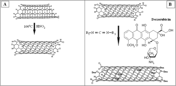
CNT-Dox 입자의 준비
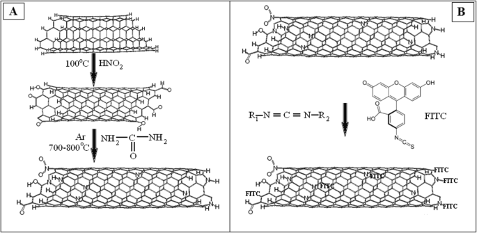
다음 단계의 임무는 MWCNT 입자 표면에 Dox 염산염(Dox, Teva Nederland B.V, 네덜란드)을 고정시키는 것이었습니다. Dox-lactose monohydrate를 포함하는 아미노로 산화된 CNT(200mg)의 표면을 기능화하기 위해 이기능성 연결제인 N-cyclohexyl-N'-(2 morpholinoethyl) carbodiimide metho-p-toluenesulfonate(C 1011, Sigma Aldrich , USA) 실온에서 15분 동안(그림 1a) 그 후, 100mg의 DOX-락토오스 일수화물을 각 나노카본 재료에 10ml의 0.15M 인산 완충액 pH 6.5에 첨가했습니다. 혼합물을 30°C에서 24시간 동안 반응시켰다. 이러한 조건에서 Dox와 CNT 사이에 공유 결합이 형성되었습니다(그림 1b). 조성물을 10,000rpm에서 15분 동안 원심분리하여 수집하고 dH2로 세척했습니다. 오 세 번. 분광 광도계에 의한 유리 DOX의 검출 농도를 위해 상청액을 사용하였다. CNT 표면에 FITS의 고정은 그림 2a, b에 표시된 방식으로 제공되었습니다.
<그림>
아 CNT와 카르보디이미드의 반응 도식. ㄴ 독소루비신에 의한 CNT의 기능화 계획
<그림>
아 CNT와 요소의 반응 방식. ㄴ FITC에 의한 CNT의 기능화 계획
MWCNT의 안정적인 정지 준비
MWCNT의 콜로이드 현탁액은 두 단계로 수행되었습니다. 첫 번째 단계에서 탄소 나노 물질은 초음파 분산기 UZDN-2 T를 사용하여 PBS(인산염 식염수 완충액)에서 초음파 처리되었습니다. 처리 모드는 I였습니다. =10mA, R =22kHz, 지속 시간 - 30분 두 번째 단계에서 생성된 하이드로졸은 실온에서 원심분리에 의해 분산되었습니다. 이 과정에는 여러 원심분리 주기가 포함됩니다. 그래서 친수성 용해 SWCNT 분획을 이렇게 선택하였다. 현탁 배양된 세포에 첨가하기 전에 MWCNT 용액을 30분 동안 끓임으로써 멸균하였다. CNT의 파생물(CNT-Dox 및 CNT-FITC)은 100배 농도의 PSGA(페니실린:스트렙토마이신:젠타마이신:암포테리신)로 멸균되었습니다.
나노튜브 현탁액의 제타 전위
샘플의 제타 전위는 전기영동 광산란(M3-PALS)에 의해 측정되었습니다. 측정을 위해 Malvern Instruments(Malvern, UK)에서 제조한 Zetasizer Nano ZS 분석기를 사용하였다. 실험은 25°C에서 7회 반복하여 수행되었습니다. 샘플을 측정하기 위해 범용 수중 전극(Universal Dip Cell) ZEN1002와 광원인 폴리스티렌 큐벳(DTS001), 즉 H-Ne 633nm 레이저를 사용했습니다.
FTIR 분광기
얻어진 나노물질의 화학적 조성을 푸리에변환적외선(FTIR) 분광법으로 조사하였다. IR 스펙트럼은 FTS 7000e Varian FTIR 분광계로 측정했습니다. 분석용 샘플은 ~ 1mg의 CNT와 150mg의 스펙트럼 순수한 KBr의 혼합물을 분쇄기에서 분쇄하여 준비했습니다. 3.0–3.5 × 10
3
의 압력으로 프레스를 사용하여 샘플을 준비했습니다. kg/cm
2
. 샘플을 600°C의 온도에서 60분 동안 가열하여 탈수했습니다. KBr의 사전 샷 스펙트럼은 예비적으로 얻은 후 샘플의 스펙트럼에서 뺍니다. 모든 스펙트럼은 Spectrometric Identification of Organic Compounds[34] 카탈로그에 따라 분석되었습니다. 비교를 위해 산화된 MWCNT(CNT-O), 독소루비신에 의해 기능화된 MWCNT(CNT-D) 및 형광 표지에 의해 기능화된 MWCNT(CNT-F)의 샘플을 사용했습니다. 샘플과 농도의 모든 처리 및 조건은 세 가지 재료에 대해 동일했습니다.
CNT 표면에서 독소루비신 방출의 비색 분석
CNM에 고정화한 후 유리 Dox의 양과 Dox 방출 효과를 평가하기 위해 495nm 파장에서 자유 Dox가 형광을 내는 능력이 사용되었습니다[35]. Dox 염산염에서 Dox의 활성 농도는 16.7% w였습니다. /와 . 보정선을 그리기 위해 Dox Teva 20mg/ml를 8 × 10
−3
으로 10회 희석합니다. mg/ml를 사용했습니다. 그런 다음, 분광광도법 플레이트 판독기 Multiscan(Labsystem, Finland)을 사용하여 CNT와 DOX 복합체의 상층액에서 유리 Dox의 형광을 측정했습니다.
CNT-DOX, CNT-FITC 입자의 기능기 농도
Dox 고정의 경우 1ml dH2에 MWCNT 1g 오가 사용되었습니다. Dox-TEVA의 양은 1ml dH2에서 100mg(활성 Dox의 16.7mg)이었습니다. O. 반응 후 상등액 중 유리 Dox의 양은 0.37 mg 또는 활성 물질의 2.2%였습니다. 따라서 우리는 16.23mg의 DOX가 1g의 CNTox에 고정되어 있다는 결론을 내렸습니다. FITC의 경우 고정화 비율이 낮았습니다. 그런 다음, 8.35mg의 FITC를 1ml dH2에서 1g의 CNTox에 고정화했습니다. O. 기능화의 효과는 1.62% w였습니다. /와 CNT-DOX 및 0.84% w용 /와 CNT-FITC용. 농도 CNT-Dox 및 CNT-FITC의 추가 계산은 이 데이터를 기반으로 합니다.
2차원 및 3차원 세포 모델에 대한 CNT 유도체의 세포독성 분석
Dox, CNT, CNT-FITC, CNT-Dox의 세포독성은 MTT assay를 이용하여 HT29 종양세포에 대해 평가하였다. 살아있는 세포에서 NAD(P)H 의존성 미토콘드리아 산화환원효소 효소에 의한 3-[4,5-dimetltiazol-2]-2,5-dipheniltetratetrazolium 염의 formazan 결정으로의 전환에 기반한 MTT 테스트. 프로토콜은 T. Mosmann [36]에 의해 설명되었습니다. 간단히 말해서 1 × 10
4
HT29를 96웰 플레이트에 접종하고 전체 배양 배지에서 12시간 동안 배양했습니다. 그런 다음, 현재 배양 배지를 CNTox(샘플 #1), MWCNT(샘플 #2), CNT-Dox(샘플 #3), Dox(샘플 #4) 및 CNT-FITC(샘플 #5)를 포함하는 배양 배지로 교체했습니다. . 샘플 #1, 2, 3, 5의 농도는 12.5–25–50–100–200μg/ml, 샘플 #4의 스톡 농도는 20μg/ml, 최종 농도는 1~10μg/ml입니다. 세포를 전체 배지에서 배양하고 대조군으로 사용하였다. 24시간의 인큐베이션 후 세포를 비색 분석에 의해 MTT로 분석했습니다. 100μl의 세포 현탁액에 20μl의 MTT 용액(5mg/ml PBS, Sigma)을 추가했습니다. 그 후 세포를 표준 조건에서 4시간 동안 MTT와 함께 인큐베이션했습니다. 그런 다음 샘플을 1500g 미만에서 5분 동안 원심분리하고 상층액을 추출했습니다. 전체적으로 MTT 결정 희석을 위해 10μl의 DMSO(Sigma)와 20μl의 25mM 글리신을 웰에 추가했습니다. 반응 용액의 흡광도는 분광광도계 플레이트 판독기 Multiscan(Labsystem, Finland)에서 540nm에서 측정되었습니다.
다세포 종양 회전타원체 생성
종양 미세전이의 모델 시스템인 HT29 세포 다세포 종양 스페로이드(MTS)(3D 배양)는 앞서 설명한 잘 정립된 방법으로 배양되었습니다[33]. 간단히 말해서, 세포 현탁액은 Trypan blue를 사용하여 계산하고 동일한 수의 세포를 심었습니다(5 × 10
4
세포/ml). 3D 세포 배양은 표준 조건(95% 습도, 5% CO2)에서 10% FBS(Sigma, USA)가 포함된 DMEM(Sigma, USA) 배지에서 유지되었습니다. 공기 중, 37°C). MTS의 생성은 우리 연구실에서 개발된 기술에 의해 수행되었습니다. 종양 세포의 배양은 0.24%의 카르복시-메틸-셀룰로오스가 포함된 배양 배지에서 1% 한천으로 코팅된 24웰 플레이트에서 24시간 동안 유지되었습니다. MTS의 크기와 개수가 CNT의 농도와 유형에 미치는 영향을 조사하기 위해 다양한 농도의 CNT가 존재할 때 MTS를 생성하였다. 세포 배양물에 대한 MTS 생성 전에, PBS 중의 CNT 용액을 MTT 분석에 대해 이전에 기재된 바와 같이 최종 농도까지 배양물에 첨가하였다. 오비탈 셰이커(80rpm)에서 플레이트를 일정하게 회전시키면서 48시간 동안 추가 배양을 수행했습니다. 다음 단계에서는 "암시야" 방식으로 마이크로 사진 이미지를 촬영했습니다. 전체적으로 120개 이상의 이미지가 완성되었습니다. 그런 다음 파일에 있는 모든 MTS의 볼륨은 Axio Vision Rel 4.7 프로그램인 Zeiss를 사용하여 계산되었습니다. 우리는 Rolf Bjerkvig의 공식을 사용했습니다:V =0.4 ∙ a ∙ 나
2
, 여기서 회전 타원체의 기하학적 크기(b <아 ) [37]. 결과의 시각화는 Zeiss의 Stemy 2000C 현미경으로 수행되었습니다.
CNT가 간 효소 시스템, 단백질 회전율 및 생체 내 세포 혈액 구성에 미치는 영향 분석
동물과 동물을 돌보는 절차는 지역 동물 실험 윤리 위원회(Protocol №1 21.10.2016)에서 승인한 유럽 지침 2010/63 EU를 준수했습니다. 얻은 물질이 유기체 항상성의 일반적인 조건에 미치는 영향을 분석하기 위해 일련의 생체 내 실험을 수행했습니다. 생체 내 연구에서는 Balb/2a 계통의 마우스를 사용했습니다. 6~8주령의 수컷과 암컷 마우스는 각 그룹에 10마리씩 그룹에서 동등했습니다. 마우스를 강철 와이어 상단과 옥수수 속대 침구가 있는 케이지에 수용하고 12/12 암/광 주기, 22°C ± 3 °C의 온도, 50-70%의 습도로 통제된 분위기에서 유지했으며 자유롭게 접근할 수 있었습니다. 음식과 신선한 물에. 따라서 4개의 마우스 그룹이 형성되었습니다. 그룹 1:온전한 동물을 대조군인 PBS 200μl로 처리했습니다. 그룹 2:마우스에 1.5mg/kg의 CNTox를 투여했습니다. 그룹 3:마우스에 1.5mg/kg의 CNT-DOX를 처리했습니다. 그리고 그룹 4에서는 마우스에게 20mg/kg의 독소루비신을 투여했습니다. 마우스는 4주 동안 3일마다 PBS 200μl에 CNT 비경구 투여를 받았습니다. 독소루비신은 체중 kg당 20mg의 농도로 3일에 한 번 복강 내 투여되었습니다.
혈청 생화학적 분석
한 달 후, 마우스는 실험에서 제외되었습니다. 심장 혈액 샘플은 죽은 동물로부터 즉시 수집되었습니다. 오전 10시부터 11시까지 동시에 동물에서 혈액을 채취했습니다. 혈장의 경우 혈액을 37°C에서 40분 동안 배양한 후 원심분리했습니다(20분, 2000rpm). 그런 다음 생화학적 매개변수, 총 단백질, 알부민, 아스파르테이트 아미노트랜스퍼라제(AST) 및 알라닌 아미노트랜스퍼라제(ALT), 알칼리성 포스파타제(ALP)가 진단 키트(Cormay, Warsaw, Poland)를 사용하여 혈장에서 결정되었습니다. 실험은 반자동 생화학 분석기 FP-901M(Labsystems, Finland)에 대한 통합 실험실 프로토콜에 의해 수행되었습니다. 세포 혈액 조성은 혈액 분석기 Mindray BC-3000 Plus, China에서 측정되었습니다.
통계 분석
3D 배양의 통계 분석을 위해 모든 세포 응집체를 1 × 10
−4
크기에 따라 그룹으로 분류했습니다. mm
3
~ 1 × 10
−2
mm
3
1 × 10
−3
단계로 mm
3
. 그런 다음 각 그룹의 MTS 수와 각 그룹의 MTS 볼륨 중앙값을 추정했습니다. 모든 측정은 세 번 반복되었습니다. 미시 통계 분석 정규 분포 랜덤 변수의 경우 소규모 모집단에 대한 스튜던트 계수를 사용했습니다. 표시된 p 값은 *р였습니다. ≤ 0.05 또는 **р ≤ 0.01.
결과 및 토론
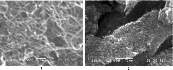
MWCNT의 주사 전자 현미경
프로토콜[38]에 따르면 CNT의 평균 직경은 10~20nm이고 아르곤 탈착에 의해 결정된 비표면적은 200~400m
2
입니다. /G. 20–40g/dm
3
이내의 부피 밀도 . 특히 20~500μm 크기의 얽힌 튜브 형태의 덩어리는 CVD 방법을 적용한 산업용 CNT 생산 과정에서 얻어진다(그림 3).
<그림>
CNT의 SEM 이미지, 1) 스케일 0.5 μm; 2) 스케일 5μm
나노튜브 현탁액의 제타 전위
CNTox 및 CNT는 응집에 대한 상당한 안정성을 나타내며, 이는 제타 전위 값에 직접적으로 의존합니다. CNT-DOX는 CNTox 및 CNT보다 제타 전위가 작고 측정의 재현성이 높아 입자의 균질성을 나타냅니다(표 1). CNT-FITC의 제타 전위의 작은 값은 측정 시 높은 노이즈 비율에서 알 수 있듯이 상당한 입자 크기 또는 작은 농도를 나타낼 수 있습니다.
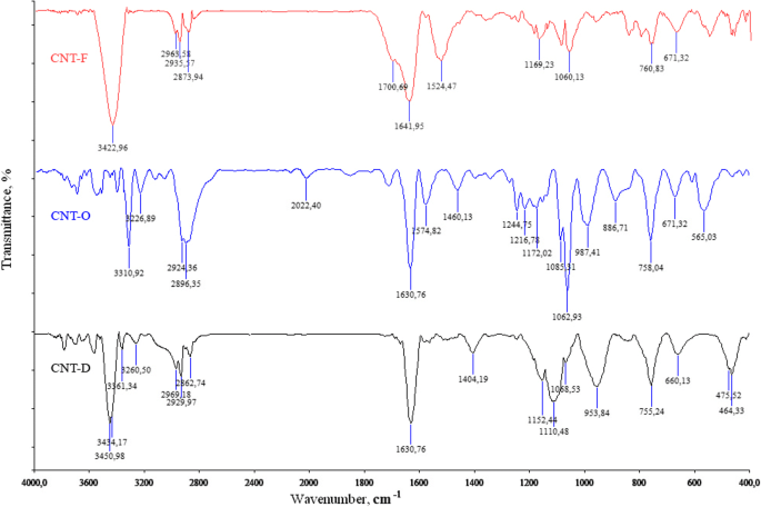
CNT, CNT-Dox 및 CNT-FITC의 FTIR 분광법
CNTox와 독소루비신(DOX) 및 플루오레세인(FITC) 간의 결합은 그림 4에 나타난 바와 같이 적외선 스펙트럼 데이터를 기반으로 추정되었습니다.
<그림>
CNTox(CNT-O), CNT-DOX(CNT-D), CNT-FITC(CNT-F)의 적외선 스펙트럼
υ =3311 cm
−1
에서 강한 밴드의 존재 (CNT-O) 및 υ =3451cm
−1
(CNT-D) CON-H 커플링 변동과 υ =1634 cm
−1
에서 강한 밴드의 존재로 인한 것 (CNT-O) 및 υ =1631cm
−1
(CNT-D)는 CNT와 Dox 사이의 아미드 결합 유형을 명확하게 나타내는 O-CNH 결합 진동에 기인합니다. 또한 IR은 2969–2834cm
-1
에 위치한 C-H 결합의 흡수를 나타냈습니다. 및 1460–1407cm의 광대역
−1
C-O-H 단편에서. 따라서 우리는 화학 반응의 결과로 CNT가 항종양 약물(doxorubicin)과 형광 표지(FITC)로 기능화되었다는 결론을 내릴 수 있습니다.
단층 및 회전타원체 세포 성장 모델에 대한 다양한 기능화 단계에서 CNT의 세포독성
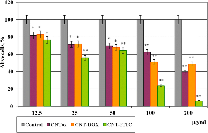
다음 단계는 산화된 CNT(CNTox), 독소루비신으로 기능화된 CNT(CNT-Dox) 및 형광 표지(CNT-FITC)의 세포독성을 결정하는 것이었다. HT29의 단층 배양에 대한 MTT 테스트 결과는 그림 5에 나와 있습니다.
<그림>
CNT 및 그 유도체(CNT-Dox 및 CNT-FITC)와 함께 48시간 동안 배양한 후 단층 배양에서 종양 세포 HT29의 생존력. 통계적 유의성:*р ≤ 0.05 또는 **р ≤ 0.01
그 결과, CNTox, CNT-Dox, CNT-FITC는 12.5μg/ml(81%) 농도에서 중간 정도의 세포독성 효과를 나타내는 것으로 나타났다. CNTox 농도를 25에서 50으로, 100μg/ml로 증가시키면 종양 세포의 생존 능력이 대조군과 비교하여 71.8-69.6-62.5%로 용량 의존적으로 감소했습니다. 최대 200μg/ml의 CNTox 농도에서 HT29의 생존율은 39.2%로 감소했습니다. 그 당시 저농도(12.5-50μg/ml)의 CNT-Dox는 CNTox와 비교하여 통계적으로 유의한 세포독성을 나타내지 않았습니다. 고농도(200μg/ml)에서 CTN-Dox는 CNTox(50%)보다 세포 독성이 훨씬 적습니다. 동시에, 플루오레세인으로 기능화된 CNT는 종양 세포에 대해 상대적으로 더 높은 세포독성 효과를 나타냈다. 25μg/ml 농도의 CNT-FITC와 함께 배양한 후 HT29 세포 생존율은 55%로, 100–200μg/ml에서는 각각 23% 및 7%로 감소했습니다. 따라서 이 경우 CNT는 오히려 불활성 세포 기질의 역할을 합니다. 그 이상으로, CNT는 Dox를 고정화하고 Dox 세포독성을 감소시켰습니다. 이전 연구[33]에서 우리는 1차 나노튜브가 소수성 특성, 중간 정도의 세포독성 효과를 가지며 많은 수의 세포 응집체 형성을 자극한다는 것을 입증했습니다. 문헌[39]에 따르면, CNT의 표면 전하는 산화 동안 변한다. CNT는 더 친수성이 되어 더 작은 응집체를 형성하고 CNT의 세포독성 효과를 증가시킵니다. 이전 연구에서 얻은 데이터는 이러한 경향을 확인시켜줍니다. CNTox는 대조군, CNT-Dox 45%, CNT-FITC 97%(200μg/ml)에 비해 종양 세포의 생존을 최소 60% 감소시켰습니다. 얻은 데이터에 대한 설명은 기능적 리간드가 대부분의 경우 CNT의 표면 제타 전위를 변경하고 CNT의 용해도를 자극하며 CNT의 세포 침투를 증가시킨다는 다른 저자의 보고서에 있을 수 있습니다[40]. 산화 및 기능화 후에 응집체를 형성하는 CNT의 능력은 감소하는 반면, 세포막을 통한 투과성은 증가하고 세포 소기관의 반응성은 증가합니다. 그 이상으로 CNT-Dox는 고농도에서 CNTox보다 세포독성 효과가 낮습니다. 동시에 얻은 데이터에 따르면 펩타이드 결합이 CNT 표면에 독소루비신을 유지한다고 가정할 수 있다. CNT와 Dox 사이의 펩타이드 결합은 세포 배양 조건에서 충분히 안정적입니다. 그것은 분해되지 않으며 비활성 형태의 독소루비신을 취합니다. 동시에, 플루오레세인-나트륨 분자(C20 H12 O5 ) 332.311g/mol 크기의 리간드는 CNT의 표면에서 매우 쉽게 해리되어 세포로 들어갑니다. 그 결과 DNA 구조, 핵 및 미토콘드리아에 돌이킬 수 없는 침해가 발생했습니다[41].
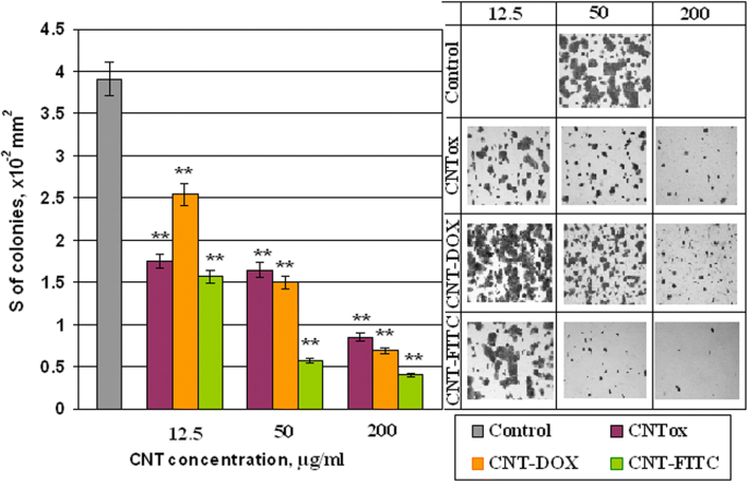
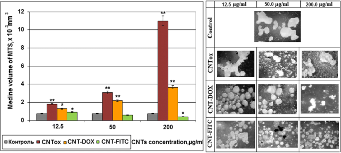
CNT 및 그 유도체의 세포독성과 종양 세포의 접착 특성에 미치는 영향을 확인하기 위해 HT29 세포 2D 집락의 면적을 분석했습니다. 그 결과를 그림 6에 나타내었다. CNT와 그 유도체는 단층 배양에서 종양 세포의 접착력과 종양 세포 콜로니 형성에 영향을 미치는 것으로 나타났다. 12.5, 50.0 및 200μg/ml 농도의 CNTox(샘플 # 1)와 함께 종양 세포의 배양은 대조군과 비교하여 55.2%, 57.8% 및 78.3%에서 2D 세포 집락의 용량 의존적 감소를 초래했습니다. 동시에 동일한 농도의 CNT-Dox 샘플(샘플 #3)에서는 이러한 효과가 발생하지 않았습니다. 2D 콜로니의 면적은 각각 34.8%, 61.6%, 82.4% 감소했습니다. CNT-FITC(샘플 #5)는 단층 배양에서 종양 세포에 가장 큰 영향을 미치는 것으로 나타났습니다. CNT-FITC 배양 후 2D 집락의 면적은 각각 59.8%, 85.2% 및 89.8%로 감소했습니다. 얻어진 결과는 MTT-assay와 동일한 경향을 갖는다는 점에 유의해야 한다. 그러나 MTT 분석의 결과는 그러한 유의한 세포독성 효과를 나타내지 않았다. 결과적인 불일치는 CNT가 세포 콜로니에 미치는 비세포독성 효과와 부분적으로는 항접착제 영향의 결과일 수 있습니다. 이전에 우리는 CNT가 항부착보다 세포독성 능력이 더 낮다는 것을 입증했습니다. 우리의 데이터에 따르면, CNT는 종양 세포의 현탁액 분획으로의 이동 및 다세포 종양 스페로이드(MTS)의 형성을 자극합니다. 단층 및 회전 타원체 배양에서 항종양제에 대한 세포의 민감도는 다릅니다. 그래서 우리는 다음 단계에서 CNTox, CNT-Dox 및 CNT-FITC가 있는 상태에서 종양 세포 HT29에 의한 MTS 형성을 조사했습니다. CNT의 농도는 이전 실험과 동일하였다. 그림 7에서 CNTox(샘플 # 1)가 MTS의 형성을 자극할 수 있음이 입증되었습니다. CNTox 농도의 증가는 MTS의 중간 부피의 용량 의존적 증가를 동반합니다. 12.5, 50, 200μg/ml의 CNTox 농도에서 MTS의 중간 부피는 1.79에서 2.18 및 10.98mm
3
로 증가합니다.; 거의 10배입니다. CNT의 유도체는 종양 세포에 이러한 효과를 일으키지 않습니다. CNT-Dox(샘플 #3)는 2.83배에서 MTS의 형성을 자극합니다. 동시에 3D 종양 세포 배양과 CNT-FITC(샘플 # 5)를 배양하면 MTS 부피가 2.4배 감소합니다.
<그림>
CNTox(#1), CNT-Dox(#3), CNT-FITC(#5)와 함께 배양된 단층 배양에서 HT29 종양 세포의 콜로니 면적. 통계적 유의성:*р ≤ 0.05 또는 **р ≤ 0.01
대부분의 실험에서 CNT-Dox는 단일 독소루비신의 효과와 비교할 수 있는 2D 및 3D 배양 모두에서 종양 세포에 대해 이러한 세포 독성 효과가 없다는 점에 주목해야 합니다. The mechanism of cytotoxic action of doxorubicin is based on penetration into the cell nucleus and intercalation between nucleotide pairs, violation of replication and DNA repair, protein synthesis and, as a result, cell death. The cause of the reduction of cytotoxic effect of the CNT-Dox may be the binding of the doxorubicin with CNT surface through peptide bonds. It prevents Dox dissociation from CNT surface and Dox penetration into the cell and cellular organelles. This fact may let to deliver the compound to certain tissues without causing a negative effect on “non-target” objects.
In this case, the next step of the study was controlled release of doxorubicin. For peptide bonds breaking, we used the commonly known peptidase trypsin as the release agent. The effect of trypsin on the Dox release from the surface of the CNT was investigated by spectral analysis. Since the free doxorubicin has a fluorescence peak at 495 nm, bounded doxorubicin has no such ability. It was analyzed how the increasing of trypsin concentration affected on concentration of free doxorubicin. The results are shown in our previous work [42]. Shortly, it was demonstrated that increasing the concentration of 0.05% trypsin from 11 to 20% in culture medium contributed to an increasing the concentration of free doxorubicin from 3.13 to 6.55 μg/ml. A further increasing the concentration of trypsin to 60% did not lead to increasing in free doxorubicin. However, the concentration of trypsin by about 66% stimulated release again and led to increasing the concentration of doxorubicin in two times, to 11.38 μg/ml. Thus, it was concluded that 0.05% trypsin has several effective concentrations. Under these conditions, peptide bonds between doxorubicin and CNT were broken and doxorubicin was released. Therefore, to analyze how CNT-Dox influence the tumor cells after Dox release, we incubated tumor cells in the w/o fetal bovine serum (FBS) nutrient medium with trypsin and in the presence of CNT-Dox. The survival of tumor cells was determined using the MTT test. The ratio of the nutrient medium, trypsin, concentration of CNT-Dox, and doxorubicin are given in Table 2. The results of incubation HT29 during 48 h are demonstrated on Fig. 8. Cell viability in the presence of trypsin—blue columns, alone doxorubicin—orange columns, and CNT-Dox with trypsin—pink columns. In results, it has been found that the simultaneous use of trypsin and CNT-Dox significantly increased the cytotoxic effect of CNT and doxorubicin compared to the separately use of these substances. So doxorubicin alone at concentrations from 0.05 to 1.0 μg/ml causes a dose-dependent decreasing the percentage of living HT29 cells from 7.18 to 45.7% relative to control (Fig. 8, orange columns). Incubation of CNT-Dox at concentrations of 20.0 to 1.25 μg/ml with trypsin at concentration from 0 t0 70% led to a dose-dependent decreasing in the percentage of living cells from 80.9 and 99.8% respectively (Fig. 8, pink columns). At the same time, incubation with trypsin alone leads to decreasing percentage of living cells only by 34.0–42.0% (Fig. 8, blue columns). Thus, we can conclude that we observe the synergistic effect of CNT-DOX and trypsin. Each individual component has a small cytotoxic effect on cells, and together the cytotoxic potential of substances increases several times.
Percentage of alive cells HT29 after 48 h of incubation in the presence of trypsin, doxorubicin, CNT-Dox. Statistical significance:*р ≤ 0.05 or **р ≤ 0.01
As functional groups (Dox) were attached to CNTs surface by peptide bonds as, in vivo Dox release will be realized in organs of the gastrointestinal tract with an increased content of proteases, peptidases. In organism, proteases are used for various metabolic processes. Acid proteases secreted into the stomach (such as pepsin) and serine proteases present in duodenum (trypsin and chymotrypsin) enable us to digest the protein in food [43]. Other proteases are present in leukocytes (elastase, cathepsin G) and play several different roles in metabolic control. This is one of the fastest “switching on” and “switching off” regulatory mechanisms in the physiology of an organism. By complex cooperative action, the proteases may proceed as cascade reactions, which result in rapid and efficient amplification of an organism’s response to a physiological signal. Therefore, the authors suggest that CNT-Dox construction will be the most effective in the case of stomach, pancreas, liver, and small intestine cancer localization in case of parenteral administration of drug.
Influence of CNTs and Their Derivatives on Cell Blood Composition, Hepatic Enzyme System, and Proteins Turnover In Vivo
To determine the impact of the CNTox and CNT-Dox on the protein metabolism and the state of the hepatic enzyme system, the level of albumin (Al), total protein (Tp), alanine aminotransferase (ALT), aspartate aminotransferase (AST), and alkaline phosphatase (ALP) was determined. Data are given in Fig. 9a, b. As a result, it was noted that CNT-Dox and Dox have influence on the hepatic enzymes activity, namely on the AST and ALP. AST level increased in mice serum from 1 group for 21.6%, 2 group for 93.9%, and 3 group for 126.4% compared with control. At the same time, the activity of ALP increased in mice serum from 1 group for 23.5%, 2 group for 119.1%, and 3 group for 147.8%. CNTox administration did not show statistically significant changes in AST, ALT, and ALP levels. In addition, it should be noted that CNTox, Doxorubicin, and CNT-Dox slightly increased the level of total protein in the blood of experimental animals. So, we found that systematic introduction of CNTox have not influence on mice serum enzyme profile. And opposite, administration of CNT-Dox and Dox had toxicity influence and induced a chronic hepatic damage. Notably, symptoms of hepatitis had more manifestation after Dox treatment than CNT-Dox. To determine possible inflammatory processes and systemic effects of CNTox, CNT-Dox on the state of blood cells composition, it was analyzed the blood cell formula of the experimental animals. The results are shown in Table 3. Obtained data are in good agreement with the well-known manifestations of doxorubicin’s hematological toxicity:anemia (decreased red blood cells, hemoglobin, hematocrit), thrombopenia (platelet count), neutropenia (decrease in the number of granulocytes). Interestingly, that it was demonstrated the same direction of the changes of practically all hematological parameters in the 1 (CNTox), 2 (CNT-Dox), and 3 (Dox) groups of animals. However, the change in the 2 and 3 groups of animals was statistically significant accordingly to control and 1 group. In the 2 group (CNT-Dox), it was found pronounced decreasing of the monocyte content related to the system of phagocyte mononuclear cells and cell’s immune response (9.5% and 0.12 × 10
9
/l in control, 4.6% and 0.06 × 10
9
/l in 2 group, and 4.55 and 0.05 × 10
9
/l in 3 group). Animals of 2 and 3 groups demonstrated reducing the content of granulocytes (neutrophils) (12.6 and 0.16 × 10
9
/l in control and 8.5% and 0.2 × 10
9
/l in 2 group and 8.05 and 0.16 × 10
9
/l in 3 group). The number of lymphocytes, both in percentage and absolute, increased in 2 and 3 groups (77.9% and 0.97 × 10
9
/l in control and 86.9% and 2.0 × 10
9
/l in 2 group and 87.8% and 2.3 × 10
9
/l in 3 group). The main function of the lymphocytes is recognition of the antigen and participation in the adequate immunological response of the body. T lymphocytes perform regulatory and effectors functions. B lymphocytes take part in humoral immunity, providing immunoglobulins in response to stimulation of other people’s antigens. So, it can be assumed that an increase in the index of lymphocyte content in experimental 1, 2, and 3 groups can be associated with the introduction of foreign antibodies (CNTs) and/or tissue distraction [44]. Some decrease in the absolute number of leukocytes in 2 (1.2 × 10
9
/l) and 3 (0.85 × 10
9
/l) groups compared with the 1 group of animals (2.0 × 10
9
/l) can be regarded as the result of a gradual accumulation of the reversal of toxic effects of Dox. It is described the reaction of WBC in experimental animal groups as well-known toxic hematologic impact from doxorubicin in the development of leucopenia. The analysis of indicators reflecting the number and state of RBC (erythrocytes and hemoglobin) also showed the same trend of misbalance in the 2 and 3 groups of animals. Statistically significant thrombocytopenia and anemia are more indicative in animals of 2 and 3 groups:the number of erythrocytes (5.54 × 10
12
/l in control, 3.97 × 10
12
/l in 2 group, and 3.12 × 10
12
/l in 3 group), the amount of hemoglobin (78 g/dl for intact animals, 55.5 g/dl for 2 group, and 46.2 g/dl for 3 group), hemoglobin (25.25% for intact animals and 16.75% for 2 group and 15.12% for 3 group). The amount of erythrocytes decreased but the average content of hemoglobin in one erythrocyte did not decrease. And, as a result, concentration of hemoglobin per one erythrocyte increased. Analysis of the RBW counts let us suggest that in 2 and 3 group of animals, there are several processes:decreasing the formation of erythrocytes in the bone marrow, the acceleration of erythrocyte destruction, the violation of the structure of membranes of red blood cells, and molecular defects (oxidation) of hemoglobin. Thrombocytopenia (27 × 10
9
/l in intact animals, 14.0 × 10
9
/l in 2 group, and 12.05 × 10
9
/l in 3 group), a dose-dependent reversible myelosuppression, leucopenia and granulocytopenia (neutropenia) are the predominant manifestations of doxorubicin hematologic toxicity and is the most common acute dose-limiting toxicity of this drug. The direction of changes in the platelet content in the blood of experimental animals treated with CNTox and CNT-Dox is the same; however, significantly more pronounced in the group of animals receiving free Dox.
Monitoring of clinical parameters following CNTox (1 group), CNT-DOX (2 group), free DOX (3 group) administration in Balb/2a mice. Mice were treated by CNTox, CNT-DOX, DOX every 3 days during 4 weeks, in concentrations:CNT—1.5 g/kg and Dox—20 mg/kg of weight. Each experiment was done in triplicate. Statistical significance:*р ≤ 0.05 or **р ≤ 0.01. ALT alanine transferase, AST aspartate transferase, AP alkaline phosphatase
Thus, according to our results from in vivo experiments, it was demonstrated that systematic introduction of CNTox have not influence on mice serum enzyme profile and protein turnover. And opposite, administration of CNT-Dox and Dox had toxicity influence and induced a chronic hepatic damage. More than that, dose-dependent reversible myelosuppression, leucopenia, and granulocytopenia (neutropenia) was shown for the group of animals receiving free Dox. This predominant manifestation of hematologic toxicity was less pronounced in the group of animals receiving free CNT-Dox and CNTs. So, we can assume that, in the case of parenteral administration of CNT-Dox under action of gastric juice peptidases, CNT-Dox particles breaks up into free CNT and Doxorubicin. After that, Doxorubicin enters to the stomach cells and partly in the bloodstream. That also causes the specified effect on blood cells composition. For modeling Dox release in vivo, trypsin were used in vitro.
One of the goals of this investigation was to minimize the side effects of carbon vehicle on the body. Previously, the authors demonstrated that CNTs may be a potential threat to tumor development due to its ability to stimulate cell migration and support cells in suspension fraction [33]. In this case, CNTs themselves play a role of artificial extra cellular matrix. Another way CNTs can stimulate suspension cells to aggregation. If this process will be coincident with antitumor drug accumulation on CNTs surface, CNTs will attract substrate-independent cells and kill them. Simultaneously, it was found that pure CNTs did not have statistically significant cytotoxicity to tumor cells. So, CNTs alone are not dangerous as cytotoxic agents and can act as carrier for antitumor drugs to target cells. Then, studies were conducted to the functionalization of the CNTs by antitumor antibodies. It was demonstrated that CNTs are able to carry on the surface not only the Dox but also specific tumor antibodies—anti EGFr [42]. Under the action of trypsin, Dox was released and realized strong cytotoxic effect on tumor cells. Recent studies have shown the synergy cytotoxic effect of CNTs and Doxorubicin after release from CNT-Dox construct in the presence of trypsin. Such effect was not demonstrated by CNTs and Dox separately. Therefore, the benefits from the use of CNTs seems in the usage CNTs as vehicle for antitumor antibody and drug. On the other hand, the negative effects of Doxorubicin, like many other early antitumor drugs, are totally cytotoxic effects. Binding of the Dox to the CNTs partially blocks it. This effect allows ensuring local accumulation of the Dox in the focus of tumor activity with subsequent release and action. Thus, greater efficacy can be achieved with a smaller dose of administration. This effect can be achieved only if there are tumor-specific antibodies on the surface of the CNTs. The possibility of creating such construct was demonstrated by the authors in previous work. Undoubtedly, that CNTs dissemination, accumulation, and Dox effective should be tested on mixed culture in vitro and on a tumor model in vivo. For this purpose, synthesized CNT-FITC was created and conducted. The ability of the CNTs to act as an extra-cellular matrix and the significance of this process for the “tumor-body” system should be tested on the animal model. The effectiveness of the drug will be investigated on the model of Ehrlich carcinoma and colon rectal carcinoma [44]. The distribution of CNTs in the tissues of the body will be investigated using the construct created by the CST-FITC, as was mentioned in this work. The accumulation of CNTs in the tissues of the organs of the gastrointestinal system, namely the stomach, liver, and intestines, will be analyzed by histological assay. These studies are already conducted by the authors.
According to Shang-Lin Wang [4] and our data, free doxorubicin causes similar side effects, but more pronounced than CNT-Dox. Other authors reported that toxicity effect of CNTs depends on way of administration, dose, and time of exposure and varies according to the size and type of the cells [45,46,47,48,49]. Settling of CNTs in the tissues of the liver, kidneys, and stomach with prolonged exposure can cause oxidative stress, DNA damages, compromise cell proliferation, necrosis of tissues, and chronic inflammation [50]. According to Manna [51], CNTs can cause oxidative stress and compromise cell proliferation. In the case of in vitro studies, the cytotoxicity of CNT highly depends on the degree of CNTs purification, functionalization, size, and surface charge [52,53,54,55]. According to our results and previous data, the obtained CNTs did not demonstrate a significant cytotoxic effect [33]. More than that, our data let us suggest that there is a combined cytotoxic effect of CNTs and DOX that occurs as in vitro. That is why we assumed that obtained CNTs can be used against tumor cells, in case of the target accumulation of CNTs/CNT-Dox in tumor tissue. However, mechanisms of the cytotoxic effect of CNTs are not completely clear. Some authors suggested that CNT particles activate NF-kappaB pathway depending on the dose and that the mechanism of activation was due to activation of stress-related kinases [51]. Other authors reported that CNTs have a direct pro-apoptotic effect in vitro in different cancer cell lines and tumor cells obtained from surgical specimens [56], CNTs able to modify fatty acids in cell membranes [57] or erythrocyte membrane damage [58]. For further investigation of accumulation CNTs in cells and tissue, we plan to use CNT-FITC composite.
Other unclear questions are as follows:what is the prolonged impact of CNTs/CNT-Dox on organism level and how does it stimulate accumulation CNTs/CNT-Dox in tumor tissue? To investigate the influence of chronical introduction of CNTs/CNT-Dox, long-time studies on model of transplanted or initiated tumors of various localizations will be realized. Other authors analyzed the distribution of nanotubes throughout the body of mice after pulmonary exposure [59]. In results, accumulation of MWCNTs was documented in several organs, including notably the white pulp of the spleen and the bone marrow. The CNTs usually deposit in the liver, spleen, or lungs after they have served their purpose from where they are expelled gradually out of the body through the renal excretion route [60]. Zhao and Liu reported that accumulation of CNTs in the body can lead to granulomatous inflammation or alveolar septal thickening. Zhuang Liu et al. reported that SWNT-PTX affords higher efficacy in suppressing tumor growth than clinical Taxol® in a murine 4T1 breast-cancer model, owing to prolonged blood circulation and tenfold higher tumor paclitaxel (PTX) uptake by SWNT delivery likely through enhanced permeability and retention [61]. Accumulation of CNTs in the target tissue can be enhanced by placing on the CNTs surface specific antibodies which over expressed by tumor cells. The possibility of using antibodies for the targeted delivery of nanostructured preparations has been described by many authors [62, 63].
Conclusion
In 2D culture CNTox concentration from 25 to 50 and 100 μg/ml led to dose-dependent decreasing the viability of tumor cells to 71.8–69.6–62.5% accordingly compared with control. At concentration of CNTox up to 200 μg/ml, the viability of HT29 decreased to 39.2%. At that time, CNT-Dox at concentrations 12.5–25–50 μg/ml did not show statistically significant cytotoxicity, compared with CNTox. At high concentrations (200 μg/ml), CTN-Dox had even less cytotoxicity than CNTox (50%). After incubation with CNT-FITC in concentration of 25 μg/ml, HT29 cell survival reduced to 55% and at 100–200 μg/ml to 23 and 7% respectively. In 3D culture, increasing of CNTox concentration is accompanied with dose-dependent increasing of median volume of MTS. At concentration CNTox from 12.5, 50, 200 μg/ml median volume of MTS increases from 1.79 to 2.18 and 10.98 mm
3
. CNTs-Dox and CNT-FITC did not cause such effect on tumor cells. Doxorubicin alone at concentrations from 0.05 to 1.0 μg/ml causes a dose-dependent decrease in the percentage of living HT29 cells from 7.18 to 45.7% relative to control. Incubation CNT-Dox at concentrations of 20.0 to 1.25 μg/ml with trypsin at concentration from 0 to 70% led to a dose-dependent decreasing the percentage of living cells from 80.9 and 99.8% respectively. At the same time, incubation with trypsin alone leads to decreasing percentage of living cells only by 34.0–42.0%. So, the synergistic effect of CNT-DOX and trypsin was observed. Each individual component has a small cytotoxic effect on cells, and together the cytotoxic potential of substances increases several times. In vivo, systematic introduction of CNTox have not influence on mice serum enzyme profile. And opposite, administration of CNT-Dox and Dox had toxicity influence and induced a chronic hepatic damage, thrombocytopenia, a dose-dependent reversible myelosuppression, leucopenia, and granulocytopenia (neutropenia). Notably, symptoms of hepatitis had more manifestation after Dox treatment than CNT-Dox.
So, the possibility of targeted delivery and controlled release of any highly toxic drug with the use of CNT depends on strategies of reducing toxicity of CNTs and realizing potential of CNTs and anti-tumor drugs “in right place in right time.” According to the data obtained by authors and literature, it is possible. Our data support the assumption that this approach allows to reduce toxicity of the doxorubicin on the general biochemical indicators of blood and violations in the blood cells composition. At the same time, doxorubicin releasing is realized under certain conditions. And combined effect of CNTs and doxorubicin let us achieve greater efficacy in suppressing tumor cell growth in vitro.
약어
Al:
Albumin
ALP:
Alkaline phosphatase
ALT:
Alanine aminotransferase
AST:
Aspartate aminotransferase
CNT-Dox:
Carbon nanotubes which were functionalized by doxorubicin
CNT-FITC:
Carbon nanotubes which were functionalized by fluorescein