동결 건조 단상 용액 방법을 사용한 글리시레틴산 리포솜의 제조:사전 제형화, 최적화 및 시험관 평가
초록
이 연구에서 글리시레틴산(GA) 리포솜은 동결건조 단상 용액 방법을 사용하여 성공적으로 제조되었습니다. 예비 제형 연구는 tert-부틸 알코올(TBA)/물 공용매에서 대두 포스파티딜콜린(SPC), 콜레스테롤 및 GA의 용해도 평가로 구성되었습니다. 승화율에 대한 TBA 부피 백분율의 영향을 조사하였다. 다른 부피 백분율을 가진 TBA/물 공용매를 사용한 동결건조 후 GA는 DSC, XRD 및 FTIR에 의해 물리화학적으로 특성화되었습니다. GA의 XRD 패턴은 명백한 무정형 특성을 보여줍니다. FTIR 분광법 결과는 화학적 구조적 변화가 발생하지 않았음을 보여줍니다. 용해도 연구는 GA의 수용해도가 향상되었음을 보여줍니다. Box-Benhnken 설계 및 동결 보호제의 선택 실험을 통해 조사한 후 508 mg SPC, 151 mg 콜레스테롤, 55% TBA 부피 비율, 4:1 트레할로스/SPC 중량비의 최적 제제 및 가공 변수를 얻었습니다. 최적의 조건에서 재구성된 리포솜의 만족스러운 캡슐화 효율(74.87%)과 평균 직경(191nm)이 얻어졌습니다. 시험관 내 약물 방출 연구는 재구성된 리포솜이 두 종류의 방출 매질에서 지속 방출 특성을 갖는 것으로 나타났습니다. 더욱이, 시험관내 세포 흡수 연구는 Hep G2 세포에 의한 약물 로딩 리포솜의 흡수 과정이 시간 의존적임을 밝혀냈습니다.
<섹션 데이터-제목="배경">
배경
트리테르펜 사포닌의 한 종류인 Glycyrrhetinic acid(GA)는 주로 한약재인 glycyrrhiza의 뿌리에서 추출됩니다[1]. 연구에 따르면 GA는 명백한 항균, 항바이러스 및 항암 효과가 있으며 만성 간염 및 간암의 임상 치료에 일반적으로 사용됩니다[2,3,4]. Biopharmaceutical Classification System에 따르면 GA는 유형 II 약물입니다. GA 분자의 낮은 극성, 높은 소수성 및 낮은 용해도로 인해 경구 생체이용률이 상대적으로 낮습니다[5]. 더욱이 GA는 고혈압과 관련된 나트륨 저류 및 칼륨 손실을 유발할 수 있으며[6], GA의 부작용은 용량 의존적인 것으로 보입니다. 따라서 적절한 제제 전략을 사용하여 GA의 흡수를 증가시키고 효과적인 농도를 유지하면 생체 이용률과 안전성이 크게 향상됩니다.
약물 운반체로서의 리포솜의 우수성은 널리 알려져 있다[7,8,9]. 그들의 기능적 이점은 주로 다음 측면을 통해 입증됩니다. (1) 리포솜은 우수한 생체 적합성과 안전성을 가지고 있습니다. (2) 리포솜은 림프절로의 표적 약물 전달을 향상시키고 항암제가 정상 세포 및 조직에 미치는 억제 효과 또는 손상을 감소시킨다. (3) 적절한 크기의 약물 운반체 리포솜은 모세혈관 투과성이 증가된 고형 종양, 감염 및 염증 부위에서 향상된 투과성 및 체류 효과를 나타내어 수동 표적화 능력을 입증한다. (4) 리포솜은 소수성 및 수용성 약물을 모두 운반할 수 있습니다. (5) 리포솜 표면이 변형되어 작용기에 연결될 수 있습니다. 이러한 유리한 특성으로 인해 많은 리포솜 약물이 승인되었습니다.
통상적인 리포좀 제조 방법에 의해 수득된 생성물은 수성 리포좀 현탁액이다. 그러나 수성 리포좀 현탁액은 상대적으로 불안정하고 저장 중 누출, 융합 및 인지질 가수분해를 겪을 수 있어 장기 저장 능력이 제한된다[10]. 현재 이러한 문제를 효과적으로 해결할 수 있는 방법은 proliposome을 준비하는 것이다[11]. 프로리포좀은 탈수된 리포좀 성분과 부형제로 만든 유동성이 좋은 분말입니다. 리포좀은 적용하기 전에 물에 프로리포좀을 분산시켜 재구성할 수 있습니다. 분무 건조와 동결 건조는 프로리포좀 제조를 위한 가장 일반적인 두 가지 방법이지만[12], 적용에 몇 가지 제한이 있습니다. 예를 들어, 분무 건조는 열에 민감한 약물에 적합하지 않으며 장비의 낮은 열 효율로 인해 벽 접착과 같은 문제를 일으킬 수 있습니다. 리포솜 이중층의 구조적 재배열은 분무 건조 과정에서 발생할 수 있습니다[13]. 일반적으로 사용되는 동결건조 방식은 현탁방식이지만 물은 동결건조 시간이 오래 걸리기 때문에 비용과 시간이 많이 소요된다.
최근 몇 년 동안 새로운 proliposomes 제조 방법(동결 건조 단상 용액 방법)이 개발되었습니다[14, 15]. 이 방법은 지질, 약물 및 수용성 동결 보호제를 tert-부틸 알코올(TBA)/물 공용매 시스템에 용해시킨 다음, 동결 건조에 의해 프로리포솜을 얻은 다음, 물을 첨가하여 균일한 리포솜 현탁액을 형성하는 것을 포함합니다. 이 방법은 다음과 같은 몇 가지 장점이 있습니다. (1) TBA를 추가하면 얼음의 승화 속도가 크게 향상되어 경제적으로 유리한 신속하고 완전한 동결건조가 가능합니다. 동시에 급속 승화는 덩어리가 무너지는 것을 방지하는 데 도움이 됩니다[16]. (2) 동결건조 단상 용액 기술은 1단계 공정으로 대규모 리포솜 제조에 매우 효과적인 방법입니다. (3) 잔류 용매에 대한 ICH 가이드라인에 나열되지 않았지만 TBA는 LD50의 유사성을 기반으로 하여 클래스 3 저독성 용매 범주에 속할 가능성이 있습니다. 다른 클래스 3 용매에 대한 독성 데이터 [17]. (4) 이 방법으로 멸균 분말을 얻을 수 있다. (5) 수용성이 좋지 않거나 물에 대한 안정성이 떨어지는 약제에 적합하다[18].
리포솜 준비를 위한 TBA/물 동결건조 시스템의 사용에 대한 몇 가지 보고가 있습니다. 그러나 이 시스템에 대한 연구는 미흡하고 여전히 많은 의문점이 남아 있다. 예를 들어, 농도가 다른 TBA/물 시스템의 승화 속도의 변화, 농도가 다른 TBA/물 시스템에 의한 동결건조 후 특정 약물의 고체 상태 변화, 동결건조 분말의 수화 및 조립 과정은 다음과 같습니다. 아직 불명. 다른 한편, 다른 비율과 온도를 가진 TBA/물 공용매에서 특정 소수성 약물의 용해도는 매우 특이적입니다. 위의 정보는 약물 운반체 리포솜의 제형 및 기술 설계에 중요합니다. 따라서 본 연구에서는 GA를 모델 약물로 사용하여 위에서 설명한 바와 같이 사전 제제 조사를 수행했습니다. 또한, 평균 직경과 포획 효율을 1차 평가 수단으로 사용하여 Box-Benhnken 설계를 사용하여 동결 건조 단상 용액 방법으로 제조된 GA-리포좀의 제형 및 처리 변수를 최적화했습니다. 동결건조 보호제 유형이 리포솜의 품질에 미치는 영향과 리포솜의 시험관 내 방출 및 간암 세포에 의한 흡수를 평가했습니다.
방법/실험
자료
글리시레틴산(> 98% 순도)은 Dalian Meilun Biology Technology Co., Ltd.(Dalian, China)에서 입수했습니다. 대두 포스파티딜콜린(Lipoid S100)은 Lipoid GmbH(독일 Ludwigshafen)에서 구입했습니다. 콜레스테롤은 J&K Scientific Ltd.(중국 베이징)에서 구입했습니다. GA의 참조 화합물은 National Institutes for Food and Drug Control(Beijing, China)에서 구입했습니다. FITC-PEG-DSPE(분자량 2000)는 Shanghai Ponsure Biotech, Inc.(Shanghai, China)에서 구입했습니다. 3차 부틸 알코올(> 98%) 및 기타 모든 시약은 달리 명시되지 않는 한 Sinopharm Chemical Reagent Co., Ltd.(Beijing, China)에서 구입했습니다. 탈이온수는 Milli-Q 정수 시스템(Millipore, Bedford, MA, USA)에 의해 준비되었습니다.
TBA/물 공용매 시스템에서 GA, SPC 및 콜레스테롤의 용해도 연구
25°C, 30°C, 35°C, 40°C 및 45°C에서 해당 비히클에서 과량의 약물을 교반하여 다른 TBA 부피 백분율을 갖는 GA의 포화 TBA-물 용액(30ml)을 제조했습니다. 72시간 동안 원심분리(3000rpm에서 15분) 후, 상층액을 0.45μm 미세다공성 필터에 통과시켰습니다. GA의 포화 용해도는 적절히 희석한 후 HPLC로 측정하였다. 각 TBA/물 공용매에서 3번의 복제를 수행했습니다. HPLC 분석은 4차 펌프, 자동 시료 주입기 및 UV 검출기에 연결된 컬럼 구획이 장착된 LabAlliance(모델 시리즈 III) HPLC 시스템(Lab Alliance, Tianjin, China)에서 수행되었습니다. C18 컬럼(4.6mm × 250mm, 5μm, Dikma Technologies, Beijing, China)에서 분리를 수행했습니다. 메탄올 및 물(90:10 V /V )를 1.0 ml/min의 유속으로 이동상으로 사용했습니다. 분석물은 250nm에서 UV 검출기로 검출되었습니다.
TBA/물 공용매 시스템에서 대두 포스파티딜콜린(SPC)(또는 콜레스테롤)의 용해도는 탁도법을 사용하여 추정되었습니다[19, 20]. 간단히 말해서, 10mg SPC(또는 콜레스테롤)를 25°C, 30°C, 35°C, 40°C 및 45°C에서 TBA에 용해하여 투명한 용액을 얻었습니다. 온도는 실험 기간 내내 유지되었습니다. 25°C의 SPC(또는 콜레스테롤)의 TBA 용액에 동일한 온도에서 점점 더 많은 양의 정제수를 탁도가 처음 발생할 때까지 첨가하고 임계 물 부피 값을 기록했습니다. 탁도는 T6 모델 UV-Vis 분광 광도계(Purkinje General Instrument Co., Ltd., Beijing)에서 블랭크 용액(정제수)에 대해 655nm(> 0.04)의 흡수 값을 감지하여 식별할 수 있습니다.
동결건조 단상 용액법을 사용한 리포솜 제조
GA, SPC 및 콜레스테롤은 45°C에서 TBA에 용해되었고 만니톨, 유당, 자당, 트레할로스와 같은 수용성 동결 보호제는 45°C 물에 용해되었습니다. 그런 다음 이 두 용액을 적절한 비율로 혼합하여 세 번째 투명한 등방성 단상 용액(총 부피 60ml)을 얻었습니다. 단상 용액을 0.22μm 기공을 통해 여과하여 멸균한 후, 이를 2.0ml의 충전 부피로 10ml 동결 건조 바이알에 채웠습니다. − 40°C에서 12시간 동안 사전 동결 후, 동결 건조기(SJIA-10N, Ningbo Shuangjia Science Technology Development Co., Ltd., 중국).
리포솜의 입자 크기 및 캡슐화 효율 측정
리포좀 현탁액은 5mg의 프로리포좀 분말을 5ml의 정제수에 첨가한 후 완전한 수화를 위해 15분 간격으로 1분 동안 2회 와류 교반하여 제조했습니다. 리포솜의 크기 분석은 레이저 입자 크기 분석기(Nano ZS90 Malvern Instruments, UK)를 사용하여 특성화되었습니다.
리포솜에서 GA의 캡슐화 효율은 한외여과-원심분리 기술에 의해 결정되었습니다. 간단히 말해서, 리포솜 분산액 1ml(정제수 5ml에 프로리포좀 500μg)을 10ml 부피 플라스크에 피펫으로 넣은 다음 정제수 5ml, 아세톤 2ml를 추가하고 정제수로 10ml로 희석합니다. 이 현탁액 0.5ml를 분자량이 50kDa인 원심분리 필터(Amicon Ultra-0.5, Millipore, Cdduounty Cork, Ireland)의 상부 챔버로 옮기고, 이를 사용하여 15°C에서 30분 동안 10,000rpm으로 원심분리합니다. 초원심분리기(CP70MX, Hitachi Koki Co., Ltd., 일본). 그런 다음 한외여과액 20μl를 UV 흡수 파장 250nm에서 HPLC 시스템에 주입하고 GA의 함량을 유리 약물 함량이라고 했습니다. 캡슐화 효율(EE)은 다음 방정식에 따라 계산되었습니다.
다양한 TBA 부피 백분율(10%, 20%, 30%, 40%, 50%, 60%, 70%, 80%, 90%)의 TBA/물 혼합물 1밀리리터를 10ml 동결 건조 바이알에 넣었습니다. , 각각. TBA/물 혼합물을 − 40°C에서 12시간 동안 미리 동결한 다음 동결건조기(SJIA-10N, Ningbo Shuangjia Science Technology Development Co., Ltd., 중국)로 - 50°C에서 동결건조했습니다. TBA/물 혼합물이 동결건조 바이알에서 완전히 사라질 때까지의 시간을 기록하였고, 승화율은 부피(μl)를 시간(분)으로 나누어 계산하였다.
TBA/물 혼합물의 포화 증기압 측정
실험 장치 및 작동 절차에 대한 자세한 내용은 다른 곳에서 설명되었습니다[21, 22]. 시스템 TBA/물(10%, 20%, 30%, 40%, 50%, 60%, 70%, 80% 및 90%)의 증기압은 정적 방법으로 측정되었습니다. 이 장치는 TBA/물 혼합물로 채워진 작동 비포계, 순수한 물로 채워진 참조 비포계, 완충 용기, 두 개의 응축기, 두 개의 온도 측정 및 압력 제어 시스템으로 구성되었습니다. 시스템의 평형 압력은 Antoine 방정식[23]으로 표시되는 온도-압력 관계의 관점에서 기준 ebulliometer에서 순수한 물의 끓는 온도에 의해 결정됩니다.
GA 용해도 측정
유리 GA의 물에 대한 용해도는 온도 조절식 수조(DF-101S, Henan Yuhua instrument Co., Ltd., 중국) 25°C에서 평형에 도달할 때까지(48시간) 샘플을 0.45μm 멤브레인 필터를 통해 여과하고 메탄올로 적절하게 희석하고 HPLC로 분석했습니다[24]. 실험은 세 번 수행되었습니다.
미리 동결된 TBA/물 혼합물의 표면 형태 관찰
5ml의 물/tert-부탄올 혼합물을 90mm 페트리 접시에 부은 다음 콜드 트랩(- 40 °C)에서 동결했습니다. 동결된 샘플은 XSP-4C 광학 현미경(Shanghai Changfang Optical Instrument Co. Ltd., Shanghai, China)을 사용하여 관찰되었습니다.
투과 전자 현미경
리포솜 외관은 100kV의 가속 전압에서 Hitachi HT7700 투과 전자 현미경(TEM)(Hitachi, Japan)으로 관찰되었습니다. 리포좀 현탁액은 실온에서 정제수 5ml에 프로리포좀 분말 5mg을 첨가하고 10초 동안 와류 혼합한 후 30초 동안 방치하여 얻었다. 마이크로피펫으로 한 방울을 빼낸 다음 탄소 코팅된 구리 그리드에 놓았다. 과량의 현탁액을 여과지로 그리드를 블로팅하여 제거하였다. 1% 인텅스텐산 용액을 사용한 음성 염색(w /와 , pH 7.1)은 침전물에 직접 만들어졌다. 초과분은 여과지로 제거하고 침전물은 분석 전에 건조되도록 둡니다.
푸리에 변환 적외선 분광기
샘플의 푸리에 변환 적외선 분광법(FTIR) 스펙트럼은 Nicolet 6700 FTIR 분광광도계(Thermo Scientific, Waltham, MA, USA)에서 얻었다. 모든 샘플과 브롬화칼륨을 마노 절구로 혼합하고 얇은 디스크로 압축했습니다. 스캔 범위는 4000–400cm
−1
였습니다. 해상도는 4cm
−1
였습니다. .
시차 주사 열량계
시차 주사 열량계(DSC) 측정은 HSC-1 DSC 주사 열량계(Hengjiu Instrument, Ltd., Beijing, China)에서 수행되었습니다. 15mg의 샘플을 알루미늄 팬에 넣고 샘플 팬 프레스에 밀봉했습니다. 프로브는 질소 분위기에서 분당 10°C의 속도로 25°C에서 350°C로 가열되었습니다.
X선 회절
샘플의 구조적 특성은 Cu-Kα 방사선과 함께 D8 Focus X-ray 회절계(Bruker, Germany)를 사용하여 얻어졌습니다. 측정은 40kV 및 40mA의 전압에서 수행되었습니다. 샘플은 5°에서 60°까지 스캔되었으며 스캔 속도는 5°/분이었습니다.
GA 프로리포솜의 안정성
GA 프로리포좀 분말을 유리병에 옮기고 질소로 채우고 밀봉한 다음 실온에서 빛을 피하여 보관했습니다. 안정성 시험은 재구성된 리포솜의 포집 효율과 입자 크기를 지표로 하여 6개월 동안 진행되었습니다.
체외 약물 방출
리포솜에서 GA의 방출은 37 ± 0.5°C에서 투석 방법을 사용하여 관찰되었습니다. PBS(pH 7.4) 또는 생리 식염수에서 리포솜을 재구성하여 GA 0.5mg/ml를 만든 후, 각 리포솜 분산액(5ml)의 분취량을 투석 백(분자량 컷오프 8000-14,000Da)에 넣고 분석했습니다. 단단히 밀봉. 그런 다음 튜브를 150ml의 이형 배지, PBS(pH 7.4) 또는 0.1%(v /v ) 싱크 조건을 유지하기 위한 트윈 80 [25, 26]. 자기 교반기를 사용하여 300rpm으로 이형 매질을 교반하면서 샘플(1.5ml)을 이형 매질에서 12시간 동안 미리 정해진 시간 간격으로 취하여 동일한 부피의 새 배지로 다시 채웠습니다. GA의 농도는 메탄올로 적절히 희석한 후 HPLC로 측정했습니다.
체외 세포 흡수
형광 리포솜은 동결건조 단상 용액법으로 제조하였다. 간단히 말해서, 30mg GA, 254mg SPC, 75.5mg 콜레스테롤 및 21.2mg FITC-PEG-DSPE의 혼합물을 TBA에 용해했습니다. 또한, 1016mg의 트레할로스를 물에 용해시켰다. 그런 다음 이 두 용액을 혼합하여 투명한 단상 용액(총 부피 30ml)을 얻었습니다. 단상 용액을 0.22μm 기공을 통해 여과하여 멸균한 후, 이를 2.0ml의 충전 부피로 10ml 동결 건조 바이알에 채운 다음 24시간 동안 동결 건조하고 사용할 때까지 물을 첨가하여 리포좀을 재구성했습니다.
HepG2 세포(Wanleibio, Co., Ltd., Shenyang, China)를 10% FBS(fetal bovine serum)가 포함된 DMEM에서 배양하였다. 6-웰 플레이트에서 90% 합류가 달성될 때까지 세포를 플레이팅하고, 세포를 37.0°C, 5.0% CO2의 가습 인큐베이터에서 배양했습니다. . 24시간 배양 후, 200μl의 FITC-GA-리포솜 현탁액을 1ml의 HepG2 세포 현탁액에 첨가했습니다(1 × 10
4
웰당 세포). 0.5시간, 1시간, 2시간 및 4시간 동안 배양한 후 세포를 pH 7.4 PBS로 3회 세척하고 세포외 형광을 0.4%(w /v ) 트리판 블루 용액. 세포는 1%(w /v ) 트리톤 X100. RF5301 형광 분광 광도계(Shimadzu, Tokyo, Japan)를 사용하여 495nm 여기 및 520nm 방출에서 세포 용해물의 형광 강도를 측정했습니다. 상대 형광 값은 세포 용해 완충액에서 측정된 인지질 농도 대 FITC 형광 강도의 표준 곡선을 기반으로 인지질 농도로 변환되었습니다. BCA 단백질 분석 키트(Pierce, Rockford, IL, USA)를 사용하여 단백질 농도를 결정했습니다. 흡수는 밀리그램 세포 단백질에 대한 인지질의 양으로 표현되었습니다[27].
결과 및 토론
예비 공식 연구
용해성 연구
리포솜은 동결건조 단상 용액법을 사용하여 제조되기 때문에, 약물 및 담체 물질이 동결건조 전에 TBA/물 용액에 용해될 수 있는지 확인하기 위해 용해도 연구를 수행했습니다.
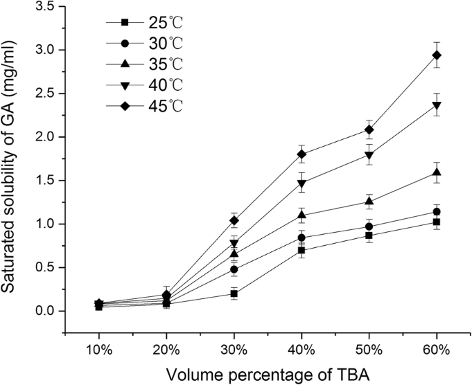
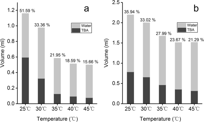
그림 1은 부피 백분율이 다른 TBA/물 공용매 시스템에서 GA의 포화 용해도 변화를 보여줍니다. 25°C ~ 45°C 내에서 GA의 포화 용해도는 TBA 부피 백분율이 10%에서 60%로 증가함에 따라 지속적으로 증가했으며 TBA 부피 백분율이> 40%일 때 GA의 포화 용해도는> 0.5mg/ml였습니다. 반면에, GA의 포화 용해도는 동일한 부피 백분율을 유지했을 때 TBA/물 용액의 온도가 증가함에 따라 증가했습니다. 상이한 온도에서 용해도의 차이는 TBA의 부피 백분율이 30%에 도달했을 때 점점 더 분명해졌습니다. TBA/물 공용매 시스템에서 대두 인지질 및 콜레스테롤의 용해도는 누적 기둥 그래프에 표시됩니다(그림 2). 그림 2a, b는 인지질과 콜레스테롤 단위(1mg)가 각기 다른 온도에서 포화 용해도에 도달하는 데 필요한 TBA/물 혼합물의 부피를 나타냅니다. 회색 영역은 물의 부피를 나타내고 검은색 영역은 TBA의 부피를 나타냅니다. 온도가 25°C에서 45°C로 점차 증가함에 따라 TBA/물 공용매의 총 부피와 1mg의 인지질을 용해하는 데 필요한 TBA의 부피 백분율(칼럼에 표시)이 점차 감소했습니다(그림 2a). 온도가 35°C 이상으로 증가함에 따라 필요한 TBA의 부피가 크게 감소하여 0.15ml 미만이었습니다. 마찬가지로 온도가 25°C에서 45°C로 점차 증가함에 따라 콜레스테롤 1mg을 용해하는 데 필요한 TBA 부피가 감소한 반면 TBA 부피 백분율은 점차 감소하는 경향을 보였습니다. 위의 결과는 온도와 TBA 부피 백분율이 인지질, 콜레스테롤 및 GA의 용해도에 큰 영향을 미친다는 것을 보여주었습니다.
<그림>
다양한 부피 백분율(mean ± SD, n)을 갖는 TBA/물 공용매에서 GA의 포화 용해도 =3)
<그림>
SPC의 용해도(a ) 및 콜레스테롤(b ) 다른 부피 백분율을 갖는 TBA/물 공용매 중
부피 비율이 다른 TBA/물 공용매 시스템의 승화율 비교
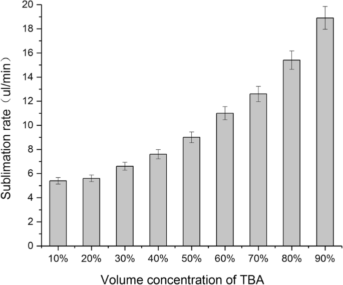
승화 속도는 동결 건조 분말의 생산 효율에 직접적인 영향을 미칩니다. 승화 속도가 빠를수록 경제적이며 재료의 붕괴를 방지할 수 있습니다[16]. 이 연구에서 우리는 TBA/물 시스템의 다양한 농도의 승화율을 조사했습니다. Fig. 3에서 보는 바와 같이 TBA의 부피%가 10%에서 90%로 증가함에 따라 혼합용매의 승화율은 점차 증가하였다. 또한, 부피 백분율이 60%를 초과함에 따라 승화 속도가 10μl/min 이상에 도달했습니다.
<그림>
다른 부피 백분율을 갖는 TBA/물 공용매의 승화율(mean ± SD, n =3)
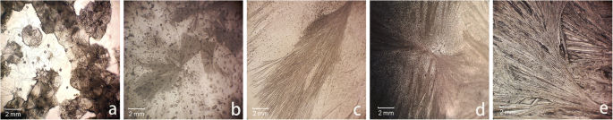
부피 백분율에 따른 TBA의 승화율 차이의 원인을 확인하기 위해 먼저 냉동 샘플의 표면 형태를 조사했습니다. 그림 4는 40%~80% 부피 비율의 TBA/물 용액의 광학 현미경 이미지를 포함합니다(<30% 부피 비율의 TBA는 광학 현미경으로 빠르게 녹기 때문에 검사할 수 없음). 40% 체적%의 TBA와 비교하여> 50% 체적의 TBA는 투명하고 흩어져 있는 바늘 모양의 구조를 가지고 있습니다. TBA의 부피 백분율이 증가할수록 침상 결정의 직경이 작아지고 비표면적이 증가하여 승화율이 증가하는 것으로 추측됩니다.
<그림>
광학현미경(× 100배율)에 의한 다양한 TBA 부피 백분율의 TBA/물 공용매의 표면 형태. 아 40%. ㄴ 50%. ㄷ 60%. d 70%. 이 80%
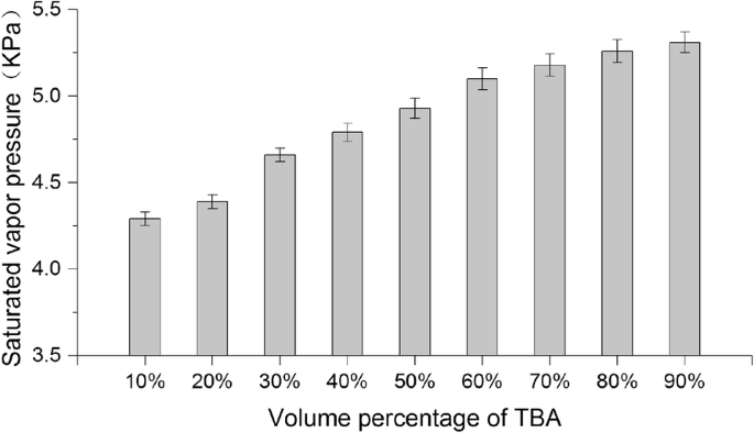
또한 25°C에서 다른 부피 백분율로 TBA/물 공용매 시스템의 포화 증기압도 측정했습니다. 그림 5는 TBA 부피 백분율이 다른 TBA/물 공용매 시스템의 포화 증기압 변화에 대한 막대 그래프입니다. 그림에서 보는 바와 같이 혼합용매의 포화증기압은 TBA의 부피비율이 증가할수록 점차 증가하는 경향이 있다. 온도는 포화 증기압과 양의 상관 관계가 있으므로 Antoine 방정식(Eq. 2)에 따라 동결 건조 온도(− 50 °C)에서 공용매 시스템의 포화 증기압이 부피 백분율로 증가할 것이라고 추론할 수 있습니다. 의 TBA가 증가하고, 이것이 승화율이 점진적으로 증가하는 이유 중 하나일 수 있습니다.
$$ {\log}_{10}p=A-\frac{B}{T} $$ (2)
여기서 p 는 증기압, T 온도, A 그리고 B 구성 요소별 상수입니다.
<그림>
다른 부피 백분율을 갖는 TBA/물 공용매의 포화 증기압(mean ± SD, n =3)
용적 비율이 다른 TBA/물 공용매 시스템에서 GA의 물리적 및 화학적 특성에 대한 동결건조 효과
TBA/물 공용매 시스템에서 동결건조가 GA의 물리화학적 특성에 미치는 영향을 조사하기 위해 다음과 같은 실험을 수행하였다. GA 10mg을 TBA 부피 백분율(40%, 50%, 60%, 70%, 80%)이 다른 8ml TBA/물 공용매에 용해했습니다. 단상 용액을 0.22μm 기공을 통해 여과하여 멸균한 후, 이를 2.0ml의 충전 부피로 10ml 동결 건조 바이알에 채웠습니다. 동결 건조기를 사용하여 - 50°C에서 24시간 동안 동결 건조를 수행했습니다.
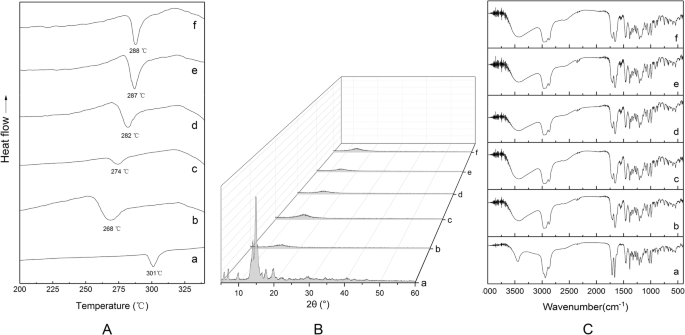
TBA 부피 백분율이 다른 TBA/물 공용매 시스템에 GA를 용해한 후 동결건조된 분말의 DSC 스펙트럼은 그림 6a에 나와 있습니다. 원료의약품의 DSC 곡선은 GA의 융점인 301°C에서 명백한 흡열 피크를 보여줍니다. TBA 부피 백분율이 다른 TBA/물 공용매 시스템의 동결건조로 인해 GA 용융 피크가 앞으로 이동했습니다. 용융 피크 이동의 크기는 TBA 부피 백분율이 감소함에 따라 증가했습니다.
이전 연구에서는 TBA의 농도가 결정질, 비정질 또는 준안정상의 복잡한 혼합물 형성에 깊은 영향을 미칠 수 있음을 이미 보여주었습니다[28]. 경우에 따라 TBA를 사용하면 결정도가 저하될 수 있으며 반대의 경우도 있습니다[29].
TBA 부피 백분율이 다른 TBA/물 공용매 시스템에 GA를 용해한 후 동결건조된 분말의 X선 회절(XRD) 스펙트럼이 그림 6b에 나와 있습니다. 원료 약물의 XRD 스펙트럼은 5°와 20° 사이에서 몇 가지 뚜렷한 결정 회절 피크를 보여줍니다. TBA 부피 백분율이 다른 TBA/물 공용매 시스템의 동결건조로 인해 샘플의 XRD 스펙트럼에서 5°~20°에서 회절 피크가 사라졌습니다. 이것은 원래의 약물 결정이 무정형이 되었음을 나타냅니다.
TBA 부피 백분율이 다른 TBA/물 공용매 시스템에 GA를 용해한 후 동결건조된 분말의 FTIR 스펙트럼은 그림 6c에 나와 있습니다. 원료 의약품의 FTIR 스펙트럼 모양은 4000–400cm
−1
내에서 TBA 부피 백분율이 다른 TBA/물 공용매 시스템의 동결건조 분말과 일치합니다. 범위. 새로운 작용기에 대한 특징적인 피크의 출현은 없었으며, 이는 GA의 화학 구조가 TBA에서 다른 부피 백분율로 동결건조 후에도 동일하게 유지되었음을 보여줍니다.
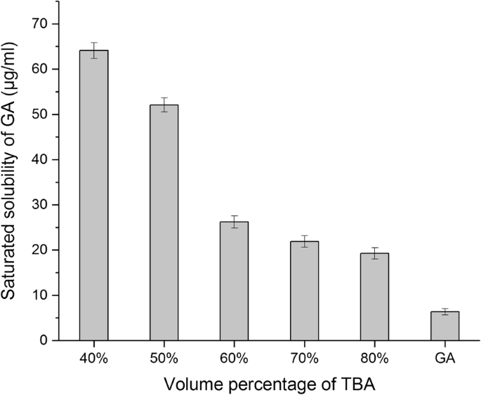
결정질에서 무정형으로의 변화는 약물 용해도를 변화시켜 수화된 재건 동안 프로리포솜 캡슐화에 영향을 미칠 수 있습니다. 이 연구에서는 25°C에서 동결건조된 GA 분말의 수용해도를 측정했습니다. TBA 부피 백분율이 40%에서 80%로 증가함에 따라 물에서 동결건조된 GA의 포화 용해도가 64.10에서 19.27μg/ml로 점진적으로 감소한다는 것을 발견했습니다. 그러나 여전히 원료의약품의 물에 대한 용해도(6.36μg/ml)보다 훨씬 높았으며, 이는 동결건조 중 결정질 구조에서 무정형 구조로의 변화가 원료의약품의 용해도에 영향을 미친다는 것을 의미합니다(그림 7). <그림>
다른 부피 백분율(mean ± SD, n)로 TBA/물 공용매에서 GA 동결건조의 수용성 =3)
단일 요인 실험
리포솜의 품질에 영향을 줄 수 있는 많은 요인이 있습니다. 인지질/약물 비율이 약물의 캡슐화 품질에 영향을 미친다는 것은 잘 알려져 있습니다[30]. 적당한 양의 콜레스테롤은 지질막의 정렬된 배열과 안정성을 증가시킬 수 있습니다. However, high content of cholesterol in the liposome can decrease the flexibility of membrane and thereby hinder the penetration of drug into the lipid bilayer [31]. In this study, we selected three factors that impact on liposome quality and performed a single-factor study to determine the appropriate values for subsequent optimization tests, including quantity of SPC, quantity of cholesterol, and volume percentage of TBA in the co-solvent. The quality of liposomes was evaluated in terms of encapsulation efficiency and mean diameter. Each experiment was performed in triplicate with all other parameters set to constant value, GA 60 mg, pre-freeze temperature − 40 °C, pre-freeze time 12 h. In this study, we compared the results via a scoring system, giving equal weight to both encapsulation rate and mean diameter. Scoring was conducted as follows:
where EE is encapsulation efficiency, MEE is maximum encapsulation efficiency of the group, MD is mean diameter, and MMD is maximum mean diameter of the group.
The experimental design and result are shown in Table 1. As can be seen in the table, within the range tested in this experiment, the highest score can be obtained separately when the amount of SPC is 480 mg (drug-SPC ratio of 1:8, w /와 ), the amount of cholesterol is120 mg (cholesterol-SPC ratio of 1:4, w /와 ), and volume percentage of TBA in the co-solvent is 50%. Therefore, these parameters were chosen as the center level of response surface optimization design, respectively.
Parameter Optimization by Box-Benhnken Design
To further study the interactions between the various factors, parameter optimization was performed by Box-Benhnken design. Based on the results of single-factor experiments, we investigated and optimized the interactions between the parameters, including quantity of SPC (X1 ), quantity of cholesterol (X2 ), volume percentage of TBA (X3 ) by Box-Benhnken design (BBD). Encapsulation efficiency (Y1 ) and mean diameter (Y2 ) were selected as responses. Optimization process was undertaken with desirability function to optimize the two responses simultaneously. We suppose that Y1 and Y2 have the same weightiness (importance). Y1 had to be maximized, while Y2 had to be minimized. The desirable ranges are from 0 to 1 (least to most desirable). Experimental design and results are shown in Table 2. To find the most important effects and interactions, analysis of variance (ANOVA) was calculated by statistical software, Design Expert trial version 8.03 (Stat-Ease, Inc., Minneapolis, USA). Two quadratic models were selected as suitable statistical model for optimization for two responses encapsulation efficiency and mean diameter. The results of ANOVA relating encapsulation efficiency as response were shown in Table 3, indicating that the model was significant for all factors investigated with F value of 12.81 (P <0.05). In this case, X1 , X2 , X1X2 , X1X1 , X2X2 were significant model terms (P < 0.05), demonstrating that the influences of the factors (X1 and X2 ) on encapsulation efficiency were not simply linear. The interaction terms were notably significant, indicating good interactions between the factors. On the contrary, the ANOVA results relating mean diameter as response (Table 3) indicated that the model was not significant for all factors investigated with F value of 1.9 (P> 0.05). In this case, X3 were significant model terms (P < 0.05), demonstrating that volume percentage of TBA have significant influence on mean diameter, while quantity of SPC (X1 ) and quantity of cholesterol (X2 ) do not have a significant effect (P> 0.05). Moreover, there were no significant interactions between the three variables.
In order to provide a better visualization of the effect of the independent variables on the two responses and desirability value, three-dimensional profiles of multiple non-linear regression models are depicted in Fig. 8. Figure 8a–f presented the interaction of X1 , X2 , and X3 under encapsulation efficiency and mean diameter as response respectively. The three-dimensional profiles demonstrated how three pairs of parameters affect the encapsulation efficiency and mean diameter of reconstituted liposomes. For encapsulation efficiency, all the three surfaces are upper convex (Fig. 8a–c), with a maximum point in the center of the experimental domain, which demonstrated that there are good interactions between the three variables. For mean diameter, the shape of Fig. 8d is similar to flat surface, indicating that X1 and X2 have less effect on mean diameter. The surface contours of Fig. 8e, f both showed a slope; the mean diameter was decreased by increasing the volume percentage of TBA, indicating that factor X3 had an obvious effect on mean diameter but there was no obvious interaction between X3 and the other two factors.
Three dimensional plots of the effect of X X (a ), X X (b ) and X X (c ) on encapsulation efficiency and the effect of X X (d), X X (e) and X X (f) on mean diameter
Based on the quadratic model, the optimal conditions for liposomes preparation calculated by software were as follows:508 mg phospholipid quantity, 151 mg cholesterol quantity, and 55% volume percentage of TBA. Under these conditions, the encapsulation efficiency and mean diameter were found to be 68.55% and 220 nm, respectively.
Selection of the Type and Dosage of Lyoprotectant
Competition for liquid water between the growing ice crystals and the hydrophilic substances (including the hydrophilic portion of the lipid membrane) during freezing leads to adhesion of ice crystals to the phospholipid groups. This can result in damage to the lipid membrane. Lipid membrane fusion following rehydration causes an increase in particle size and leakage of encapsulated drug. Lyoprotectant can reduce liposomal damage during the freeze-thaw process [32]. In this study, we investigated the effect of various types (lactose, sucrose, trehalose, mannitol) and dosage (lyoprotectant to SPC ratio was 1:2, 1:1, 2:1, 4:1, and 6:1 w /와 ) of lyoprotectant on scores of reconstituted liposome. Single-factor experiments were performed while maintaining all other variables constant:GA amount of 60 mg, SPC amount of 508 mg, cholesterol amount of 151 mg, volume percentage of TBA in the co-solvent of 55%, pre-freeze temperature of − 40 °C, pre-freeze time of 12 h. Experimental results are shown in Fig. 9. The encapsulation efficiency increases firstly and then decreases by decreasing lyoprotectant/SPC weight ratio from 1:2 to 1:6, wherein lactose, sucrose, and mannitol are respectively used as lyoprotectant. However, the encapsulation efficiency of the trehalose group increases constantly with decreasing lyoprotectant/SPC weight ratio (Fig. 9a). In terms of the mean diameter (Fig. 9b), it was found that the mean diameter was greater than 218 nm for lactose, sucrose, and mannitol group in the range from 1:2 to1:6. Nevertheless, the mean diameter of trehalose group can be reduced to less than 190 nm when lyoprotectant/SPC weight ratio is more than 4:1; obviously, the protective effect of trehalose is better than other lyoprotectants tested. Trehalose has a good protection ability for membrane, perhaps because of the formation of hydrogen bonds with the polar head groups of lipids, and disruption of the tetrahedral hydrogen bond network of water [33]. According to scores (Fig. 9c), the highest score (0.24) was obtained when trehalose/SPC weight ratio is 4:1 and 6:1. Finally, we choose trehalose and 4:1 (trehalose/SPC weight ratio) for following experiments from the perspective of cost and increasing drug loading.
The effect of mass ratio between cryoprotectant and SPC on encapsulation efficiency (a ), mean diameter (b ) and scores (c ) of reconstituted liposomes (mean ± SD, n =3)
Through the above Box-Benhnken design and lyoprotectant screening experiment, the experimental conditions were determinated:GA amount of 60 mg, SPC amount of 508 mg, cholesterol amount of 151 mg, volume percentage of TBA in the co-solvent of 55%, weight ratio of trehalose to SPC was 4:1. Under these conditions, the encapsulation efficiency and mean diameter were 74.87% and 191 nm, respectively.
Transmission Electron Microscopy
In this study, TEM of liposomes suspension was taken at the same time point (same hydration time). We have observed different states in the sample, which could explain the self-assembly behavior of the liposomes. Figure 10a shows the initial state of hydration; it can be seen that a large amount of GA (black dots) is wrapped in dispersed phospholipids (translucent material), and spontaneous aggregation of the phospholipid fragments occurs. Figure 10b shows the morphology of fully assembled liposomes (average diameter of about 200 nm), which were nearly spherical with a phospholipid bilayer structure (the light-gray portion). Moreover, the drug particles (dark gray dots) were entrapped in the lipid bilayer.
Transmission electron micrographs of reconstituted liposomes, (a ) initial state of hydration of proliposomes, (b ) fully assembled liposomes
Stability of GA Proliposome
After 6 months, the proliposome powders have a good mobility and an unaltered appearance. The liposome suspension formed automatically when in contact with purified water. The entrapment efficiency and particle size of the reconstituted liposome were 72.82% and 198 nm. There is no significant difference from the data of the reconstituted liposome 6 months before. Therefore, the GA proliposome could be considered stable at 25 °C for over 6 months.
In Vitro Drug Release Studies
Evaluation of in vitro drug release from encapsulated liposome was done by dialysis method. The in vitro release profiles of GA from GA-loaded liposomes at 37 °C in PBS (pH 7.4) and physiological saline solution are shown in Fig. 11. The release profile of both group showed a fast release (the larger slope) within 1 h, then curve slope becomes smaller after 1 h, the release rate begins to slow down. The drug-release curve shapes of physiological saline solution group are similar to PBS group. The in vitro release of GA from the GA-loaded liposomes was 65.25 ± 4.82% and 69.46 ± 4.32% from PBS and physiological saline solution in 12 h. No significant difference (P = 0.088, paired t test, SPSS software17.0) was found for the release of GA at different release medium over the entire study period, which demonstrated that the reconstituted liposomes have both sustained-release performance in two kinds of release medium.
In vitro dissolution profiles of GA from GA-loaded liposomes in a PBS and b physiological saline solution (mean ± SD, n =3)
In Vitro Cell Uptake
Figure 12a showed that the uptake process of GA-liposomes by Hep G2 cells is time-dependent under the experimental concentrations. After incubation for 30 min, the uptake amounts of drug-loaded liposomes (unit mass protein) by Hep G2 cells were 1480 ng. In the range from 30 to 240 min, the uptake amounts of drug-loaded liposomes (unit mass protein) were gradually increased from 1480 to 2030 ng. Figure 12b–e showed fluorescence microscopy images of Hep G2 cells at 30, 60, 120, and 240 min after ingestion of drug-loaded liposomes, and it is observed that the fluorescence intensity is also gradually increased over time. This result indicates that the reconstituted liposomes prepared by monophase solution method can be effectively uptaken by the hepatoma cells.
In vitro cellular uptake of GA-loaded liposomes by Hep G2 cells. 아 The uptake amount versus incubation time. ㄴ –이 Fluorescence microscopy images at 30, 60, 120, and 240 min
Conclusions
In the present work, preformulation investigation, formulation design along with in vitro characterization of GA-loaded liposomes by lyophilization monophase solution method have been done. After carrying out a preformulation study, we found that solubility of GA, cholesterol, and SPC in TBA/water co-solvent was substantially increased when temperature was over 40 °C. Sublimation rate of co-solvent gradually increased with increasing TBA volume percentage, which perhaps relate to surface morphology of the frozen co-solvent and saturated vapor pressure. After lyophilization using TBA/water co-solvent system, GA became amorphous structure; moreover, water solubility increased. This may have an effect on proliposome encapsulation during hydrated reconstruction. After optimization by Box-Benhnken design and screening of lyoprotectant, the optimum conditions (508 mg SPC, 151 mg cholesterol, 55% volume percentage of TBA, 4:1 trehalose/SPC weight ratio) for lyophilization monophase solution process were achieved. Under the optimum conditions, satisfactory encapsulation efficiency (74.87%) and mean diameter (191 nm) of reconstituted liposomes were obtained. The reconstituted liposomes resulted in initial assemble and final spherical shape, as confirmed by TEM analysis. The in vitro release profile of the produced GA-loaded liposome was investigated in the two media and it both showed prolonged release during 12 h. Cellular uptake studies showed that the uptake process of reconstituted liposomes by Hep G2 cells is time-dependent.