밀 스케일 폐기물에서 자철광 나노 흡착제를 사용하여 수용액에서 구리(II) 이온의 흡착 제거:합성, 특성화, 흡착 및 동역학 모델링 연구
초록
이 연구에서는 밀 스케일 폐기물에서 자철석 나노 흡착제(MNA)를 추출하고 합성하여 Cu
2+
제거에 적용했습니다. 수용액에서. 밀링 스케일 폐기물은 기존 밀링을 사용하여 분쇄되고 다양한 3, 5 및 7 밀링 시간 동안 고에너지 볼 밀링(HEBM)을 사용하여 충격을 받았습니다. 이와 관련하여 제조된 MNA는 X선 회절(XRD), 고해상도 투과 전자 현미경(HRTEM), 전계 방출 주사 전자 현미경-에너지 분산 X선 분광법(FESEM-EDS), UV-Vis를 사용하여 조사되었습니다. 분광학, 푸리에 변환 적외선(FTIR), Brunauer-Emmett-Teller(BET) 및 제타 전위. 결과 MNA-7 h 밀링 시간은 11.23nm의 불규칙한 모양, 5.98m
2
의 비표면적을 갖는 결정 구조를 나타냈습니다. g
−1
, 포화 자화, Ms 8.35개 중
−1
, 및 pH 5.4에서 등전점 전하. 최적의 흡착력 qe 4.42 mg.g
−1
Cu
2+
제거용 120분의 접촉 시간에서 이온에 도달했습니다. 실험 데이터는 Temkin 등온선 모델에 가장 잘 맞았습니다. 실험적 운동 연구와 이론적 측면을 비교하면 유사 2차 상관 계수가 (R
2
> 0.99). 또한 3주기의 재사용성 연구를 통해 70.87%의 재생 효율을 달성했습니다. MNA는 밀 스케일 폐기물을 재사용하고 Cu
2+
를 제거하기 위한 초고속 분리를 제공하는 실용적이고 효율적인 저비용 접근 방식을 제공합니다. 물에서.
소개
물은 자연을 기반으로 하는 가장 귀중한 천연자원 중 하나로 모든 생명체의 존재에 근본적인 역할을 한다[1]. 세계 경제의 혁명과 현대 산업화의 급속한 확장으로 수질 오염 문제는 더욱 심각해지고 있습니다[2]. 산업 및 인구 경로에서 배출되는 엄청난 양의 배출로 인해 중금속을 통한 생태 환경의 유해성은 전 세계적으로 강력하게 증가하고 있습니다. 생분해되지 않는 중금속은 매우 유독하며 인간의 장기 및 기타 거주 유기체에 축적되는 경향이 있으며 수질 공급을 저해하고 수중 생물에 다양한 어려움을 일으키고 수많은 질병 및 장애를 유발합니다 [3,4,5] . 특히, 구리(II) 이온(Cu
2+
) 인위적인 경로로 인해 국내 수역으로 유입되는 것은 공중 보건 및 환경에 문제가 있는 것으로 판명되었습니다[6,7,8]. Cu
2+
지만 인체 및 동물의 다양한 생물학적 경로 및 대사에서 중요한 미량 원소[9], 높은 농도에서 막대한 소비는 경련, 경련, 구토 또는 동시에 사망과 같은 심각한 독성학적 영향을 초래할 수 있습니다. 예를 들어, 2가 Cu로 인한 오염은 극화, 각질화, 손과 발의 따끔거림을 유발하고 발암 및 돌연변이 유발 영향을 나타냅니다[10-12]. 또한 과도한 구리 섭취는 산화적 외상과 근위축성 측삭 경화증, 멘크스 장애, 알츠하이머병 및 윌슨병을 포함한 급성 신경퇴행성 증후군을 유발할 수 있습니다[13, 14]. Cu
2+
에 의한 수질 오염 Cu
2+
이후 가장 널리 퍼진 환경 골칫거리 중 하나로 간주됩니다. 화합물은 과잉 산업 활동에 포함되어 있습니다[15]. 반대로 Cu
2+
결핍 동물 영양에서 설사, 빈혈 및 신경 장애를 유발할 수 있습니다[16]. 세계보건기구(WHO)는 Cu
2+
의 허용 수준을 규정했습니다. 식수에서 2mgL
−1
[17, 18]. 또한 USEPA(미국 환경 보호국)는 물에 허용되는 최대 구리 농도를 1.3mgL
−1
로 고정했습니다. [19]. 따라서 Cu
2+
의 수준은 매일의 식단, 특히 식수와 폐수에서 모니터링하고 주변으로 방출하기 전에 최소량으로 낮추어야 합니다[20].
환경에 대한 이러한 유해한 영향을 억제하기 위해 침전[21], 이온 교환[22], 화학적 침전, 공동 침전[23], 막 공정[24, 25, 82]과 같은 수많은 폐수 정화 방법이 활용되었습니다. ], 응고 [26] 및 흡착 [3, 27–30] Cu
2+
제거 폐수에서. 이러한 접근 방식 중에서 흡착은 매우 편리하고 비용 효율적이며 작동 용이성, 유연성, 단순한 설계 절차, 단순성, 우수한 제거 효율, 더 넓은 실용성 및 재활용성으로 인해 매우 바람직합니다[10, 31-33]. 따라서 중심 초점은 Cu
2+
를 가속화할 수 있는 다양한 작용기로 구성된 새로운 흡착제를 개발하는 것으로 옮겨졌습니다. 제거.
활성탄[34], 농업용 바이오매스[35], 금속 산화물[36], 실리카 나노물질[37, 38], 점토 광물[31, 39, 40], 기타 [41]. 그러나 이러한 흡착제의 복잡한 조건이나 특정 장비의 수정 및 낮은 흡착으로 인해 더 많은 응용 프로그램이 제한됩니다. 또한, 기존의 흡착제는 확산 부적절, 최소 결합 용량 및 활성 표면 부위의 불충분으로 인해 큰 부피의 용액에서 표적 금속 이온의 약한 회수를 나타냅니다[42]. 따라서, 더 높은 흡착 효율, 상당한 흡착 표면적, 최소 확산 저항, 우수한 흡착 용량 및 막대한 양의 용액에 대한 신속한 분리를 갖는 저가의 신규 나노 흡착제를 탐색할 필요가 필수적입니다.
최근에는 나노소재, 메조포러스 소재, 탄소나노튜브(CNT), 이온코팅 소재, 자성 나노입자 등 다양한 새로운 흡수제가 활용되고 있다[11, 43, 44, 45]. 이 중 나노 기반 물질은 생물학적, 물리적, 화학적 특성, 큰 표면 대 부피비, 우수한 흡착능과 함께 금속 이온의 높은 흡수성으로 인해 큰 주목을 받고 있다[46,47,48] 자성 나노 흡착제는 기체 또는 수성 액체 폐기물에서 오염 물질을 흡착하는 경향이 강하며, 이러한 자성 흡착제를 사용하여 많은 환경 오염 문제를 해결하는 것이 최근 몇 년 동안 주목을 받고 있습니다[11]. 마그네타이트 흡착제는 높은 표면적, 초상자성, 높은 이방성, 높은 보자력, 높은 활성 낮은 퀴리 온도, 분리 용이성, 높은 자화율, 우수한 재활용 및 재사용 기능, 자성과 같은 몇 가지 고유한 특성을 가진 유망한 저비용 전구체입니다. 매력 속성 [49, 50, 83]. 또한 NP는 일부 금속 이온, 음이온, 리간드, 양이온 및 염료의 흡착에 효율적인 것으로 간주되므로 일부 이온의 흡착, 회수 또는 제거의 혁신적인 분야에서 그 응용이 매력적입니다[51,52,53] .
이에 비추어 볼 때, 고에너지 볼 밀링 기술을 사용하여 Cu
2+
를 제거하기 위해 현지에서 얻을 수 있는 산업용 밀링 칩에서 마그네타이트 나노 흡착제를 사용한 연구를 유추할 수 있습니다. 물에서 얻는 것은 다소 제한적이며 인상적인 잠재력에도 불구하고 거의 적용되지 않았습니다. 이와 관련하여 본 연구는 고에너지 볼 밀링 방법을 사용하여 밀 스케일 폐기물로부터 신규한 자철석 나노 흡착제(MNA)를 합성하고 이를 모델로부터 물 속의 중금속 구리(II) 이온의 경제적 제거에 적용하는 데 중점을 둡니다. 수용액의. 또한 폐수 처리에서 금속 흡착제로 사용되는 밀 스케일 폐기물에서 재활용된 MNA의 표면 동역학 연구도 조사했습니다. MNA 반응의 동역학 모델 방정식은 신중하게 개발되고 연구되었습니다.
자료 및 방법
재료 및 화학 물질
이 작업에서 가공되지 않은 밀스케일 폐기물 칩은 말레이시아 테렝가누에 위치한 철강 공장에서 공급되었습니다. 배치 실험에 사용된 탈이온수(DI)는 정제 시스템 Milli-Q water에서 얻었습니다. 질산구리(Cu(NO3 )2 ) 구리 표준 용액(1000ppm)의 제조를 위해 Aldrich(Chemical Industry Stock Co., Ltd., 중국)에서 조달했습니다. pH 측정 및 교반을 위한 실험에는 pH 5-SS(Spear pH Tester)가 사용되었습니다. UV-Vis 분광 광도계(HACH DR4000U)를 사용하여 600nm 파장에서 구리 농도를 분석했습니다. 초기 구리 농도는 표준 절차(APHA, 2005)[54]를 사용하여 측정되었으며 나머지 샘플은 4℃의 냉각기에 보관되었습니다.
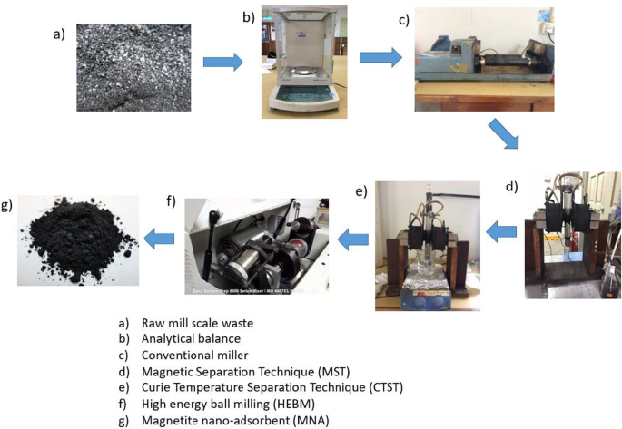
자철광 나노 흡착제(MNA) 합성
원밀 스케일 폐기물 칩에는 자성 입자와 불순물(비자성 입자)이 포함되어 있습니다. 샘플 오염을 방지하기 위해 불순물을 제거했습니다. 그림 1은 밀링된 칩에서 자철석 나노 흡착제(MNA)를 합성하는 데 사용되는 방법을 설명합니다. 먼저, 밀링된 칩을 탈이온수를 사용하여 광범위하게 세척하고 104℃에서 24시간 동안 건조한 후 기존 밀링 머신을 사용하여 마이크론 크기로 파쇄했습니다. 이 절차는 48시간 동안 꾸준히 수행되었으며, 그 결과 생성된 마이크로 크기의 자철광(Fe3 O4 )는 MST(Magnetic Separation Technique)로 연속적으로 세척되었습니다. MST는 비자성 입자와 자성 입자의 분리를 촉진합니다. 그 후, 세척된 마이크론 마그네타이트를 104℃에서 48시간 동안 오븐 건조시킨 후 밀폐 용기에 옮겼다. 또한 그림 1과 같이 CTST(Curie Temperature Separation Technique)에 의해 강한 자성 입자와 약한 자성 입자를 분리하였다. 이 방법은 [55-57]에서 채택한 절차에 따른다. 분리된 강력한 자성 입자는 이후 24시간 동안 공기 건조된 다음 3, 5, 7시간의 3가지 밀링 시간 동안 고에너지 볼 밀링(HEBM)을 거쳐 나노 크기의 자철석을 얻습니다[58].
<그림>
HEBM법을 이용한 마그네타이트 나노흡착제의 합성과정
준비된 나노자철광 흡착제(MNA)의 특성
합성된 흡착제의 구조적 형태는 Hitachi Co., Japan Model No. S3400N을 사용하여 TEM/EDS로 분석하였다. FTIR은 합성된 흡착제에 존재하는 작용기에 대한 정보를 제공합니다. 스펙트럼 범위 400–4000cm
−1
에서 표준 KBr 펠릿 방법을 사용하는 Bruker-Tensor 27 IR 기기 2cm
−1
해상도는 나노 자철광 흡착제의 FTIR 스펙트럼을 식별했습니다. X선 회절(XRD) 기술은 2θ 범위에서 얻은 Cu Kα 방사선(λ =0.154 nm)을 사용하는 X선 회절(XRD) Philips Expert Diffractometer를 사용하여 합성된 MNA의 결정 구조 및 위상을 분석하기 위해 적용되었습니다. 20 ~ 80°, 2θ =0.033의 스캔 단계 크기로 단계당 5초를 계산합니다. 관찰된 XRD 스펙트럼을 표준 ICSD 데이터베이스와 비교했습니다. MNA 샘플의 구조 및 형태학적 조성은 고분해능 투과 전자 현미경(HRTEM)이 있는 전계 방출 주사 전자 현미경 사진(FESEM), JEM JEOL 2100, USA를 사용하여 얻었다. Brunauer-Emmett-Teller (BET):Micromeritics II PLUS, USA는 NOVA2020e 자동 표면적 및 다공성 분석기를 사용하여 질소 흡탈착에 의한 MNA의 비표면적을 확인하기 위해 수행되었습니다. 분석 전에 MNA는 100°C에서 탈기되었습니다. 제타 전위 측정은 제타 사이저(Malvern ZS, UK)를 사용하여 수행되었습니다. 제타 사이저는 여러 pH 값의 적정을 제공했습니다. MNA 분말 샘플의 자기 특성에 대한 조사는 진동 샘플 자력계(VSM) 모델:LAKESHORE 7404를 사용하여 수행되었으며 0–13 kOe(kG)의 외부 필드가 적용되었습니다.
흡착 연구
수착 테스트는 배치 시스템을 사용하여 수행되었습니다. 질산구리 원액(Cu(NO3 )2 ) (50 mgL
−1
)을 준비하고 적절한 농도가 되도록 희석하였다. pH는 0.1 molL
-1
을 사용하여 조정되었습니다. HCl 또는 0.1 molL
−1
NaOH. Cu
2+
의 초기 및 최종 농도 준비된 보정 곡선을 기반으로 λ =600 nm에서 자외선-가시광선 분광광도계(UV-Vis)(모델:HACH DR4000U)를 사용하여 측정했습니다. 모든 흡착 시험은 적절한 양의 흡착제와 100mL의 이온 용액을 첨가한 250mL 플라스크를 사용하여 수행되었습니다. 역학 연구는 0.5g의 Fe3와 함께 1mg/L의 Cu(II) 이온 초기 농도를 사용하여 수행되었습니다. O4 . 온도와 pH는 각각 25°C와 pH 7로 일정하게 유지되었습니다. Cu(II) 이온의 초기 농도는 200mL 중 10, 20, 30, 40, 50mg/L였습니다. HCl 및 NaOH를 첨가하여 pH를 2, 4, 6, 8, 10 및 12에서 변화시켰다. 예비 연구에서는 흡착 과정이 180분 만에 평형 상태에 도달했음을 보여줍니다. 흡착 용량에 영향을 미치는 용량을 연구하기 위해 10~50mg 범위의 다양한 MNA 용량을 사용했습니다. 제거율(%RE ) 및 흡착 용량(qe )의 Cu
2+
이온은 식을 사용하여 결정되었습니다. 각각 1과 2[59, 60].
여기서 C오 및 Ce , 초기 및 최종 농도를 나타냄(mgL
−1
) 각각의 솔루션입니다. V 용액의 부피(리터), m 그램(g) 단위의 흡착제의 질량입니다. qe (mg/g)은 시간 t에서 흡착제의 단위 질량당 흡착물의 양입니다.
운동학 연구
실험은 28℃의 일정한 온도와 pH 5.4에서 200mL 구리 용액과 함께 Jar Tester를 사용하여 수행되었습니다. 샘플은 0, 10, 20, 30, 40, 50분의 다른 시간 간격으로 채취하여 UV-Vis를 사용하여 분석했습니다. 운동 모델은 Lagergren 의사 1차 및 의사 2차를 사용하여 연구되었습니다. Lagergren의 pseudo-first-order는 Eq. (3):
여기서 qe (mg/g) 및 qt (mg/g)은 평형 및 시간 t에서 흡착된 흡착물의 양입니다. , 각각 및 k1 (최소
−1
)는 유사 1차 흡착의 속도 상수입니다. 2차 메커니즘의 흡착 속도에 대해 유사 2차 운동 속도 방정식은 다음과 같이 표현될 수 있습니다. (4):
MNA는 활성 기공 부위가 평형에 도달했을 때 용매 탈착 접근법에 의해 회수되었습니다. MNA는 수용액으로부터 외부 자석에 의해 분리되고, 후속적으로 HCL 용액에 침지되고 26℃에서 180분 동안 혼합되었다. 그런 다음 생성된 MNA를 증류수로 헹구어 중성 pH를 얻은 다음 60℃에서 1시간 동안 유지했습니다. 재생된 MNA는 이전 연구와 함께 재사용되었습니다[54, 55]. 재사용성 효율성(RE%)은 식을 사용하여 계산되었습니다. 5:
실험 데이터는 완전 무작위 설계를 거쳐 얻은 데이터는 SAS 소프트웨어 9.4 버전(SAS Institute Inc., Cary, NC)에서 GLM(general linear model) 절차에 의한 일원 분산 분석(ANOVA)을 사용하여 분석되었습니다. , 미국). Duncan 다중 범위 검정은 p에서 평균을 분리하는 데 사용되었습니다. <0.05 유의 수준.
결과 및 토론
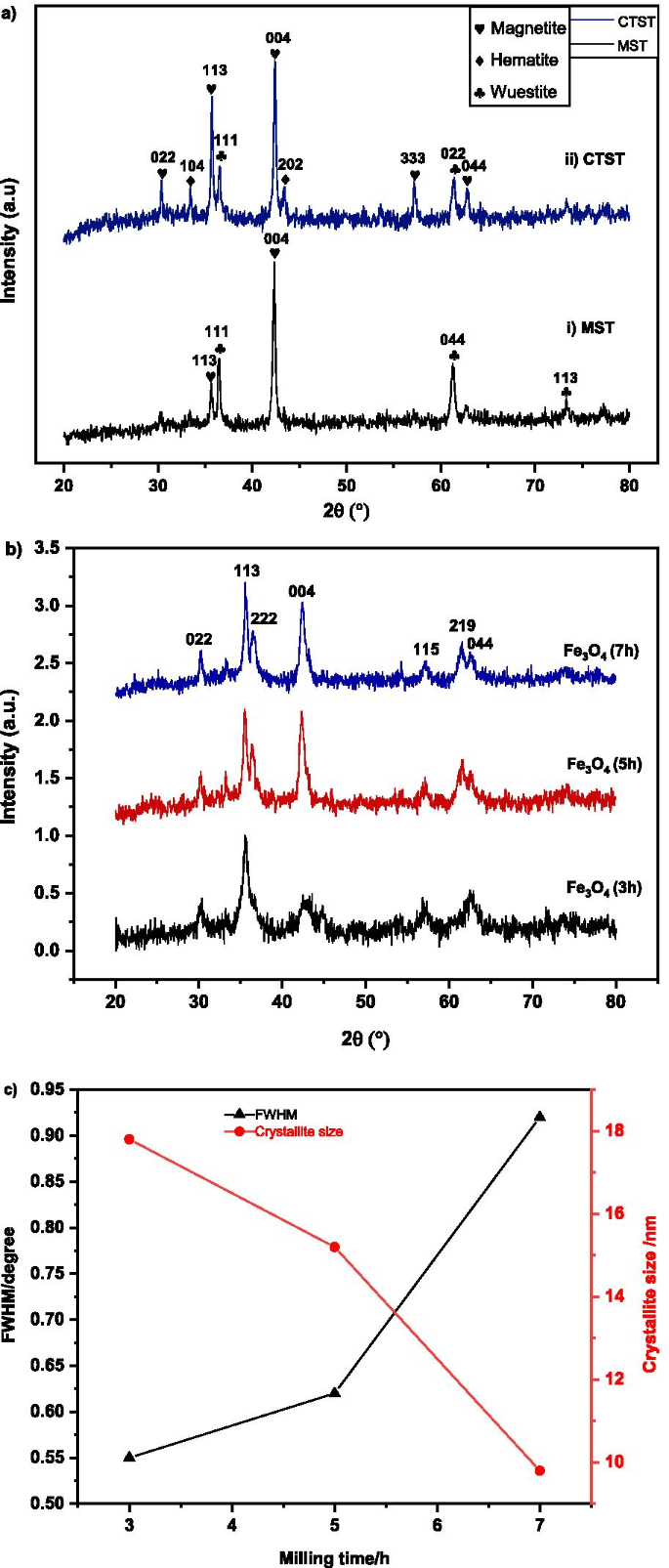
구조 및 단계 분석
MST 및 CTST 후 폐기물 분쇄기 스케일의 XRD 검사 결과는 그림 2a에 나와 있습니다. MST 공정 후(그림 2ai), XRD는 wuestite(FeO)와 자철광(Fe3 O4 ). Westite의 브래그 회절은 ICSD:98-001-2335와 일치하는 36.33°(111), 61.40°(044), 73.25°(113)의 2θ에서 관찰되었습니다. 자철석 상은 35.61°(113) 및 43.28°(004)의 2θ에서 관찰되었으며, 기준 Fe3에 동의했습니다. O4 ICSD 파일 98–010-9826.
<그림>
아 MST 및 CTST를 거친 후 밀 스케일의 X-선 회절 스펙트럼, b 3, 5, 7시간의 다양한 밀링 시간에서 자철광의 인덱스 스펙트럼 c 밀링 시간에 따른 MNA의 반치폭(FWHM) 및 결정자 크기
CTST 공정 후의 XRD 스펙트럼(Fig. 2aii)에서는 자철광, 적철광 및 우에타이트 상의 존재가 관찰되었다. 모든 피크의 브래그 회절각(2θ)은 필수 식별이며 피크가 30.23°(022), 35.61°(113), 43.28°(004)인 마그네타이트에 대한 참조 ICSD 파일 98-010-9826에 동의했습니다. 57.24°(333), 62.86°(044). 적철광 피크는 ICSD 98-004-6407과 일치하여 33.44°(104), 48.48°(202)에서도 관찰되었습니다. ICSD:98-001-2335와 일치하는 36.33°(111) 및 61.27°(022)의 웨타이트 회절각. 결과는 이전 문헌[84]에서 보고된 것과 같이 동의되었고 유사했습니다.
그림 2b는 3, 5, 7시간의 서로 다른 시간 간격에서 고에너지 볼 밀링 공정 후 밀 스케일 분말의 XRD 회절 패턴을 보여줍니다. 합성된 샘플의 회절 스펙트럼은 자철광(Fe3 O4 ) 모든 밀링 시간 동안 직경이 나노 크기인 상. 30.24°, 36.66°, 36.57°, 42.45°, 57.26°, 61.54° 및 62.76°의 2θ에서의 회절 각도는 (022), (113), (222), (004), (22)로 인덱싱될 수 있습니다. (115) 및 (044) 입방 단위 셀 Fe3의 서명 피크 확인 O4 , 각각. XRD 스펙트럼은 공간 그룹이 Fd인 자철광의 참조 ICSD 98–01-11,241과 일치했습니다. -3 m 및 격자 매개변수(a =ㄴ =ㄷ )의 8.3440Å입니다. 나노자철광 흡착제는 그림 2b와 같이 밀링 시간이 증가함에 따라 고순도를 나타냅니다. 유리병에서 충돌하는 강구에 의해 생성된 높은 에너지는 산소 결합을 깨고 적철광(Fe2 O3 ) 자철광(Fe3) O4 ) 단계. 밀링 시간이 증가함에 따라 나노결정질 마그네타이트의 형성이 XRD 피크의 확장에 의해 결정되는 것으로 관찰되었다. 밀링 시간이 증가함에 따라 XRD 피크 넓어짐이 증가하는 것으로 관찰되었으며, 이는 입자 크기의 감소를 나타냅니다. 밀링 시간이 증가함에 따라 XRD 피크 강도도 감소하는 것으로 관찰되었습니다. 패턴은 샘플의 입자 크기 감소를 나타냅니다[61]. 입자 크기가 감소함에 따라 밀링 과정에서 유도된 변형으로 인해 피크 강도가 감소하고 회절 피크가 넓어졌습니다. 평균 결정자 크기 D Eq.에서와 같이 Debye-Scherrer 공식을 사용하여 샘플의 수를 계산했습니다. (6) [62].
여기서 D 평균 결정자 크기, λ 는 X선 파장(0.1541nm), β 반값의 전체 너비(FWHM), θ 회절각이다. XRD 스펙트럼은 X'pert Highscore Plus 소프트웨어를 사용하여 자동으로 분석되었습니다. FWHM과 결정자 크기의 관계는 그림 2c에 나와 있습니다. 분석 결과 FWHM의 변화와 MNA의 평균 결정자 크기의 변화에 따라 3, 5, 7시간에서 밀링 시간의 증가에 따른 미세한 MNA 분말의 변화가 그림 2c와 같이 나타났습니다. FWHM의 변화 경향은 밀링 시간이 3, 5, 7시간에서 증가함에 따라 FWHM이 증가하는 경향을 나타내는 것으로 나타났습니다. 밀링 시간이 3시간, 5시간, 7시간에서 증가함에 따라 평균 결정자 크기는 각각 최소값인 17.8nm, 15.2nm, 9.8nm에서 감소했습니다.
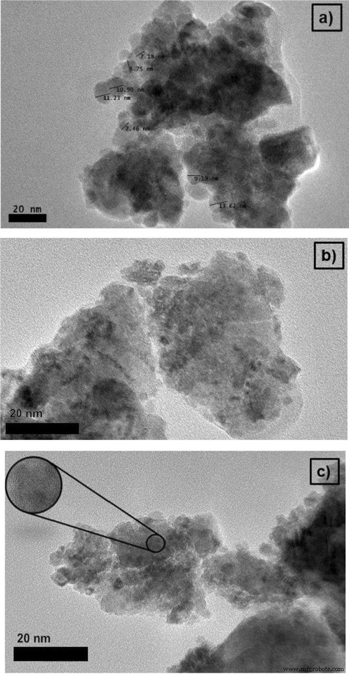
형태 및 미세 구조 구성
3, 5, 7시간의 서로 다른 밀링 시간에 밀링된 MNA의 HRTEM 현미경 사진이 그림 3에 나와 있습니다. 현미경 사진은 그림 3과 같이 MNA 입자가 3개의 밀링 기간 동안 불규칙한 모양을 나타냄을 보여줍니다. 또한, 5.53nm의 평균 MNA 입자 크기는 14.45nm(5시간) 및 19.16nm(7시간)에 비해 밀링 시간 3시간에서 확인되었습니다. 이것은 더 짧은 밀링 시간에 더 작은 MNA 입자 크기를 얻을 수 있음을 의미합니다. 3, 5, 7시간 동안의 평균 입자 크기는 10~22nm 범위에서 얻어졌습니다. 밀링 시간이 증가함에 따라 샘플의 미세 변형도 증가합니다[63]. 따라서 밀링 시간이 길어지면 샘플에 더 많은 변형이 발생합니다. 밀링 시간에 따른 격자 변형률의 증가는 밀링 과정에서 도입된 격자의 원자 전위 및 확산으로 인한 강한 왜곡 효과 때문입니다. 그러나 MNA 시료에서는 자철석 분말의 자기 인력 거동으로 인해 그림 3과 같이 응집 효과가 관찰되었습니다.
<그림>
a에서 MNA의 20nm 스케일 막대가 있는 HRTEM 이미지 3시간 b 5시간 및 c 7시간 밀링 시간
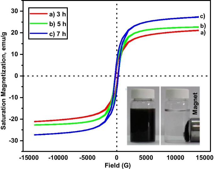
자기 특성 분석
샘플의 자기적 특성은 상온 실험에서 VSM을 사용하여 조사되었습니다. 자화 응답(M ) 적용된 외부 자기장(H )는 그림 4에 나와 있습니다. 포화 자화(Ms ), 잔존(Mr ) 및 보자력(Hㄷ ) 샘플의 )는 표 1에 요약되어 있습니다. MNA의 보자력 값은 200–270G 범위, 잔류물은 1.5–6.6emu/g 사이, 포화 자화 값은 21–27emu/g 사이입니다. 20nm 미만의 입자 크기로 인해 샘플은 초상자성 특성을 갖습니다. 자화 매개변수(표 1)에서 이는 샘플이 흡착 용량 증가에 기여하는 초상자성 및 강자성 화합물의 혼합물로 구성되어 있음을 나타냅니다. MNA-7 h 샘플은 Cu 흡착 연구(그림 5)에서 가장 높은 흡착 용량에 기여한 가장 높은 자기 매개변수(그림 4)를 보여줍니다.
<그림>
M-H 다양한 밀링 시간에서 샘플의 히스테리시스 그래프 a 3시간, b 5시간 및 c 7시간
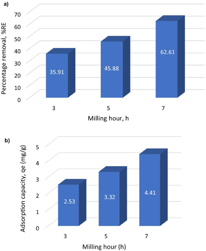
<그림>
아 제거율의 막대 차트; ㄴ Cu
2+
에 대한 MNP의 금속 흡수/흡착 용량 다른 밀링 시간에
흡착 매개변수의 영향
3, 5, 7시간의 MNA 배치 흡착 연구에 대한 추가 분석이 조사되었습니다. 그림 5는 3, 5, 7시간의 다양한 밀링 시간에서 MNA에 대해 수행된 흡수 연구를 보여줍니다. 그래프는 가장 높은 흡착 용량(금속 흡수)을 보여줍니다(qe ) 및 가장 높은 제거율(%RE) ). MNA-7 h 밀링 시간은 가장 높은 흡착 용량과 수용액에서 가장 높은 제거율을 나타냅니다. 따라서 MNA-7 h는 접촉 시간, 초기 농도, 흡수제 용량, 표면적, pH 및 온도의 여러 매개변수에 대한 추가 배치 흡수 분석을 위해 MNA 나노 흡수제로 선택되었습니다.
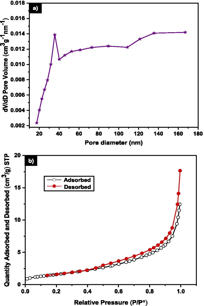
표면적 분석
BET를 사용한 질소 흡착은 7시간 밀링에 대한 MNA의 표면적 및 기공 특성을 평가하는 데 사용되었습니다. 그림 6a는 평균 기공 부피가 0.011cm
3
인 MNA-7 h에 대한 BET 결과를 보여줍니다. g
−1
5.98m
2
의 비표면적 g
−1
. N2 MNA-7 h의 흡착-탈착 곡선은 Sing et. 알. (1985) [65]. BET 결과는 흡착된 분자의 흡착제-흡착물 상호작용이 MNA 표면 주위에 밀집되어 있음을 설명합니다[66]. 따라서 유형 III의 흡착은 기체 분자가 MNA에 물리적으로 흡착되었음을 나타냅니다[67].
<사진>
아 7h MNA의 기공 직경 분포 b 질소 흡탈착 등온선
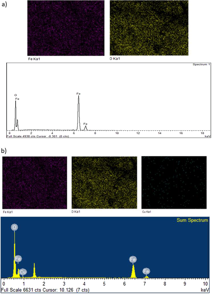
EDS 분석
MNA-7 h의 원소 성분은 그림 7에 나와 있습니다. FESEM 및 EDS 분석은 흡착 공정 전 MNA-7 h에 대해 각각 78.25% 및 21.75%의 비율로 Fe 및 O 원소의 존재를 나타냈습니다(그림 1). 7a). 그림 7b는 흡착 과정 후 Fe, Cu 및 O 원소의 존재를 나타내는 EDS 스펙트럼을 보여줍니다. 스펙트럼에 구리의 존재는 Cu
2+
의 흡착을 나타냅니다. MNA에 의해. 이 경향은 또한 Lingamdinne et al.에 의해 보고된 연구에 표시된 스펙트럼과 일치합니다. (2016) [64] 산화철 나노입자를 중금속 흡착 제거에 활용하였다.
<그림>
EDS 스펙트럼 분석(a ) 흡착 전 및 (b ) Cu(II) 이온 흡착 후
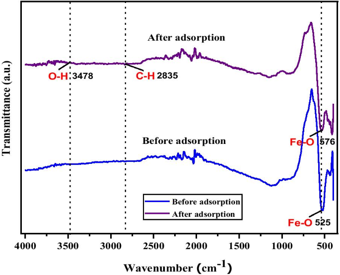
FTIR 분석
MNA-7 h에 대한 Cu 흡착에 대한 추가 확인도 FTIR 분석을 통해 결정되었습니다. Cu
2+
의 작용기와 부착을 확인하기 위해 FTIR을 조사했습니다. MNA에. 그림 8은 Cu(II) 이온 흡착 전후의 MNA-7 h의 FTIR 스펙트럼을 보여줍니다. FTIR 스펙트럼은 Fe3의 강력한 특성 피크 밴드를 나타냅니다. O4 나노 입자. 흡착 후 FTIR 스펙트럼은 500–600cm
−1
범위에서 밴드 강도의 변화를 나타냅니다. 및 2800–3600cm
−1
Cu
2+
로 이어집니다. 수착. 또한 525cm
−1
의 흡착 밴드 및 576cm
−1
Fe-O 밴드 마그네타이트 나노 입자의 사면체 및 팔면체 사이트를 나타냅니다 [68]. 3478cm
−1
에서 강력하고 광범위한 흡착 스펙트럼 MNA 표면의 수산기(- OH) 및 미량의 물 분자에 해당합니다[69]. FTIR 분광법은 MNA가 MNA의 표면에 흡착된 몇 가지 화학 물질의 존재로 인해 결정 구조를 가지고 있음을 보여줍니다.
<사진>
Cu(II) 이온의 흡착 전후에 MNA-7 h의 푸리에 변환 적외선 스펙트럼
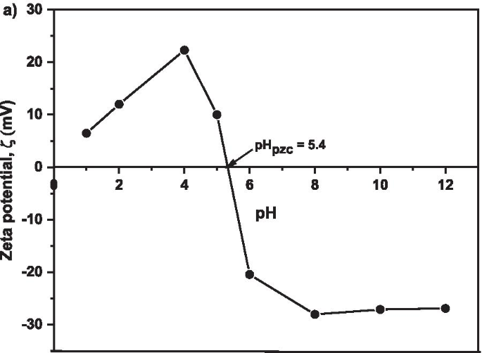
제타 전위 분석
그림 9는 MNA-7 h의 표면 제타 전위를 보여줍니다. 흡착 과정에서 발생하는 중화 전하를 정확하고 정밀하게 측정하기 위해 제타 전위를 조사했습니다. 제타 전위 데이터는 Cu 흡착 과정에서 최적의 흡착제 용량을 모니터링하기 위해 측정 가능한 값을 제공합니다. 결과는 등전점(pHpzc ) Cu
2+
가 발생하는 pH 5.4에서 발생 발생한 MNA에 대한 흡착이 최적입니다. 수용액에서 산화철의 표면은 OH
-
로 덮여 있습니다. 표면의 FeOH가 FeO 또는 FeOH2와 같은 다른 Fe 작용기로 변할 수 있도록 하는 그룹 , 양성자화 또는 탈양성자화 과정으로 인해 [70, 71]. 양성자화와 탈양성자화의 균형은 용액의 pH와 pHpzc에 따라 다릅니다. 흡수제의. 제타 전위 결과는 흡착이 pH 5.4에서 효율적임을 시사합니다.
<그림>
MNA-7 h에 대한 1, 2, 4, 6, 8, 10, 12의 pH 값에 대한 제타 전위
Batch Adsorption Analyses
Effect of Contact Time
Figure 10a shows the effect of contact time on adsorption capacity and rate of Cu
2+
uptake onto MNA after 250 min. It is evident that at longer contact time, the adsorption capacity reach equilibrium as the pH was kept at 5.4 and adsorbent dosage at 0.05 g. The maximum removal efficiency attained was 62.61% as shown in Fig. 10b. The Cu
2+
removal efficiency surges rapidly from the early first 5 min, and later slower and stable throughout the adsorption process. This is attributed to the fact that the rate of the adsorption capacity was high due to the abundant free binding and active sites of the Cu
2+
. Based on Fig. 10, it was noticed that the percentage removal and adsorption capacity increased rapidly with the increase in contact time at the initial stage. The contact time has a substantial influence on the efficacy of Cu
2+
removal and adsorption capacity. Increase in contact time from 0 to 240 min led to an increase in the removal efficiency of Cu
2+
from 0.81% to 62.61%. For contact time greater than 120 min, the removal efficiency of Cu
2+
remains steady, as the active sites has been saturated on the surface of the adsorbent. Similarly, the highest adsorption capacity of 4.41 mg/g was attained at 120 min of contact time. Thus, the equilibrium time was attained at 120 min.
아 Adsorption capacity; ㄴ Copper removal efficiency under various contact times of MNA-7 h (metal solution:200 mL; temperature:25 °C; initial pH:5.4; initial concentration:50 mg/L; adsorbent dosage:0.05 g)
The results of copper removal efficiency follow a definite trend (Table 2). It shows that the higher the time, the more the removal efficiency. There were significant differences (p < 0.05) among the removal efficiencies under varying contact times. Generally, as contact time progresses, the removal efficacy also improves. Table 3 shows the percentage of the copper removal at different adsorbent dosages. The result shows that as the time progresses, the removal percentage increases. 0.05 g adsorbent dosage recorded the highest copper removal efficiency (62.58 g) after 120 min contact time.
Effect of Initial Concentration
Figure 11 shows as the initial concentration increases, the equilibrium adsorption capacity also increases. Thus, the higher initial concentration due to 0.05 g adsorbent in the Cu
2+
solution to fill the active sites on the adsorbent and the quantity of copper adsorbed increases with the increase in Cu
2+
concentration [83]. The initial concentration of Cu
2+
increased from 10 mg/L to 50 mg/L with corresponding increase in adsorption capacity from 0.04 mg/g to 4.41 mg/g, which in turn provide a higher driving force for the ions from the solution to the adsorbents, resulting in more collisions between Cu
2+
and active sites on the MNA-7 h. Since nearly all the adsorption sites of MNA-7 h existed on their exterior, it is easy for the adsorbate to access these active sites, thereby facilitating a rapid attainment of equilibrium condition.
Copper removal efficiency under various initial concentration of MNA-7 h (metal solution:200 mL; temperature:25 °C; initial pH:5.4; adsorbent dosage:0.05 g)
Effect of MNA Dosage
Adsorbent dosage plays an important role during the adsorption process, as it controls the ability of the adsorbent for a given solution. The more the dosage, the more obtainable site for sorption to occur [67]. Figure 12a shows the adsorption capacity, qe of Cu
2+
with respect to different dosages of MNA-7 h at 0.05 g, 0.2 g, 0.5 g, and 0.8 g, respectively. The adsorption capacity was observed to be dependent on adsorbent dosage, which determines the availability of the active sites and the amount of the surface area for adsorption. This is due to the increase in surface area and the probability of collision and interaction between the particles of nano-adsorbent and Cu
2+
[72]. As shown in Fig. 12b, at 0.05 g dosage, 62.61% copper removal efficiency and 4.41 mg/g of adsorption capacity were recorded. The Cu
2+
removal increases sharply and becomes stable as the adsorbent dosage increases. As the adsorbent dosage increases, the larger surface interaction and the agglomeration effects develop. Thus, it causes a decrease in free specific area per unit mass of MNA surface, causing a reduction in contact surface with the adsorbate surface. This will lead to the decrease in qe and %RE. Besides, the decrease in qe and %RE , perhaps was due to the saturation of Cu
2+
in solution with respect to available adsorption binding sites [73]. Thus, a higher amount of adsorbent causes an aggregation which decreases the total surface area of the MNA, thereby leading to a decrease in adsorption capacity [74–76]. The aggregation could result to a decrease in total surface area of the adsorbent and an increase in diffusion path length [75].
아 Adsorption capacity; ㄴ Copper removal efficiency under various adsorbent dosage of MNA-7 h (metal solution:200 mL; initial concentration:50 mg/L; temperature:25 °C; initial pH:5.4)
Effect of pH
The removal of Cu
2+
from the aqueous solution through adsorption is highly dependent on the solution pH which determines the surface charge of the adsorbent and the adsorbate speciation [77]. Adsorption is regarded to be minimal at acidic state owing to higher concentration of H3 O
+
which competes with the positively charged ions for the actively binding site on the adsorbent surface, and this usually led to low contaminant removal [77, 78]. The influence of pH on the adsorption of Cu
2+
on MNA was evaluated between the pH range of (2–12). Figure 13 shows the effect of pH on the adsorption capacity and removal efficiency of Cu
2+
. It was observed that increase in pH from 2 to 5.4, results in an increase in adsorption capacity from 0.58 mg/g to 4.408 mg/g and percentage removal of copper from 10.71% to 62.61%, respectively. However, the equilibrium adsorption capacity of Cu
2+
is low at a strong acidic condition recording 0.58 mg/g at pH 2 due to the presence of a high percentage of H3 O
+
ion which competes with Cu
2+
at the sorption sites of MNA. Besides, when pH is higher than 5.4, the adsorption capacity decreases from 49.32% to 44.69%. At a higher pH, higher concentration of OH
−
causes a decrease in the adsorption rate. Figure 13a and b shows that pH has a significant impact on Cu
2+
adsorption capacity and removal percentage (%RE ) of Cu. The removal rate for Cu
2+
increases with an increase in pH, from 10.71% to 28.04% and to 62.61% when pH is at 2, 4 and 5.4, respectively, before declining to 49.32%, 42.56% and 44.69 at pH 8, 10 and 12, respectively (Fig. 13a).
아 Adsorption capacity; ㄴ copper removal efficiency under various pH of MNA-7 h (metal solution:200 mL; initial concentration:50 mg/L; temperature:25 °C; adsorbent MNA dosage:0.05 g)
Similarly, the adsorption capacity also increases from 10.71% to 62.61% with the increase in pH from 2 to 5.4 and decreases until it reaches pH 12. At pH 2 and 4, the amount of protonation of the adsorbent surfaces results in a decrease in Cu
2+
adsorption. The results also are in agreement with zeta potential graph as indicated in Fig. 9. Also, Fig. 9 describes the net charge of the MNA adsorbent surface at different pHs, with the point of zero charges (pHpzc ). As the pH increases, the H
+
ion is lower and causes the surface of the adsorbent to become negatively charged, with the increase in %RE of Cu
2+
, thereby increasing the electrostatic attraction force between the adsorbents in the solution [79]. Therefore, pH influences the surface zeta potential of MNA. The surface functionality of iron oxides varies depending on the nature of iron oxides and the pH value.
Copper Adsorption Kinetics
Kinetic studies are essential in the adsorption process to describe the uptake rate performance of MNA-7 h and influence the residual time for the entire adsorption process. The adsorption kinetics of Cu
2+
on MNA-7 h was determined using similar procedures to those used in the batch adsorption studies [55]. The Lagergren’s first-order kinetic model and second-order kinetic model for the removal of Cu
2+
at various initial concentrations from the aqueous solution using MNA at 0.05 g/L of the MNA dosage are shown in Fig. 14. The calculated qe values are in agreement with the theoretical values, and the graph shows good linearity with R
2
above 0.96. Therefore, the adsorption kinetics follows the pseudo-second-order model. The pseudo-second-order model represents the adsorption kinetics, involving donation or electron exchange between adsorbate and adsorbent. Table 4 shows the fitted parameter summary of Cu
2+
kinetics at different initial Cu
2+
concentrations (qe :mg/g, k1 :min
−1
, k2 :g/mg/min) of MNA-7 h. For the parameters of initial concentration, Ci (mg/L), adsorption capacity, qe (mg.g
−1
), k2 is the rate constant of pseudo-first-order, k2 is rate constant of pseudo-second-order, and the R
2
is the correlation coefficient. Two kinetic models:Lagergren’s first-order and pseudo-second-order order were applied to further study the rate of adsorption process for Cu
2+
. The kinetic parameters of pseudo-first-order and pseudo-second-order are presented in Table 4. According to Table 4, pseudo-second-order was best fitted for the adsorption of Cu
2+
. Pseudo-second-order revealed a higher correlation coefficient of R
2
= 0.999, for Cu
2+
removal.
아 Lagergren’s first-order kinetic model; ㄴ Pseudo-second-order kinetic model for the removal of Cu (II) ions at various initial concentrations from the aqueous solution using MNA at 0.05 g/L dosage of MNA-7 h
Copper Adsorption Isotherms
The adsorption isotherms experimental data were investigated using Freundlich isotherm model (Eq. 7) and Temkin isotherm model (Eq. 8). The adsorption isotherm is illustrated in Fig. 15a and b. The Freundlich isotherm is expressed as:
where qe is adsorbent capacity at equilibrium (mg/g); B :(RT /b ) is the Temkin constant related to heat of adsorption (J/mol) R is universal gas constant (8.314 J/mol K); T is absolute temperature (K); 1/b indicates the adsorption potential of the adsorbent; kt is equilibrium binding constant corresponding to the maximum binding energy (L/mg); and Ce is adsorbate concentration at equilibrium (mg/L). From the graph, the straight line emerged, and the values of qm and KL constants can be calculated using the slope and the intercept of the straight line. Freundlich and Temkin models was used to examine the relationship between the adsorbent and adsorbate. As shown in Fig. 15, the initial concentrate correlation coefficient with an R2
value of 0.914 and manifested Temkin adsorption isotherm to be more favourable for the removal of copper. The synopsis of the isotherm parameter of Freundlich, Temkin parameter with correlation coefficient, R
2
for adsorption of Cu (II) on MNA-7 h at room temperature is depicted in Table 5. 1/n constant reciprocal implies natural sorption; therefore, the adsorption process is beneficial. The values of n, 1/n , KF , and R
2
for the current work are also presented in Table 5. Temkin isotherm adsorption plot shows maximum Cu
2+
removal by MNA-7 h at optimal conditions and reveals the feasibility of the process. The data prove that the Temkin model well fitted the experimental data than Freundlich based on the correlation coefficient, R2
(Table 5). This could be due to the fact that the Temkin isotherm model considers the effect of indirect adsorbate on the adsorption process and assumes that the heat of the adsorption of molecules decreases linearly in the adsorption layer [80].
아 Linearized Freundlich; ㄴ Linearized Temkin isotherm models for Cu
2+
adsorption by MNA-7 h at various adsorbent dosages:contact time 240 min; initial pH 5.4; room temperature
Regeneration and Desorption Study
The reusability of adsorbent is a prime issue since periodically regenerating adsorbent is strongly desirable for industrial applications [81]. In the reusability tests, the adsorption and desorption cycle of Cu
2+
onto MNA was repeated three times. The adsorption capacity of MNA was recycled, and the regeneration of the MNA of the adsorption–desorption cycle of Cu
2+
was repeated three times using the same MNA. The desorption process was studied for 3 cycles of MNA-7 h. The results proved that the magnetic nanoparticles have higher sustainability for industrial applications. The results also revealed that Cu
2+
could be desorbed from the adsorbent in the presence of deionized water as the desorbing agent. For repeated use of MNA, the adsorbed Cu
2+
were desorbed under suitable conditions. In this work, the percentage of desorption by MNA was obtained by 0.1 M HCl as shown in Table 6. Hence, MNA exhibits an enhanced recovery efficiency of 70.87%. Figure 16 displays the desorption efficiency of MNA. The adsorption capacity decreased by 10% during three adsorption–desorption cycles which indicates the stability and reusability of MNA.
Desorption of copper onto MNA-7 h in three cycles
Conclusions
Novel magnetite nano adsorbent (MNA) from mill scale waste has been successfully synthesized via conventional milling technique and impacted by high-energy ball milling procedure at varying milling time. The high-energy ball milling (HEBM) at 3, 5 and 7 h successfully produced the MNA in the range of 10–25 nm, as confirmed by HRTEM. The HEBM technique was used to reduce the microcrystalline size to nano-sized particles showing the potentials of MNA as an efficient precursor for Cu
2+
removal in an aqueous solution.
VSM results showing the MNA-7 h possess the highest magnetization property and indicate the best absorbent, with the specific surface area of 5.98 m
2
g
−1
and the average pore size of 8.01 nm thereby showing the better adsorption capacity. The adsorption of Cu
2+
on MNA-7 h was confirmed by EDS and FTIR analysis. For the adsorption studies, pH at 5.4, dosage of 0.05 g and 240 min of contact time, the highest adsorption capacity, qe and removal efficiency of 4.408 mgg
−1
and 62.61% were achieved. Also, at the initial concentration of 50 mgL
−1
Cu
2+
, the qe of 4.41 mgg
−1
was recorded. The reusability efficiency of 70.87% was attained even after three cycles of reapplications and desorption. The Temkin adsorption isotherm fits best with a correlation coefficient, R2
of 0.91. Based on these findings, it can be inferred that MNA is a promising precursor for Cu
2+
removal.
데이터 및 자료의 가용성
The datasets generated during and/or analysed during the current study are available from the corresponding author on reasonable request.