의학은 질병에 대한 새롭고 개선된 치료법을 끊임없이 찾고 있으며, 이는 높은 효능과 비용 효율성을 필요로 하며, 그러한 새로운 치료법을 발견하기 위한 과학적 연구에 대한 큰 수요를 만들고 있습니다. 모든 치료의 한 가지 중요한 측면은 질병만을 대상으로 하고 신체의 다른 건강한 부분에 해를 끼치지 않는 능력입니다. 이러한 이유로 금속 나노 입자는 의료 영상, 항균 및 항바이러스 응용을 포함하여 가능한 의료 용도로 광범위하게 연구되어 왔으며 현재도 연구되고 있습니다. 초상자성 금속 나노입자는 자기장으로 신체 주위를 유도하거나 자기 임플란트로 유도할 수 있는 특성을 가지고 있어 다양한 바이오 카고를 나노입자에 접합할 수 있는 가능성을 열어 체내에서 치료를 유도할 수 있습니다. 여기에서는 단일 금속 나노입자, 기능화된 금속 나노입자 및 Fe3 코어를 사용하는 코어-쉘 금속 나노입자를 포함한 다양한 금속 나노입자의 현재 생물의학 응용에 대해 보고합니다. O4 뿐만 아니라 이러한 코어-쉘 나노입자의 합성 방법.
소개
금속 나노 물질은 의학의 미래를 위한 중요한 관문입니다. 의학에서 금속 나노입자의 장기적인 안전성에 대해서는 아직 많이 알려져 있지 않지만[1], 이러한 입자는 이미 생체 내 부위 특이적 영상화[2,3,4], 암 탐지와 같은 다양한 생물 의학 응용 분야에서 그 위치를 찾았습니다. [5,6], 암치료[7,8,9,10], 신경퇴행성질환치료[11,12,13], HIV/AIDS 치료[14,15,16], 안과질환치료[17,18,19 ], 호흡기 질환 치료 [20, 21]. 그림 1은 의학에서 나노 입자의 현재 사용 범위를 보여줍니다. 최근 나노의학의 발전에도 불구하고 나노치료의 방법에는 여전히 많은 장애물이 존재하는데, 예를 들어 쉽게 반복 가능한 결과를 생성하는 합성 경로를 달성하기 어려울 수 있고 많은 나노입자 합성 방법이 두 가지 크기 모두에서 범위를 생성할 수 있기 때문이다[22 ,23,24] 및 나노입자의 모양[25,26,27,28] 및/또는 경제적으로 실행 가능하도록 충분히 많은 양의 나노물질을 생산하지 못한다[29]. 또 다른 핵심 요소는 연구 분야가 비교적 새로운 분야이기 때문에 일정 기간 동안 일부 나노 입자의 장기 독성에 대해 상대적으로 알려져 있지 않다는 것입니다[30,31,32]. 금속 나노입자의 많은 가능한 용도 중에는 약물 전달 영역이 있습니다[33, 34]. 나노 입자가 제공하는 넓은 표면적 때문에[35], 그들은 많은 양의 약물이나 기타 의료 화물을 전달할 수 있는 능력을 가지고 있습니다[36]. 단일 금속 나노입자에 대한 대안은 쉘 물질에 대한 대체 특성을 갖는 나노입자에 코어를 통합하는 것이며, 이것의 한 예는 자기 코어를 통합하는 것입니다. 여전히 존재하는 한 가지 과제는 코어-쉘 나노 입자를 합성하는 것입니다. 나노 입자를 합성하는 방법에는 여러 가지가 있지만[37], 코어-쉘 나노 입자를 합성하려고 할 때 새로운 도전이 나타납니다[38].
<그림>
의학에서 나노입자의 현재 사용 중 일부
이 리뷰는 먼저 Fe3 코어를 사용하는 데 초점을 맞춘 코어-쉘 나노입자를 생성하는 데 현재 사용되는 몇 가지 방법에 중점을 둡니다. O4 및 금 또는 은의 코팅. 그런 다음 단일 금속 나노 입자의 현재 바이오 의학적 응용과 그 한계, 자기 코어의 적용으로 이를 극복하는 방법을 조사합니다.
코어-쉘 나노입자 합성
금속 나노입자의 합성 방법은 예를 들어 Stevenson et al. HAuCl4 환원을 통한 금 나노입자 합성 발표 1951년 [39]. 그 이후로 가스 증착[40], 졸-겔[41] 및 에어로졸/증기상[42]과 같은 나노 입자 합성을 위한 많은 다른 경로가 있었습니다. 그러나 하나의 금속이 코어를 형성하고 두 번째 금속이 쉘을 형성하는 코어-쉘 구조로 구성된 금속 나노입자를 합성하려고 시도할 때 새로운 도전이 나타납니다. 예를 들어 Fe 입자는 물에서 분해되는 반면 HAuCl4 강한 산화제이다[38]. 더 논의될 한 가지 예는 Fe3를 사용하는 것입니다. O4 (산화철) 코어 및 코팅 쉘로 금. 이러한 코어-쉘 금속 나노입자의 제조에서 가장 큰 두 가지 문제는 코팅 속도를 제어하고 코팅의 균일성을 제어하여 모두 매우 유사한 모양과 크기의 나노입자 용액을 만드는 것입니다[43]. 산화철 코어에 금 또는 은을 코팅하는 것은 두 가지 주요 범주로 나눌 수 있습니다. 즉, 금/은을 철에 직접 코팅[44]하거나 중간층을 사용하여 금과 철 층 사이의 접착제 역할을 합니다[45]. 전자의 범주는 여기에서 논의될 것입니다. 다음 텍스트는 금 및 은으로 코팅된 Fe3를 합성하기 위해 고안된 몇 가지 방법을 설명합니다. O4 나노 입자.
역 미셀 합성
금속 나노입자 합성을 위한 대중적인 경로는 역미셀법(reverse micelle method) 또는 마이크로에멀젼(microemulsion) 경로라고 불리는 방법을 사용하는 것이다[46]. 이 방법은 로듐, 백금, 팔라듐 나노입자의 콜로이드 용액이 처음 합성된 1980년대에 처음 도입되었습니다[47].
미셀은 소수성 및 친수성 구성 부분을 가진 분자가 수성 또는 소수성 상과 접촉할 때 형성됩니다[48]. 미셀은 친수성 부분이 수상과 접촉하고 소수성 성분이 소수성 상에 대면하도록 하는 방식으로 조직화됩니다[49]. 본질적으로 회전 타원체는 내부 차폐 위상으로 형성되며, 여기에는 화물을 더 포함할 수 있습니다[43, 50,51,52].
마이크로에멀젼 경로에는 다양한 접근 방식이 있으며 여기에는 유중수(w/o) [53] 및 초임계 CO2가 포함됩니다. (w/sc-CO2 ) [54]. w/o 에멀젼은 물이 탄화수소 기반 연속상에 분산될 때 발생하고[53], 열역학적으로 구동되는 계면 활성제 자체 조립이 가장 일반적인 모양인 구형 미셀과 함께 역 미셀을 생성합니다[43]. 이 혼합물에 첨가된 극성 또는 이온성 물질은 미셀 내에서 구획화되고 미셀 막이 브라운 운동에 의해 서로 수축될 때 나노입자가 형성된다[55]. A w/sc-CO2 에멀젼은 유체(CO2 ) 초임계 상태, 즉 임계 압력과 온도보다 높은 상태[56]. 이 방법은 독성 유기 용매가 필요하지 않기 때문에 나노 입자 합성에 대한 보다 "친환경적" 접근 방식이므로 특히 관심이 있습니다. 또한 단순히 압력을 낮추고 유체를 CO2로 방출하여 제품 회수가 더 쉽습니다. 가스 [57].
역 미셀 경로는 금속 나노 입자 합성에서 이전에 합성된 나노 입자 코팅에 적응되었습니다[58]. 최초의 금도금 산화철(Au-Fe3 O4 ) 역 미셀에서 합성된 나노 입자는 거의 20 년 전에 그렇게 되었습니다[59]. 이 Au-Fe3 합성 O4 나노 입자는 H2를 사용하여 수행되었습니다. 수소화붕소나트륨(NaBH4)으로 미셀을 생성하기 위한 O/CTAB(세틸트리메틸 암모늄브로마이드) 시스템 ) 환원제로 염화금(HAuCl4 환원) ) 철심에. 이 합성은 평균 크기가 12 nm인 나노입자 분산액을 생성했습니다. Au-Fe3의 첫 작품이기 때문에 O4 마이크로에멀젼을 사용하는 NP, Au-Fe3 범위가 있습니다. O4 NPs 합성 경로가 발견되었습니다[46, 60,61,62,63]. 그림 2는 역미셀 경로를 사용하여 나노입자가 어떻게 형성되는지를 일반적으로 나타낸 것입니다.
<그림>
염을 함유한 역마이셀의 상호작용에 대한 일반적인 표현은 금속 나노입자를 형성하기 위해 반응합니다.
Linet al. Fe3를 코팅하기 위해 약간 수정된 방법을 발표했습니다. O4 역 미셀 방법을 사용하여 금으로 [60]. 합성은 또한 역 미셀을 형성하기 위해 계면활성제로 CTAB를 사용하는 시스템을 사용하지만, 1-부탄올을 보조 계면활성제로, 옥탄을 오일 상으로 사용하여 NaBH4 HAuCl4 감소 산화철 나노입자 표면에 코팅된 입자의 보고된 광학 결과는 금 콜로이드(526 nm)에서 Au-Fe3로 UV/vis 스펙트럼의 흡광도 피크의 이동을 보여주었습니다. O4 (555 nm). 코팅된 입자의 TEM 결과는 평균 크기가 10 nm인 5–15 nm의 크기 분포를 나타냅니다. 이 방법은 Pana et al.에 의해 반복되었습니다. 5–35nm 크기의 Au-Fe3의 약간 더 큰 크기 분포 O4 나노입자[63]. 또한 Seip 등은 매우 유사한 시스템을 사용했습니다. HAuCl4을 줄이기 위해 히드라진을 사용하는 것을 제외하고 [64].
Fe3 코팅 O4 나노 입자는 금에만 국한되지 않습니다. Lopez Perez et al. 시클로헥산/Brij-97(공동계면활성제) 및 FeSO4의 철 염이 있는 수상을 포함하는 시스템을 사용하여 산화철 나노입자 합성에 대해 보고했습니다. .7H2 O 및 FeCl3 .6H2 오 [65]. 이 시스템은 은[58]과 금[46]으로 코팅되어 13nm 입자를 생성합니다. 다른 방법은 Tamer et al.에 의해 보고되었습니다. Au-Fe3 합성용 O4 나노입자[62]. 이 방법은 NaOH에 철염을 공침시킨 다음 HClO4로 세척합니다. 산화된 Fe3 생성 O4 나노 입자. Fe3에 금 코팅 O4 NP는 HAuCl4의 감소를 통해 발생했습니다. CTAB 미셀에 의해 시스템에 전달된 NaOH에 의해. 오페3 O4 NP는 23.5 nm의 평균 크기로 생성되었습니다. 특성화 후 입자는 다양한 작용기로 수정되어 SAM(자가 조립 단층)을 형성하고 Escherichia coli의 포획 및 검출에 추가로 사용됩니다. .
역 미셀 합성의 수정된 버전은 Zhang et al. 철 나노입자를 금으로 코팅하기 위한 개시제로 레이저를 사용하는 것을 포함한다[66]. 이 공정은 CTAB 미셀에 캡슐화된 철 나노 입자, 물에 금 나노 분말, 옥탄가의 반응 혼합물을 만든 다음 반응을 격렬하게 교반하면서 펄스 레이저를 조사하는 과정을 포함합니다. 레이저 조사는 금 나노 입자의 열분해를 촉진합니다. 철 나노 입자 주위에 금 원자와 클러스터가 형성되어 금으로 코팅 된 철 나노 입자를 형성합니다. 이러한 방식으로 합성된 Au-Fe 나노입자에 대한 TEM 결과는 ±36 nm의 크기 분포와 18 nm의 평균 크기를 제공했습니다.
열 합성
금 껍질-철 코어 나노입자 합성의 다양한 방법 중에는 반응 혼합물을 끓는점 이상으로 가열하고[67], 때로는 환류[68, 69]하는 반응이 포함되는 열적 경로가 있습니다. 이러한 합성 유형에는 두 가지 주요 범주가 있습니다. 열수(수성 용매)[70, 71] 및 용매열(유기 기반 용매)[68, 72]입니다. 열 경로를 통해 금속 나노 입자를 합성하는 기술은 많이 있지만[73,74,75,76,77,78], 원 포트 반응에서 코어의 합성과 금 코팅을 달성하는 것은 불가능합니다[68, 69, 72, 74, 77, 79,80,81] 및 경우에 따라 Fe3 O4 코어는 역 미셀 경로[70] 또는 콜로이드 경로[78]를 통해 합성된 다음 입자가 수력 또는 용매열 기술을 사용하여 코팅됩니다[70, 76, 78]. 이러한 합성 방법에 사용되는 다양한 용매 시스템이 있지만 대부분의 경로는 끓는 HAuCl4에 산화철 나노 입자를 추가하는 것을 포함합니다. 또는 HAuCl4의 역 산화철 나노입자의 끓는 용액에 첨가[74, 79].
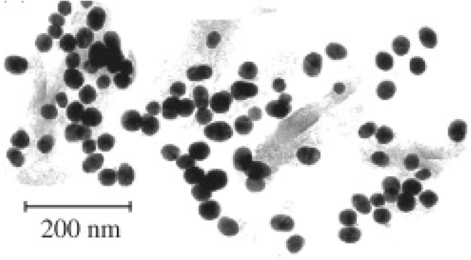
Au-Fe3 합성 방법 O4 나노 입자는 Rudakovskaya et al. 열수 기술을 통해 [76]. 이 방법의 원리는 Fe3의 추가를 따릅니다. O4 끓는 HAuCl4에 나노입자 해결책. 이들 나노입자의 TEM 분석은 30 nm의 평균 크기를 나타내었고, 일반적인 구형 모양과 20에서 35 nm 사이의 크기 분포를 나타냅니다. 이 이미지는 그림 3에서 볼 수 있습니다.
<그림>
Rudakovskaya et al.에 의해 합성된 나노입자의 TEM 이미지. 알 수 있는 바와 같이, 나노입자는 평균 크기가 30 nm인 대략 구형이다[76]
콜로이드 합성
콜로이드 합성 기술은 금속 나노입자를 합성하는 간단하면서도 효과적인 방법을 제공합니다[82]. 콜로이드 기술은 다른 용매가 필요하지 않거나 실온에서 수행할 수 있다는 점에서 나노입자 합성을 위한 다른 기술보다 단순한 수준을 제공하는 경우가 많습니다[83, 84]. 합성의 기본 원리는 다양한 금속 이온을 수상에 분산시키고 혼합물에 환원제를 첨가한 다음 제어된 온도에서 혼합하여 불용성 나노입자를 형성하는 것입니다[39]. 콜로이드 합성 경로는 합성에 잠재적인 독성 용매를 포함할 필요가 없다는 이점을 제공합니다(나노입자가 생물학적 용도로 의도된 경우 이상적). 그러나 최종 합성된 나노 입자의 크기 분포를 제어하기 어려울 수 있고[85] 나노 입자의 모양이 시약 농도에 크게 영향을 받을 수 있는 것과 같은 콜로이드 경로에는 몇 가지 제한이 있습니다[85]. 그러나 긍정적인 측면에서는 더 많은 양의 나노입자를 생산하는 것이 더 쉬울 수 있습니다[86]. 금속 나노입자 합성을 위한 이 방법은 은[87] 및 금[39, 88]과 같은 다양한 유형의 나노입자 합성에 사용되어 수년 동안 사용되어 왔습니다.
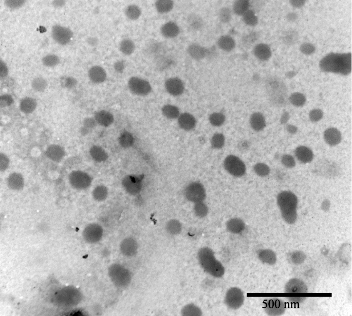
이 기본 방법은 금으로 코팅된 산화철 나노 입자의 형성을 위한 다양한 합성 경로를 생성하기 위해 발전되고 개발되었습니다[83, 84, 89,90,91,92,93,94,95,96,97]. 금으로 코팅된 산화철의 합성 방법은 대부분 HAuCl4을 환원하기 위해 다양한 환원제를 사용하는 방식입니다. 산화철 표면에 Nadagoudaet al. HAuCl4을 줄이기 위해 아스코르브산을 사용하여 제안된 "친환경" 합성 경로 제공 [84]. 그러나 이 방법은 합성에 사용되는 캡핑제(나노입자의 추가 "성장"을 멈추게 하는 나노입자 외부에 결합하는 작용제)가 없기 때문에 코팅된 나노입자의 크기나 모양을 거의 또는 전혀 제어하지 못하는 것으로 보입니다. [98]. 합성 코팅된 입자의 모양과 크기를 더 잘 제어하는 방법은 Pal et al. 이 방법은 6nm Fe3의 표면으로 환원되는 금염으로 gold acetate를 사용한다. O4 7nm 크기의 Au–Fe3를 생성하는 나노 입자 O4 모양이 구형인 입자. Fe3 코팅을 위한 신속한 방법 O4 나노 입자는 Rawal et al. 이것은 Fe3를 분산시키는 것을 포함합니다. O4 HAuCl4 용액의 나노 입자 , 그 다음 에탄올과 혼합[83]. 실온에서 15분 후, 반응을 멈추고 Au-Fe3 O4 그런 다음 나노 입자를 자석으로 분리했습니다. 정제된 용액의 TEM 분석은 생성된 입자의 크기가 30~100 nm 범위였으며 샘플 전체에 걸쳐 다양한 모양을 가짐을 보여주었습니다. 이 이미지는 그림 4에서 볼 수 있습니다. 이 합성 기술은 코팅된 나노입자를 빠르게 생성하지만 균일한 모양과 크기의 입자를 생성하는 데에는 그다지 효율적인 합성으로 보이지 않습니다[83].
<그림>
Rawal et al.에 의해 합성된 나노입자의 TEM 이미지. 이 나노 입자는 20–100 nm의 크기 분포를 가지고 있습니다[83]
일부 기술은 금 염의 환원만을 제공하지만 다른 기술은 하이드록실아민과 같은 환원제를 철 표면에 두는 것을 선호합니다[90, 93]. 많은 경우 Fe3 O4 나노 입자는 금으로 코팅되어 있으며 금 염의 환원은 표준 금 나노 입자를 생성하므로 [74], 철 나노 입자의 표면에 환원제를 첨가하면 코팅의 효율을 향상시키는 것이 목적이며 코팅의 효율을 낮추기위한 것입니다. 부산물로 생성되는 금 나노입자의 양[93].
또 다른 기술은 자기 나노입자의 표면에 금을 시딩하는 것인데, 이는 나노입자의 자기 코어 주위에서 금이 핵을 형성하도록 하는 보다 직접적인 경로를 제공합니다[91, 92, 97]. 이 기술은 용액 내 산화철 나노입자보다 작은 금씨드를 산화철 표면에 결합시키는 기술이다. HAuCl4 용액에서 Au
+
가 감소합니다. 이온은 산화철에 씨를 뿌리고 산화철 나노입자 주위에 껍질을 형성합니다. 이 금 씨뿌리기는 여러 그룹에서 성공적으로 사용되었습니다. Goon et al. Fe3 표면에 금이 뿌려지는 것을 제어하기 위해 폴리에틸렌이민을 사용했습니다. O4 , 완전히 코팅된 나노 입자를 생산합니다. 그러나 합성된 Au-Fe3 O4 입자는 40~110 nm 범위의 입자 크기로 높은 다분산성을 나타냅니다. Levin et al. 골드 시드에 결합하기 위해 유기 실란 분자로 기능화된 코어를 사용하여 50–70 nm 크기 범위의 금 껍질 자기 코어 나노 입자를 생산할 수 있었습니다[92]. 철 코어에 금 나노 입자를 시딩하는 것은 다양한 코어 모양으로 시연될 수 있습니다(예:Wang et al. 벼 모양의 "나노 쌀" Fe3에 금 파종 시연 O4 금이 표면으로 환원되었을 때 완전한 두꺼운 금 껍질로 이어졌습니다[97].
금속 나노입자의 생물 의학 응용
항균제
박테리아 감염은 1928년 Alexander Fleming에 의해 페니실린이 발견된 이후 항생제가 주요 치료 방법으로 사용되는 매우 일반적입니다[99]. 나노의학은 미래의 치료를 위해 금속 나노입자를 탐색하는 새롭고 광범위한 가능한 치료 양식을 제공합니다[100]. 표 1은 항균제 적용을 위해 탐색된 일부 나노입자를 나열합니다. 잠재적인 용도에 대해 조사된 한 가지 재료는 은으로, Sreekumar et al. 항균 섬유 네트워크의 일부로 은 나노 입자를 활용했습니다. 나노입자의 크기는 20~120 nm로 다양하며 Escherichia coli에 대한 항균 효과가 있습니다. 은 나노입자가 없는 섬유에 비해 94.3%나 높다[102]. 암피실린과 같은 항생제는 E에서 ≤ 99.9%의 사멸률을 달성할 수 있는 것으로 나타났습니다. 대장균 [103], 같은 연구에서는 E의 특정 균주에서 암피실린에 대한 내성의 출현도 보고했습니다. 대장균 . 이와 동일한 메모에서 E. 대장균 은 나노 입자에 대한 내성을 개발할 수 있습니다. 그러나 이 저항은 유전적 변화가 아니라 콜로이드성 나노입자를 응집시키려는 물리적 반응이다[104]. 또한 항균 특성을 위해 은을 사용하는 Holtz et al. 은 직경이 1-20 nm인 은 나노입자로 '장식된' 60nm 은 바나데이트 나노와이어 시스템을 설계했습니다[105]. 이 시스템은 세 가지 황색포도상구균에 대해 유망한 것으로 나타났습니다. 또한 흥미롭게도 메티실린 내성 황색 포도구균에 대해 훨씬 낮은 성장 억제 농도를 보였습니다. (MRSA) 항생제 옥사실린보다.
은 나노입자 합성은 Verma et al.에 의해 보고되었습니다. 박테리아에 대해 나노입자를 사용한 곳:Pseudomonas fluorescens , E. 대장균 및 곰팡이 Candida albicans [106]. 은 나노입자는 최소 억제 성장 농도가 각각 4.0μg/ml 및 16.0μg/ml인 암피실린 및 네오마이신과 같은 일부 일반적으로 사용되는 항생제와 비교하여 세 가지 균주에 걸쳐 5.83μg/ml의 평균 최소 억제 성장 농도를 가졌습니다. E의 균주에 대해. 대장균 [110]. 잠재적인 관심 대상은 나노입자가 P에 대해 표시되는 특성입니다. 형광등C. 알비칸 , 둘 다 면역 저하 환자에서 질병을 일으키는 것과 관련이 있습니다[111]. 추가 조사는 은 나노입자가 광범위한 부작용이 있는 암포테리신 B와 같이 가장 일반적으로 사용되는 일부 항생제보다 병원체를 치료하는 더 효율적인 방법임을 발견할 수 있습니다[112].
티오구아닌으로 덮인 금 나노입자의 합성은 Selvaraj et al. 여기서 다음을 포함한 여러 박테리아에 대한 향상된 항균 효과:E. 대장균 , 아스페르길루스 푸미가투스 , 및 녹농균 [107]. 티오구아닌으로 덮인 금 나노입자는 비접합 티오구아닌보다 항암제 및 항균제로서 더 효과적이며 그 활성이 암 약물의 운반체로서의 잠재적인 용도를 나타내는 것으로 밝혀졌습니다. 유사한 방식으로 금 나노 입자는 코리네박테리움 슈도결핵에 항균 효과가 있는 것으로 보고되었습니다. [108], 평균 크기가 25nm인 나노 입자는 50 μg/ml의 용량을 사용하여 20분 노출 후 95%의 박테리아 성장 억제를 나타냈다. 유사하게, 네이키드 금 나노 입자는 S를 포함한 다양한 그람 음성 및 그람 양성 박테리아에 항균 효과가 있는 것으로 나타났습니다. 구균 , 클렙시엘라 폐렴 , 및 고초균 [109]. 1.35 μg/ml의 AuNPs의 투여량은 S에 대해 46.4±0.4%, 38.3±0.2% 및 57.8±0.2%의 성장 억제를 나타냈다. 구균 , 케이. 폐렴 , 및 B. 자막 , 각각.
항바이러스제
항균 적용과 마찬가지로 금속 나노입자는 항바이러스 적용에서 유망한 것으로 나타났습니다. 표 2는 항바이러스 특성이 있는 것으로 밝혀졌으며 바이러스 치료에 잠재적으로 적용될 수 있는 다양한 나노입자를 보여줍니다. 노출된 은 나노입자와 코팅된 은 나노입자[113,114,115,116]는 나노 규모 범위에 있을 때 항바이러스 적용 범위가 넓은 것으로 나타났습니다.
B형 간염(HBV)은 현재 전 세계적으로 2억 5,700만 명의 사람들에게 영향을 미치는 바이러스 감염이며 세계 보건 기구에 따르면 2015년에 887,000명이 사망했습니다[121]. 작은(10-50 nm) 은나노입자가 HBV에 대한 가능한 치료법으로 테스트되었으며[113], HBV에 효율적으로 결합하고 HBV RNA 생성을 더욱 억제하는 것으로 나타났습니다. 작용 방식은 AgNP가 HBV dsDNA(이중 가닥 DNA)에 결합하기 때문인 것으로 가정됩니다. Rogers et al. 원숭이폭스 바이러스(MPV) [114]에 대한 항바이러스제로서 노출되거나 다당류 코팅이 된 은 나노입자의 사용이 입증되었습니다. 나노 입자는 12.5–100 μg/ml 사이의 농도 범위에서 MPV에 대해 시험관 내에서 테스트되었습니다. 연구 결과에 따르면 사용된 다당류 코팅 은 나노입자(Ag-PS-NPs)의 모든 농도는 시험관 내에서 MPV로 인한 플라크 형성을 감소시킬 수 있었습니다.
은 나노입자는 인간 면역결핍 바이러스(HIV)의 치료에서 역할을 할 수도 있습니다[115, 116]. HIV는 주요 건강 문제이며 WHO는 2016년 현재 3,670만 명이 HIV와 함께 살고 있다고 추정합니다[122]. HIV 치료법을 빠르고 효율적으로 발견하고 시행하는 것이 중요합니다. Lara et al. 은 나노입자(30–50 nm)가 HIV-1 분리주의 모든 균주의 억제를 나타내는 HIV-1 분리주에 미치는 영향을 입증했습니다[116]. 네이키드 나노입자는 전체 IC50를 나타냈습니다. HIV-1에 대해 0.44 mg/ml ± 0.3, 바이러스 억제 메커니즘이 바이러스-숙주 세포 결합 억제인 것으로 나타났으며, 특히 은 나노입자는 gp120 단백질(외피 당단백질)과 표적 세포 사이의 상호작용을 억제합니다. 막 수용체. 또한 같은 그룹에 의해 폴리비닐피롤리돈(PVP)으로 코팅된 은 나노입자가 HIV-1이 인간의 자궁 경부 조직 이식 모델로 형질감염되는 것을 방지하는 능력이 입증되었습니다[115]. 구체적으로, 0.15 mg/ml PVP 코팅된 은 나노입자(PVP-AgNPs)는 HIV-IIIB에 의한 감염을 억제했습니다. 및 HIV-AZT-RV 분리합니다. 이 농도의 PVP-AgNPs는 또한 대조군과 비교하여 감염 부위로 림프구(면역 세포)의 증식을 유도했습니다[115].
바이러스에 대해 사용된 것은 은 및 코팅된 은 나노입자뿐이 아닙니다. 양친매성 황산염 리간드로 코팅된 2nm 금 나노입자도 HIV-1에 대해 효과적인 것으로 나타났습니다[118]. 이 입자는 바이러스의 융합 과정을 표적으로 하는 것으로 나타났고 시험관 내에서 gp120 단백질에 결합하고 HIV-1 감염을 직접 중화시키는 것으로 나타났습니다. Mercaptoethanesulfonate-coated gold nanoparticles (Au-MES)는 아마도 숙주 세포에 대한 바이러스 결합, 세포 간 바이러스 확산 또는 바이러스에 대한 세포 감수성의 변경을 억제함으로써 단순 헤르페스 바이러스 1형(HSV-1) 감염을 억제하는 것으로 나타났습니다. 나노입자의 존재로 인한 감염 [117].
구리-요오드화물 나노입자(CuI-NPs)는 고양이 칼리시바이러스(FCV)[119]와 더 흥미롭게도 돼지 기원의 인플루엔자 A 바이러스(H1N1)[120]와 같은 여러 다른 바이러스에 대해 항바이러스 특성을 갖는 것으로 나타났습니다. 100~400nm CuI-NPs는 FCV에 대해 사용될 때 항바이러스 특성을 나타냈고, 1가 Cu 이온이 후속 캡시드 단백질 산화를 유발하여 FCV 비활성화를 유발하는 활성 산소 종(ROS)의 생성에 책임이 있다고 가정했습니다. H1N1 바이러스는 또한 매우 유사한 방식으로 CuI-NPs에 의해 억제되는 것으로 나타났습니다. 즉, 하이드록실 라디칼 생성이 단백질 분해를 유발합니다. 그러나 이러한 라디칼은 감염되지 않은 조직에도 독성이 있는 것으로 판명될 수 있으며, 이는 치료가 사용 승인을 받기 전에 결정하는 것이 중요합니다[123].
이미징
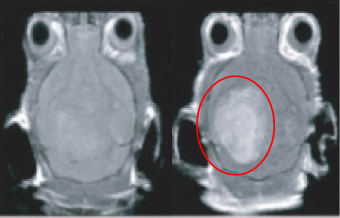
자기공명영상(MRI) 스캐닝은 의료 진단에 매우 유용한 도구이며 선명한 해부학적 영상을 제공합니다. MRI를 사용하면 혈류, 이화학적 특성, 신체 조직 및 기관의 상태를 시각화할 수 있습니다[124]. 조영제는 진단 감도를 향상시키기 위해 MRI에 자주 사용됩니다[125]. 기존에 사용되는 조영제는 킬레이트 계열이지만, 현재 조영제의 가장 큰 단점은 생물학적 안정성과 세포에 축적되었을 때의 독성 수준이다[126]. 예를 들어, 일부 조영제는 요오드 기반이며, 요오드화 조영제 노출은 이후에 발생한 갑상선 기능 항진증 및 발생하는 명백한 갑상선 기능 저하증의 발병과 관련이 있다고 보고되었습니다[127]. 조영제가 신체에 미칠 수 있는 부정적인 영향을 줄임으로써 향상된 스캐닝 효율을 제공하기 위한 대안이 개발되었습니다[128]. 대안은 MRI 스캐닝을 위한 조영제와 유사한 방식으로 작용하는 제제와 접합될 수 있는 금속 나노입자를 포함합니다[129]. 그림 5는 AuNP의 치료 전후에 쥐 대뇌 피질의 MRI 대조 이미지입니다[130].
<그림>
쥐 대뇌 피질의 치료 전(왼쪽)과 치료 후(오른쪽)의 MRI 대조 이미지. AuNP가 포함된 영역은 빨간색으로 표시됩니다.
표 3은 의료 영상에 사용하기 위해 탐색된 일부 나노 입자를 보여줍니다. 일부 컴퓨터 단층촬영(CT) 조영제는 짧은 순환 반감기[131] 및 잠재적인 조직 손상[130]을 포함한 문제가 있습니다. 이로 인해 금속 나노 입자는 CT 영상에 사용하기 위해 연구되었습니다[132]. Au 나노 입자는 X선 감쇠로 인해 이미징에 유망한 용도를 보여줍니다[133]. Kojimaet al. 은 시험관 내에서 우수한 조영제를 위해 만들어진 PEG-AuNPs(PEG-AuNPs)와 결합된 금 나노입자를 보여주었습니다. X선 컴퓨터 단층촬영의 경우 시판되는 요오드 제제인 iopamidal과 비교[134]. PEG-AuNPs는 시중에서 판매되는 iopamidal보다 더 높은 명암비 효율을 보여 체내에서 빠르게 배설됩니다[135]. 저자들은 또한 PEG-AuNP가 광열 치료를 가능하게 하는 광세포독성 특성을 가지고 있다고 언급했습니다.
Li et al. 죽상 동맥 경화증에 대한 이미징 도구로 코팅 된 AuNP의 사용을 입증했습니다. AuNP는 "단일 광자 방출 컴퓨터 단층 촬영"(SPECT)이라고 하는 의료 영상 유형에 적용되었습니다[136]. 이러한 유형의 이미징은 감마 카메라를 사용하는 것과 매우 유사하지만 보다 정확한 분석 기술을 달성하기 위해 슬라이스, 회전 및 조작할 수 있는 진정한 3D 이미지를 제공할 수 있습니다[136]. 변형된 나노입자는 세포사멸성 대식세포를 포함하는 죽상동맥경화증 플라크를 특이적으로 표적화했으며, 이는 죽상동맥경화증 플라크를 침습적으로 정확하게 감지하는 데 유용한 도구임을 나타냅니다[136].
AuNP는 이전에 높은 공간 분해능과 감도를 나타내는 광음향 영상(PA)에 대한 가능한 에이전트로 입증되었습니다[137]. PA relies on the detection of ultrasonic waves which are emitted from tissues when exposed to non-ionizing pulsed laser irradiation [140]. The intensity/magnitude of the ultrasonic emission is responsible for the image contrast, therefore any agent that can both absorb the laser pulses and then give off heat as a result will increase the magnitude of the ultrasonic emission and AuNPs possess the ability to do both of these [141, 142]. AuNPs are potentially better than organic dyes due to the organic dyes’ susceptibility to photo-bleaching and rapid clearing from the blood [143]. AuNPs also have use in cell imaging for examining movement of nanoparticles within cells when conjugated with various cargoes. Figure 6 is a darkfield imaging of A431 lung cancer cells treated with AuNPs that target epidermal growth factor receptor, and the bright areas within the cells are the nanoparticles indicating their locations within the cells [144].
Darkfield imaging of A431 lung cancer cells treated with AuNPs; the bright yellow/orange dots are nanoparticles within the cells
Biomedical Cargo Delivery
Nanoparticles make for an ideal molecule for drug delivery due to the huge surface area to the volume ratio they provide when compared to their bulk material [145]. In addition, it is possible to engineer nanoparticles to either avoid or interact with the immune system in specific ways [146, 147]. For example, it has been demonstrated that an increased hydrophobicity of nanoparticles/sub-groups conjugated to the nanoparticles illicit and increased immune response by measuring cytokine mRNA levels in mice [146]. Focusing in the opposite direction, it has been suggested that nanoparticles can be conjugated with various ligands to directly activate the immune system to target the destruction of a tumor [148] or by accumulation in the liver or spleen for the generation of tolerance or immunity, respectively [147].
Gold nanoparticles have been extensively studied for their delivery of medical cargo, for example, Bhumkar et al. have explored the application of AuNPs for trans-mucosal delivery of insulin. Gold nanoparticles were synthesized in the presence of chitosan, which acts as a polymeric stabilizer [149]. These nanoparticles were then loaded with insulin and administered both nasally and orally to diabetic rats. The results showed an overall reduction in the rat’s blood glucose levels, an indication of successful movement of the nanoparticles through the mucosal membranes and into the bloodstream.
More recently “smart” AuNPs have been employed in PA [138]. These nanoparticles are roughly 10 nm in diameter and are functionalized with citraconic amide moieties which are susceptible to hydrolysis. The citraconic amides are converted into positively charged primary amino acids at a mildly acidic pH, while the surface molecules adopt negative charges at physiological pH [138]. Combined, these two properties cause the “smart” nanoparticles to adopt both positive and negative charges allowing them to aggregate rapidly due to electrostatic attraction. These nanoparticles are referred to as “smart” due to the nanoparticles presenting cancer-specific properties and accumulate rapidly and efficiently in cancer tissues and show a much lower accumulation in normal tissues [150].
Gold nanoparticles can also be used as a delivery system for nucleic acids [153], including oligonucleotides [151] and small interfering RNA (siRNA) [154]. Many different methods have been developed to functionalize AuNPs with nucleic acids, for example, Yonezawa et al. have synthesized gold nanoparticles modified with thiocholine, which then bound to DNA and formed a fusion of wire-like structures throughout the DNA [155]. Sandström et al. demonstrated the ability to bind nucleic acids onto gold nanoparticles [151], and a similar modification has been done by Rosi et al. where tetrathiol-modified antisense oligonucleotides were bound to 13-nm gold nanoparticles [152]. Being able to conjugate nucleic acids to nanoparticles opens up the possibility of targeted gene delivery, which could, for example, lead to genes coding for a specific protein to be delivered to a cell that was either deficient in that protein or could not produce the protein themselves [156]. It has also been exhibited that gold nanoparticles modified with DNA can transfect cancer cells [157] (Table 4).
Anticancer Drug Delivery
Cancer is one of the world’s leading killers with large areas of scientific research being dedicated to the fight against cancer, and nanoparticles offer a new doorway into methods to target and treat cancer. Table 5 presents a selection of nanoparticle/drug conjugates that have been tested for anticancer treatments. Paciotti et al. have investigated the application of PEGylated AuNPs as a carrier for tumor necrosis factor (TNF) which is a cell-signaling protein that possess the ability to induce apoptosis in healthy cells [158]. The Au-PEG-TNF nanoparticles were injected intravenously and agglomerated significantly more in MC-38 colon carcinoma cells compared to other healthy cells/tissues. The TNF not only gave therapeutic action on the MC-38 cells, but also seemed to possess a targeting property, indicated by the lack of agglomeration in healthy cells. Another interesting observation reported was the ability for the Au-PEG-TNF nanoparticles to diminish a tumor mass compared to “free” TNF.
Doxorubicin is a widely used cancer therapeutic agent but has dose-limiting associations with cardiotoxicity. A gold nanoparticle-doxorubicin conjugate has been developed that demonstrates little no to cardiotoxicity to mice while being able to treat cancer [160]. Dixit et al. demonstrated the selective delivery of folic acid-coated AuNPs into folate receptor (FR) positive cancer cells, whereas when compared with a cell line that did not have folate receptors, uptake was shown to be minimal [159]. These results demonstrated the use of folate to target metal nanoparticles to FR-positive cancer cells for tumor imaging and ablation.
Limitations of Single Metal Nanoparticles and Overcoming Them
The principal obstacle with nanoparticle drug delivery is the ability to direct the nanoparticle to the target area [162, 163]. There are several methods in use for metal nanoparticle targeting such as antibodies [164,165,166] and homing peptides [167, 168]. There are however limitations to these methods, with the biggest being that before they even reach the desired target cells they have to pass through a variety of other barriers, such as blood vessels and the blood-brain barrier [169]. One way to overcome this targeting limitation is to use magnetic nanoparticles [170]. A magnetic nanoparticle-targeting system works by directing the nanoparticles to a target site using an external magnetic field, it has already been demonstrated that the magnetic anisotropy of the nanoparticle is a very important factor for medical treatments [171], with a change in anisotropy being able to the change the efficacy of hypothermia treatments for example [172]. Superparamagnetic metal nanoparticles have this property (they only present magnetic properties while in the presence of a magnetic field) [173]. However, the benefit of magnetic nanoparticles also presents a potential limitation, due to the toxicity of many magnetic materials [31, 174, 175]. Despite iron being approved for various imaging uses [5, 6, 31], it has been suggested in several studies that naked iron oxide nanoparticles may have some adverse effects when used in cell labeling [176,177,178]. One method that can be used to overcome any potential toxicity limitations is to coat the iron core [179]. A range of materials can be used as the coating material:silica [180,181,182], polymers [183, 184], gold [62, 93, 95, 185], or silver [58, 186]. Gold has low pharmaceutical activity [187] and silver has been used in biomedical applications for many years [188, 189],
The combination of a superparamagnetic core with an inert and safe metal coating produces metal nanoparticles with superior characteristics to non-magnetic metal particles [190]. As well as reducing toxicity, the coating also provides the potential for the conjugation of functionalized molecules onto the surface, such as drugs and biomolecules for application in the medical field [97, 140, 152]. It is of note that a core-shell nanoparticle still possesses the properties and uses of a nanoparticle made from the same material as just the shell, but the superparamagnetic core gives the ability to direct the nanoparticle in the body [191]. For example, a gold nanoparticle with an antibody is classified as a targeting nanoparticle, introducing the core would classify the nanoparticle as a directed targeting nanoparticle [173].
Current Medicinal Uses of Gold-Coated Iron Oxide Nanoparticles
Core-shell superparamagnetic nanoparticles have already been assessed for their biomedical uses, with a wide range of uses already being applied [192] and with a majority of research investigating into the use of gold as a shell for the nanoparticles, in part due to its biocompatibility and ability to easily bind to a variety of materials. As such, this section will deal exclusively with gold shell nanoparticles. One of these uses is as a magnetic carrier for drug targeting [192,193,194,195,196]. Kayal and Ramanujan have tested an in vitro apparatus that simulates the human circulatory system as a test for the magnetic delivery of gold-coated iron oxide nanoparticles (Au-Fe3 O4 ) loaded with doxorubicin [194]. Their system had various magnetic fields of increasing strength next to a capillary through which the doxorubicin-loaded particles were passed. A significant percentage of these nanoparticles were captured within the magnetic fields, strongly indicating the potential for the use of magnetic nanoparticles in drug delivery. Another use for a targeted system is the application of Au-Fe3 O4 nanoparticles in photothermal therapy. Bhana et al. demonstrated the use of a core-shell system used in combination therapy deployed against two different cancer cell lines; head and neck (KB-3-1) and breast (SK-BR-3) with a reported decrease in cell viability of 64% when they exposed cell lines to a combined photothermal and photodynamic therapy, compared to each modality used on its own [197]. In photothermal therapy, gold nanoparticles are coated with a ligand, such as PEG [142], and these nanoparticles are irradiated with a laser, with a wavelength that matches the UV-vis λ -max of the gold nanoparticles [194]. The nanoparticles vibrate at the laser’s frequency which causes heat to be released causing the death of the surrounding tissue [198], introducing a core which is superparamagnetic can allow for a more accurate targeting for use in this therapy. Similarly, it has been reported by Kirui et al. that gold hybrid nanoparticles were deployed against SW1222 colorectal cancer in photothermal therapy, showing an increased case of cellular apoptosis after therapy, with their conclusion being that the cells showed an increased uptake, leading to a reduced laser power required to reach threshold therapeutic levels [199]. The use of core-shell nanoparticles for photothermal therapy of cancer has also been reported by other groups [200, 201].
Metal nanoparticles have already shown to have a place in contrast imaging, for example core-shell nanoparticles can also be used in T1- and T2 -weighted imaging in MRI [202]. Research by Cho et al. demonstrated that gold-coated iron nanoparticles can be successfully used in MRI imaging, as well as opening the route for conjugating various ligands for use in biosensors [202]. A magnetic carrier capable of imaging and photothermal therapy has been reported by Cheng et al. They demonstrated the magnetic targeting of multi-functional nanoparticles to a tumor in a mouse model, which could be imaged inside the tumor and showed a reduction in the tumor size when combined with photothermal therapy [203]. It is also of note that in this work, both the nanoparticle dosage (1.6 mg/kg) and laser power (1 W/cm
2
) are among the lowest applied for in vivo photothermal therapy. Moreover, there was no obvious toxicity from the nanoparticles reported. Table 6 presents some of the currently reported uses of core-shell nanoparticles.
Another medical area where such core-shell metal nanoparticles have been suggested to make an impact is in directed enzyme prodrug therapy (DEPT) [170, 191]. DEPT is a promising method of cancer treatment, with several therapies making it through to clinical trials [207, 208]. The main principal of DEPT is the targeted delivery of a prodrug-activating enzyme to a tumor site. Upon arrival at the tumor site, the enzyme enters the target cells where it can later activate an administered prodrug. However, the efficacy of the therapy depends on the ability to direct the enzyme to the tumor site, with current directional techniques relying on passive targeting methods such as viruses [207, 209] or antibodies [210, 211], rather than an active targeting system for enzyme delivery. A novel therapy proposed by Gwenin et al. potentially overcomes the targeting issue [170, 212]. This approach involves conjugating a genetically modified prodrug-activating enzyme onto the surface of a gold-coated iron oxide superparamagnetic nanoparticle (AuMNP), then directing the AuMNP-enzyme conjugate to the target site using a magnetic field to increase the efficacy of the targeted therapy. Figure 7 presents some of the uses of a core-shell nanoparticle.
A pictorial representation of the applications of core/shell nanoparticles
결론
In brief, single metal nanoparticles have been shown to currently possess a wide range of biomedical applications, with more application for these nanoparticles being discovered. One of the limiting factors that these nanoparticles face in medical treatments is to find a way for precise accurate targeting of areas within the body, be it for targeting of a drug delivery or for therapies involving the nanoparticles directly. A way to overcome this is to employ a magnetic core to create core-shell nanoparticles that can then be directed around a body using a magnetic field. There are a variety of methods that can be used to synthesize these core-shell nanoparticles, with each method having its own advantages and disadvantages. There remain many obstacles for core-shell nanoparticles before they can be routinely applied in the medical field and these include
<리> 1)
Achieving a synthesis route which produces easily repeatable results;
<리> 2)
Producing particles of a set size [22,23,24] and shape [25,26,27,28]; and
<리> 3)
Producing large enough quantities to make it economically viable [29].
Another key factor is the relatively unknown toxicity of some nanoparticles over an extended period of time due to how relatively new the field of research is.