빠르고 통제할 수 없는 세포 성장이 합병증과 조직 기능 장애를 일으키는 질병인 암의 유병률 증가는 과학자와 의사의 심각하고 긴장된 우려 중 하나입니다. 오늘날 암 진단과 특히 그 효과적인 치료는 지난 세기 건강과 의학의 가장 큰 과제 중 하나로 여겨져 왔습니다. 약물 발견 및 전달의 상당한 발전에도 불구하고 일반적으로 건강한 조직 및 기관에 손상을 일으키는 많은 부작용과 부적절한 특이성 및 민감도가 약물 사용에 큰 장벽이 되어 왔습니다. 이들 치료제의 투여 기간과 양에 대한 제한도 도전적이다. 반면에, 화학 요법 및 방사선 요법과 같은 일반적인 암 치료 방법에 내성을 갖는 종양 세포의 발생은 항종양 약물 특성의 혁신, 개선 및 개발에 대한 강렬한 필요성을 강조합니다. 리포솜은 물리적, 화학적 특성이 다른 약물을 저장할 수 있는 능력 때문에 나노의학 분야에서 약물 전달 및 암 치료에 적합한 후보로 제안되었습니다. 또한, 다양한 고분자, 리간드 및 분자를 결합하여 화학적 변형을 위한 리포좀 구조의 높은 유연성과 잠재력은 약리학적 장점을 향상시킬 뿐만 아니라 항암제의 효과를 향상시키는 리포좀의 중요한 장점입니다. 리포솜은 체내에서 이러한 항악성 세포제의 민감도, 특이성 및 내구성을 증가시킬 수 있으며 나노 의약품에 적용할 놀라운 이점을 제공할 수 있습니다. 우리는 다양한 종류의 암과 질병을 치료하기 위한 임상 적용을 중심으로 리포솜의 발견과 개발을 검토했습니다. 리포솜 약물의 특성을 어떻게 개선할 수 있는지, 암 치료에 대한 기회와 과제도 고려하고 논의했습니다.
그래픽 개요
<그림>
소개
암은 신체의 건강한 세포가 정상적인 상태를 벗어나 통제할 수 없이 분열하는 질병으로, 현 세기의 큰 의학적 도전 과제로 인식되고 있습니다. 이러한 합병증은 환경적 발암물질의 축적이나 유전적 변이[1]로 인해 발생하며, 현 세기에 큰 의학적 도전으로 인식되고 있다. 매년 수백만 명이 암으로 사망하고 신규 환자 수와 사망률이 지속적으로 증가하고 있다[2]. 세계보건기구(WHO) 보고서에 따르면 암은 2018년 사망의 두 번째 주요 원인이었고 추정에 따르면 그 해에 다양한 암으로 약 960만 명이 사망한 것으로 나타났습니다. 2018년에 사망 6명 중 1명은 대략 암으로 인한 것이었습니다. 암 사망의 약 70%는 개발도상국과 저소득 국가에서 발생합니다. 그러나 선진국의 암 발병률과 치사율도 고려해야 한다[3].
항암제를 이용한 화학요법은 암의 중요한 치료법으로 알려져 있다[4]. 무료 약물을 사용한 화학 요법은 적절한 민감도와 특이도가 부족하기 때문에 제한적입니다. 결과적으로 이러한 제한은 부작용으로 인한 정확한 치료를 방해하고 충분한 항종양 효과 발휘를 저해하였다[5]. 병용 요법인 화학 면역 요법은 기존 약물에 내성이 있는 종양 세포를 명시적으로 치료하는 암 요법에 대한 효과적이고 유망한 방법으로도 제안되었습니다. 최근 몇 년 동안 다양한 기존 및 고급 치료법이 발견되어 암 치료에 적용되고 있습니다. 예를 들어, 기존의 항암제, 특히 화학 요법제의 부작용을 줄이기 위해 바이러스 나노 입자(VNP)[7, 8], 양자점[9], 고분자 나노 물질[10], 리포좀을 비롯한 다양한 나노 의약품[6] [11]이(가) 적용되었습니다.
다양한 나노 의약품 중에서 구형 나노 입자(NP)로서의 리포솜은 특정 구조를 가지고 있습니다. 리포솜 성분에 두 가지 수성 및 유기상의 존재는 친수성 및 소수성 제제의 두 종류 모두를 포획할 수 있게 하고 많은 나노캐리어에 비해 리포솜에 대해 현저한 이점을 생성합니다. 항종양제의 특이성, 생체이용률, 생체적합성을 높이는 방법 중 하나는 다양한 종류의 리포솜에 이들을 포획하는 것이다[5]. 지난 20년 동안 치료 목적으로 리포솜을 이용하기 위한 상당한 노력이 있었습니다. DaunoXome® 및 Caelyx®와 같은 이러한 약물 중 일부는 일반 및 임상 적용을 위해 승인된 반면 다른 약물은 최종 생산 및 승인 단계에 있습니다[12].
일반적으로 면역리포솜과 pH 민감성 리포솜과 같은 다양한 종류의 치료용 리포솜이 있습니다. 면역리포좀은 연구 및 연구에서 상당한 항악성 효과를 보인 표적 약물 전달 시스템(DDS)이라고도 하는 대규모 나노 의료 장치 그룹입니다[13]. pH 민감성 리포좀은 pH 변화에 의해 구조와 구성 분자가 변경되어 약물 함량이 방출되는 다형성 리포좀 그룹으로도 알려져 있습니다[14]. 또한, 리포솜은 다른 나노의약과 마찬가지로 약물 전달 시스템 외에도 조직 복구 및 재생, 영상화 및 진단에 활용될 가능성이 있습니다. 다양한 측면에서 리포솜을 사용하면 질병과 암의 식별, 관리 및 치료가 간편해집니다[15].
이 글에서는 리포솜의 발견과 구조, 리포솜의 다양한 성질, 시중에 나와 있는 암 치료용 리포솜 약물과 그 개발에 대한 연구 결과를 요약한다. 궁극적으로 리포솜 나노의약품 활용의 기회와 과제에 대한 보고서가 마무리될 예정이며, 이는 과학자들의 향후 연구에서 주목해야 할 중요한 이슈로 부각되어 한계를 해소하고 긍정적인 점을 강화할 수 있을 것입니다.
본문
리포솜의 과학적 역사:발견 및 정의
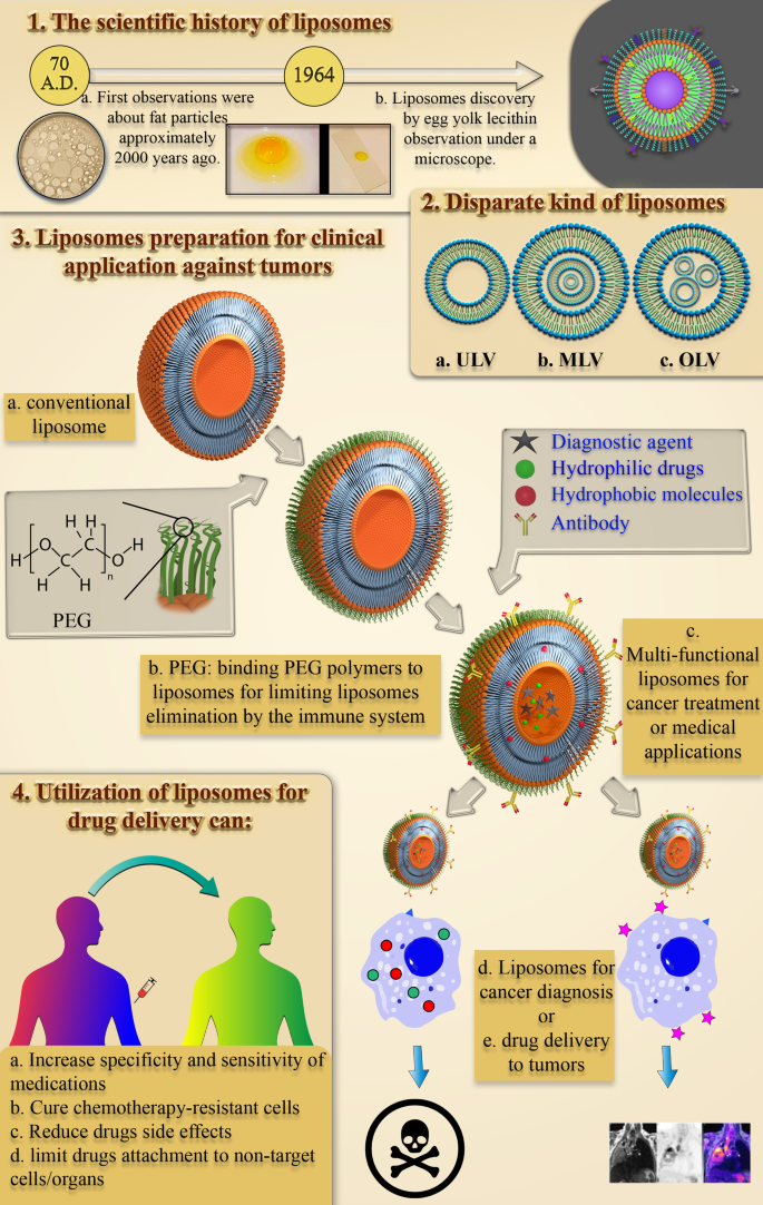
수성 환경에서 작은 지질 입자의 구조와 거동에 대한 초기 연구에서 미국 FDA 승인을 받은 최초의 지질 기반 약물 전달 나노입자가 나오기까지는 약 1950년이 걸렸습니다. 수중 지질 및 지방 입자의 행동을 연구하는 과정은 거의 2000년 전 Pliny the Elder가 처음으로 관찰한 것으로 시작되었습니다[16]. 17세기 후반 Anthony Van Hook의 세포 발견은 세포의 구조에 대해 많은 의문을 제기했습니다[17]. 그런 다음 Gorter와 Grendel은 세포막에서 인지질 이중층의 존재를 발견했습니다[18]. 이후 Singer와 Nicolson은 세포막 인지질의 거동을 설명하기 위해 이중층 모자이크 막 모델을 설명했습니다[19]. 이러한 과학적 관찰과 가설은 지방 유래 나노입자에 대한 다른 과학자들의 관심을 끌었습니다. 1960년대에 Babraham Institute[20, 21]에서 혈액 응고 과정에 대한 지질, 특히 인지질의 영향을 연구한 Alec D. Bangham은 우연히 첫 번째 리포솜을 관찰하고 자연적으로 구형 입자가 형성되는 것을 보고 놀랐습니다. 물 [22]. 이후 뱅햄의 연구 결과를 알고 알렉 뱅햄의 연구실을 방문한 제럴드 와이스만(Gerald Weissmann)은 알렉이 관찰한 스멕틱 중간상을 '뱅호솜'이 아닌 '리포솜'이라고 부르며 노벨상을 수상했다[22]. 리포솜 발견의 과학적 역사는 그림 1에 요약되어 있습니다.
<사진>
리포솜의 발견으로 이어진 관찰 도표. 물에서 지질 및 지방 입자의 거동을 연구하는 역사적, 과학적 경향과 리포솜 발견으로 이어지는 관찰, 이벤트에 관련된 과학자의 이미지와 함께 Pliny the Elder [23], Anthony Van Hook [24], Alec D. Bangham [25] 및 Gerald Weissmann [26], 각각 왼쪽에서 오른쪽으로
요즘 리포솜 나노입자로 알려진 구조는 무엇입니까?
리포솜 NP를 정의하고 그 특성을 합리적으로 발견하기 위한 강렬한 노력이 있습니다. 오늘날 리포솜은 지질 이중층 막과 친수성 코어로 구성된 자발적으로 형성되는 구형 단편으로 정의됩니다.
리포솜의 크기는 약 10nm에서 2500nm(또는 2.5µm)까지 다양합니다[15]. 그러나 약물 전달을 위해 투여되는 대부분의 리포솜은 일반적으로 크기가 약 50~450nm입니다. 확실히, 훨씬 더 큰 치수를 가진 리포솜은 의료 용도로도 사용될 수 있습니다[27]. 또한, 리포솜은 주로 인지질로 구성됩니다. 인지질은 흥미롭게도 트리글리세리드와 유사한 지질 유형입니다. 인지질의 구조에는 친수성 극과 두 개의 소수성 사슬이 있습니다. 따라서 인지질은 양친매성 분자로 간주됩니다.
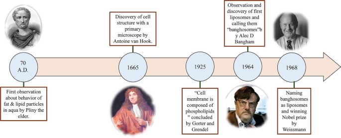
인지질의 리포솜 막은 대부분 포스파티딜콜린(PC), 스핑고미엘린(SM), 포스파티딜세린(PS), 포스파티딜에탄올아민(PE)을 포함하며, 이들은 양친매성이며 물에서 특정 구조를 형성하는 경향이 강합니다[28]. 이 현상의 물리적인 이유는 인지질에 친수성 머리(인산염 분자)와 두 개의 소수성 꼬리(지방산)가 공존하기 때문입니다. 인산기는 H2와 상호작용합니다. O 극성 분자, 소수성 꼬리는 물 분자에서 빠져 나와 서로 상호 작용합니다 [29]. 이 경우 비극성 사슬은 서로 반대 방향으로 배치되어 이중층을 만들어 그 사이에 친유성 공간을 만듭니다. 따라서, 이러한 리포솜의 친유성 부분 구조는 소수성 제제 및 물질을 저장하는데 적용될 수 있다. 더욱이 인지질의 친수성 부분은 수소 결합, 반 데르 발스 등과 같은 분자력을 통해 물 분자로 향하게 되며, 이는 그들 사이에 나타납니다. 이들은 리포솜 내부에 친수성 영역을 형성합니다. 레시틴 분자의 구조는 난황에 풍부하고 물과 리포솜의 다양한 영역에서 리포솜을 형성할 수 있는 천연 인지질로서 그림 2에 나와 있습니다.
<사진>
레시틴에서 유래한 리포솜의 개략도. 친수성 코어와 소수성 이중층을 포함한 리포솜의 다양한 영역이 설명됩니다. 레시틴 분자의 구조, 친수성 극 및 소수성 사슬이 지정됩니다.
또한 물에 용해된 후 리포솜의 구형 구조는 분자의 유형, 수성 매질의 온도, 몰 농도 및 이온과 같은 다른 물질의 존재 여부에 따라 궁극적인 모양을 결정합니다[30]. 리포솜의 주된 물리적 및 화학적 특성은 지질, 특히 인지질과 이를 구성하는 기타 분자의 순 특성입니다. 이러한 속성에는 투과성, 표면 전하 밀도 및 전체 크기가 포함됩니다[31].
다양한 유형의 리포솜 분류
리포솜 발견 이후 이러한 구조는 항상 생물학, 생물물리학, 생화학 또는 제약 연구의 필수 부분으로 활용되어 왔습니다.
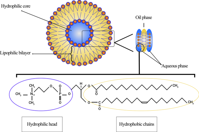
오늘날 리포솜은 크기, 인지질 이중층의 수, 합성 절차 및 준비 메커니즘에 따라 분류할 수 있습니다. 크기에 따라 리포솜은 소형, 중형, 대형의 세 그룹으로 나눌 수 있습니다. 막 층의 수를 고려하면 단층 소포(ULV), 올리고층 소포(OLV) 및 다층 소포(MLV)가 될 수 있습니다. 이와 관련하여 ULV는 약 50~250nm 측정되는 하나의 인지질 이중층으로 구성된 리포솜인 반면 MLV는 훨씬 더 큰 약 0.5~1.5μm이며 여러 인지질 이중층 막을 포함합니다[32]. 다른 합성 방법으로 인해 이 두 그룹 사이에 여백이 생깁니다. 적용 측면에서 ULV는 내부적으로 큰 친수성 환경을 가지므로 친수성 약물의 포획에 적합합니다. ULV와 같은 작은 단층 소포(SUV)는 하나의 인지질 이중층으로 구성되어 있지만 크기가 100nm 미만입니다[33, 34]. 형태학적 관점에서, OLV는 동일하거나 다른 크기를 가질 수 있는 2-5개의 소포로 구성된 리포솜입니다. OLV의 구조에서 소포는 모두 서로 내부에 있지 않고 하나의 큰 인지질 이중층으로 둘러싸여 있습니다. OLV는 일반적으로 약 0.1–1 µm입니다[33,34,35]. ULV와 달리 MLV는 친수성 물질의 전달에 이상적이지 않습니다. MLV는 소수성 제제의 전달에 주로 이용됩니다[36]. 다양한 유형의 리포솜이 그림 3에 나와 있습니다.
<그림>
다양한 기준에 따른 리포솜 분류:a 리포솜은 크기면에서 세 가지 범주로 나뉩니다. ㄴ 작은 단층 소포(SUV) 구조는 단층 소포(ULV)의 구성원으로서 눈에 띄게 작은 크기를 가지고 있습니다.
리포솜 준비 방법 및 차세대 개발
경질 나노입자인 금 나노입자와 달리 리포솜은 연성 나노입자[37]이며 다양한 방법으로 합성할 수 있습니다. 예를 들어, MLV와 ULV는 준비 메커니즘이 다릅니다. 이러한 방법의 대부분에서 특정 용매(클로로포름 또는 메탄올 등)는 둥근 바닥 플라스크(RBF)에서 리포솜 막을 형성하기 위한 지질(원하는 몰비)을 해결하는 데 사용됩니다. 예를 들어, 악수는 MLV를 합성하는 기본 절차입니다[38]. 지질막 수화라고도 알려진 이 과정에서 지질이 유기 용매에 첨가됩니다. 그런 다음 회전 장치로 용매를 증발시키고 고체 생성물을 지방분해시킨다. 궁극적으로 리포솜은 수화 및 압출 방법에 따라 합성됩니다[39]. 리포솜 합성의 다른 방법으로는 초음파 처리, 역상 증발, 프렌치 압력 셀, 동결 건조 및 막 압출이 있습니다[38, 40].
또한, 리포솜은 시간 경과에 따른 발견 및 개발에 따라 다양한 범주로 주문할 수도 있습니다. 1세대 리포솜은 일반적으로 기존 리포솜 또는 고전 리포솜으로 명명됩니다. 기존의 리포좀을 치료적 나노입자로 사용할 때 관찰되었던 문제가 생체 내에서 매우 빠르게 확인되었습니다. 초기에 조사된 문제 중 하나는 리포솜으로의 약물 포획의 한계였습니다. 즉, 많은 약물이 1세대 리포솜 내부에 저장될 수 없었습니다[41]. 안정성, 치료 효능 및 임상 적용 가능성과 같은 리포솜의 구조와 특성을 조사하려는 큰 열망과 함께 이러한 과제는 구성 지질, 표면 전하, 순중량, 총 부피[42]. 정확히 말하면, 2세대 리포좀은 주로 기존의 리포좀에 일부 친수성 고분자를 추가하여 체액에서 저장 수명을 연장하여 약물 전달 시스템에 적합한 후보로 만드는 방식으로 합성됩니다. 이러한 종류의 리포솜은 비특이적 장기 순환 리포솜과 리간드 표적 장기 순환 리포솜의 두 그룹으로 나눌 수 있습니다[43].
새로운 세대의 리포솜인 Archaeosome은 고세균막 지질과 합성 인지질 유사체로 구성됩니다. 지난 10년 동안, 약물 및 백신 전달에 사용되는 archaeosome의 잠재력을 조사하기 위해 광범위하고 상당한 노력이 있었습니다. 고세균형 지질의 구조 핵은 약 20~40개의 탄소를 포함하는 포화 피타닐 사슬을 가진 디에테르 또는 테트라에테르 분자입니다. 이 탄소 사슬은 고고학 또는 칼다르카올에서 발견되는 백본 글리세롤의 sn-2,3 탄소의 에테르 결합에 부착됩니다. 위에서 언급한 바와 같이 이러한 입자는 또한 백신 접종뿐만 아니라 종양 합병증, 알레르기 및 감염에 대한 약물 전달에 엄청나게 사용될 수 있습니다[44].
리포솜의 생체물질 특성 및 물리화학적 특성 평가
앞서 언급한 바와 같이 의학의 광범위한 발전에도 불구하고 일부 질병, 특히 암의 치료는 비효율적인 치료제와 방법으로 인해 여전히 어려운 문제에 직면해 있습니다. 종양에 영향을 미치기 위해 주사된 약물 용량을 조정하는 것은 항암제의 치료 범위가 좁기 때문에 긴박한 문제입니다. 즉, 치료 용량과 독성 용량 사이의 약간의 거리, 부적절한 민감도 및 특이성으로 인해 고급 치료 절차에 대한 큰 수요가 발생했습니다[42].
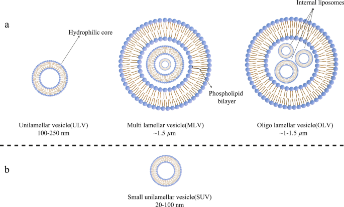
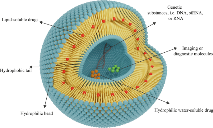
또한 최근에는 조직으로의 약물 전달을 위한 나노물질의 활용이 주목받고 있다. 생체 적합성과 생분해성은 전달 시스템에서 나노 물질을 사용하기 위한 생체 물질 특성의 두 가지 필수 기능입니다. 치료용 나노입자가 신체 조직 및 시스템을 손상시키는 것을 억제하기 위해서는 생체적합성이 필요하며, 나노입자를 무독성 화합물로 분해하고 단순히 장기에서 제거하기 위해서는 생분해성이 시급하다[15]. 리포솜 검출 후 과학자들은 이를 약물 전달용 나노 물질로 적용하기 시작했습니다. 언급된 바와 같이, 리포솜은 치료 목적을 위해 생체 적합성과 생분해성의 두 가지 필수 생체 재료 특성을 가지고 있습니다[36]. 또한, 리포솜 NP는 이러한 목적에 적합하게 만드는 다른 특성을 가지고 있습니다. 예를 들어, 리포솜의 특정 구조로 인해 친수성(수용성) 및 소수성(지용성) 약물 그룹 모두가 리포솜에 캡슐화될 수 있습니다. 또한, 리포솜에 인지질 이중층막이 존재함으로써 효소 분해, 면역 구조에 의한 생물학적 불활성화, 생체 내 화학적 변화 등의 다양한 현상 및 손상으로부터 리포솜에 저장된 제제를 보호한다. 이 점에는 두 가지 중요한 장점이 있습니다. 첫째, 리포솜에 갇힌 분자의 구조가 표적 조직에 도달하기 전에 보존되고 변형되지 않으며, 둘째, 다른 건강하고 표적이 아닌 조직이 리포솜에 노출되지 않도록 보호됩니다. 약물은 리포솜 막으로 인해 영향을 받지 않으며 이러한 약제의 영향을 받지 않습니다[42]. 리포솜은 또한 DNA, RNA 등과 같은 유전 물질을 전달하고 유전자 치료 목적으로 적용될 수 있습니다. 이 목적에 사용되는 리포솜은 양이온, 음이온, 중성 지질 및 인지질 또는 이들의 혼합물로 구성될 수 있습니다[45]. 탄소점과 같은 일부 진단 및 영상화제는 리포솜의 조합 또는 단독으로 암 탐지 및 영상화에 사용될 수 있습니다[46]. 카본 도트가 임상 적용을 위해 부분적으로 승인되고 조사에서 활용되지만, 세포 독성은 광범위한 적용을 위한 도전적인 장벽으로 남아 있습니다[47]. 리포솜 약물 NP의 일반적인 구조는 그림 4에 나와 있습니다.
<그림>
리포솜의 일반적인 구조는 인지질 층으로 구성됩니다. 약물의 친수성-소수성에 따라 전달을 위한 적절한 종류의 리포솜이 결정됩니다. 친수성 약물은 중심 친수성 핵에 갇히고 소수성 약물은 친유성 영역에 위치합니다. 리포솜은 유전자 전달에도 활용될 수 있습니다.
리포솜에 존재하는 막 형성 인지질은 무독성 화합물이며 광범위한 크기로 합성될 수 있습니다. 리포솜의 물리화학적 특성은 구성 성분에 따라 다릅니다. 따라서 원하는 특성을 가진 리포좀은 콜레스테롤, 폴리에틸렌 글리콜(PEG) 등과 같은 특정 화합물을 추가하여 합성할 수 있습니다. 게다가 리포좀의 막은 거대 분자에 대해 불투과성이므로 리포좀 내에 물질을 더 잘 유지하는 데 도움이 됩니다[48 ]. 언급된 리포솜의 모든 특징은 다양한 질병, 특히 암을 치료하기 위한 치료제 전달에 활용하기 위한 적절한 나노물질로 리포솜을 소개합니다.
이 분야의 선구자 중 한 명인 Gregory Gregoriadis는 약물 전달 시스템에 리포좀을 사용한다는 가설을 제안하고 약물 화합물이 리포좀에 포함될 수 있다고 말했습니다[49]. 리포솜의 적절한 생체 재료 및 물리화학적 특성이 보고되었습니다. 예를 들어, 동물 모델에서 사용된 항종양 약물 시토신 아라비노사이드를 함유하는 리포솜에 대한 조사는 L1210 백혈병이 있는 마우스의 수명이 유의하게 증가하는 것으로 나타났습니다[50]. 리포솜을 적용하면 충분한 양의 활성 형태의 약물이 보호된 방식으로 표적 부위에 전달될 수 있습니다[42].
치료 용도를 위한 리포솜 NP의 특이성 및 감도 향상
앞서 언급한 바와 같이 다양한 분자와 고분자를 이용하여 리포좀의 구조와 막의 변화를 가능하게 하고 이를 통해 리포좀에 새로운 기능을 추가하거나 특성을 변형시킬 수 있다[51]. 혈액 내 리포솜의 순환을 연장하고 EPR 효과를 통해 특정 종양 조직 또는 병리학적 부위에 축적되는 능력을 증가시키는 것은 리포솜의 높은 제거율 때문에 고려해야 하는 첫 번째 중요한 특징입니다. 화학적 접합을 통한 리포솜 막에 대한 PEG 분자의 접합은 이 기능을 리포솜에 추가하기 위해 순차적으로 사용되었습니다[52]. 리포솜, 특히 혈액과 같은 체액에서 리포솜 치료 나노입자의 반감기를 증가시키는 데 에틸렌 글리콜 중합체의 중요성과 역할은 약 20년 전에 표현되었습니다[53].
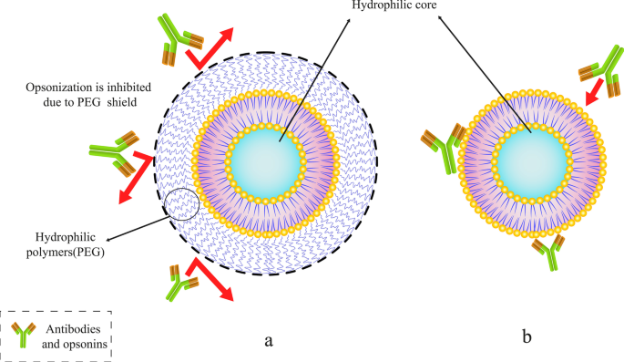
Abuchowski와 McCoy는 PEG를 구조에 결합하여 혈류에서 리포솜의 반감기를 연장하려는 첫 번째 시도를 했습니다. 결과적으로 그들의 노력은 일반적으로 리포솜의 순환 시간과 혈류에서의 반감기를 증가시켰다[54]. 몇 달 후, 다른 연구자들은 MPS(단핵 식세포 작용 시스템 세포)에 의한 리포솜 제거의 고속 감소 가능성을 조사했습니다. 리포솜의 표면 분자에 PEG를 부착함으로써[53], 리포솜의 혈액 내 순환이 개선될 것으로 기대된다. 이 분야에는 수많은 기사가 있습니다. 또한, 기존의 리포솜과 달리 PEG로 코팅된 리포솜은 용량과 무관한 약력학 특성을 보여주었습니다[55]. 다양한 고분자 중 PEG 분자는 리포솜 표면에 부착되어 생체 내에서 유통기한을 연장할 수 있는 고분자 중 하나이다. 이 목적을 위해 다른 폴리머도 사용할 수 있습니다[56]. 그림 5는 PEG 폴리머 분자가 어떻게 항체로부터 리포솜을 보호하고 혈류 내에서 리포솜의 수명을 연장할 수 있는지 보여줍니다.
<그림>
폴리에틸렌 글리콜(PEG)과 같은 특정 중합체를 리포솜에 접합합니다. 아 PEG 폴리머 분자가 있는 PEG화된 리포솜이 보호합니다. ㄴ 항체와 옵소닌에 의해 갇힌 기존 리포솜
언급한 바와 같이, PEG 외에 다른 분자를 이용하여 리포솜의 순환을 연장할 수 있다는 점은 주목할 만합니다. 또한, 폴리옥사졸린 폴리머는 반감기를 향상시키기 위해 리포솜 막을 수정하는 데 사용되는 물질 중 하나입니다. 이와 관련하여 Woodle et al. 스텔스 리포솜을 합성하기 위해 폴리[2-에틸-2-옥사졸린](PEOZ)을 적용한 첫 번째 그룹이었습니다. 그들의 결과는 간-비장 세포에 의해 쥐에 주입된 폴리[2-에틸-2-옥사졸릴화] PETOXylated-리포좀의 제거 및 흡수 감소를 입증했습니다[57]. 그들의 결과는 폴리[2-에틸-2-옥사졸린] 및 폴리[2-메틸-2-옥사졸린](PMOZ)과 같은 다른 중합체의 접합이 반감기를 증가시키고 연장시키는 데 PEG와 유사한 효과를 가질 수 있음을 나타냅니다. 생체 내 리포솜 순환. 그들은 또한 다양한 기관과 시스템에서 PEG-, PEOZ- 및 PMOZ-접합 리포솜의 생체 분포를 비교했습니다. 결과는 또한 혈액과 비장에서 이러한 모든 리포솜의 생체 분포가 거의 동일하지만 간에서 PMOZ의 분포가 다른 것보다 훨씬 낮다는 것을 보여주었습니다[57].
Pain et al. ULV의 표면에 결합된 덱스트란 분자. 그들의 결과는 dextran-conjugated liposomes가 기존의 liposomes와 비교하여 순환이 더 확장되고 간과 비장에 의한 흡수 및 흡수가 더 낮다는 것을 보여주었습니다. 이 결과는 덱스트란 분자가 체내에서 리포솜의 저장 수명을 연장하는 것 외에도 안정성을 높이고 리포솜에서 약물 방출 속도를 조절하는 데 적용될 수 있음을 입증했습니다[58].
주목해야 할 두 번째 문제는 리포솜의 유동성과 안정성입니다. 콜레스테롤을 포함한 다른 지질은 리포솜의 틀에 사용될 수 있습니다. 콜레스테롤은 리포솜의 일부 특성을 향상시키기 위해 인지질 이중층의 일부 화합물을 때때로 대체할 수 있습니다. 그럼에도 불구하고, 리포솜 이중층의 함량을 수정하고 인지질 분자의 일부를 특정 화합물, 특히 콜레스테롤로 대체하면 리포솜의 유동성을 감소시킬 수 있다는 것이 입증되었습니다[59]. 게다가, 리포솜 막에 콜레스테롤이 존재하면 구조의 안정성이 증가합니다(생체 내 및 시험관 내 실험 모두). 또한 투과성과 갇힌 물질의 누출 가능성을 줄입니다. 콜레스테롤은 인지질 이중층 사이의 소수성 사슬과 상호작용하여 리포솜 막에 존재할 때 구조를 안정화시키는 소수성 스테로이드입니다. 콜레스테롤의 이러한 작용은 체내에서 리포솜이 고밀도 지단백질(HDL) 및 저밀도 지단백질(LDL)로 전환되는 것을 방지하기 때문에 생체 내에서 리포솜을 임상적으로 이용할 때 상당합니다. 더욱이, 혈액과 세포내액에 존재하는 지질 구조는 리포솜에 영향을 줄 수 있습니다. LDL 및 HDL과 같은 지단백질은 주입된 리포솜에 영향을 미치고 지질 이동 및 막의 재배열을 유발합니다. 또한 약물 함유 리포솜 나노입자의 안정성을 크게 감소시킵니다[12]. DNA 및 치료용 리포솜 막에 사용되는 기타 분자와 같은 다른 물질은 막의 콜레스테롤에 고정되어야 합니다. 리포솜 막에 다양한 물질을 추가하는 것은 리포솜에서 긍정적인 특징을 만드는 한 가지 방법입니다[42].
고려해야 할 세 번째 필수 기능은 정확한 식별 및 표적 세포에 대한 특이적 결합을 위한 리포솜의 민감도 및 특이성입니다. 단일클론 항체와 같은 결합 화합물에 의해, Fab 단편 및 트랜스페린 및 엽산과 같은 기타 접합 분자의 경우 리포솜의 특이성을 향상시켜 종양 세포에 특이적으로 결합할 수 있습니다[60]. 또한, 약물 나노운반체, 특히 리포솜의 특이성 및 민감도의 향상은 이전에 조사되었습니다. 예를 들어, Mohammad J. Akbar et al. 소세포 폐암(SCLC)을 치료하기 위해 펩티드-PEG-지질-접합 리포솜을 연구했습니다. 그들의 결과는 GRPR(Gastrin Releasing Peptide Receptor) 길항제 펩타이드를 리포솜에 결합하는 것이 GRPR 발현 세포에서 이러한 리포솜의 특이성과 축적을 증가시킬 수 있음을 보여주었습니다. 그들은 또한 펩타이드에 부착된 이러한 리포솜이 GRPR 발현 유전자의 상향 조절로 인해 폐암 치료에 적용될 수 있다고 주장했습니다[61].
결국 PEG화 리포좀에 적절한 특성 때문에 약물과 약물이 첨가되었고, 현재는 이러한 리포좀 구조가 확립되어 산업-임상 활용이 가능하다[62]. 항체는 표적 세포에 결합하는 리포솜 능력을 증가시키기 위한 초기 연구에서도 사용되었습니다[63]. 이 경우 수용체 매개 엔도사이토시스(endocytosis)는 리포좀에 의해 세포에 들어가기 위해 수행되었다[64]. 한편, 항체를 리포솜에 결합시키는 다양한 방법이 개발되었다[65]. 항체 결합 리포솜에 대한 연구에서 배양된 종양 세포에 대한 항암제의 독성이 리포솜 표면에 항체 결합과 함께 증가한다는 것이 입증되었습니다[66]. 항체가 PEG 리포솜의 표면에 적용될 때 표적 수용체에 부착되는 항체의 유물은 특히 PEG에 부착된 측쇄가 긴 경우 PEG 중합체에 의해 가려졌습니다[67]. 따라서 리포좀 치료제에 PEG와 항체를 동시에 사용하는 것과 그 단점을 과학자들이 고려해야 한다.
리포솜의 치료적 적용에서 네 번째 중요한 요소는 리포솜에 갇힌 약물의 방출 과정입니다. 손상된 조직의 비정상적 상태에 의해 영향을 받는 약물의 배출을 위한 리포좀의 조절은 리포좀을 임상적으로 투여하는 데 있어 중요한 문제 중 하나이다. 또한, 표적 세포의 조직 및 막 표면에 결합할 수 있는 리포솜 표면의 온도 민감성 화합물, pH 또는 특정 대사 산물을 사용하여 이러한 약물을 정확하게 방출하는 방법입니다. 이 방법을 활용하면 표적 세포의 막 표면에 리포솜의 특정 효과를 가져올 수 있고 또한 세포 내부의 약물 함량을 방출할 수 있습니다[68].
The releasing rapidity of compounds entrapped in liposomal NPs is the fifth substantial criterion for adjusting the dose of drugs available at the target site. One of the essential objects that should be considered for the proper usage of all kinds of drug delivery systems, including liposomes, is the releasing rate of drugs and regulation. With regard to liposomal drug delivery systems and NPs, it is worth mentioning that the encapsulated substances in the liposomes are not biologically available and can only be bioavailable while it is released from the initial state. Therefore, drug-containing liposomes can provide the ability to increase the concentration of bioavailable drugs for cancerous tissues and to improve the quality of treatment and therapeutic efficacy can be achieved on condition that the rate of drug release from the liposome is adjusted [69]. Furthermore, it has been proven that changing the liposome bilayer content and replacing some phospholipids with certain compounds, especially steroid molecules like cholesterol, can decrease the permeability and unintended leakage of the compounds stored in them [70]. Consequently, this advantage can be exploited to adjust the release rate of the encapsulated compound. Once released, the drugs must penetrate sufficiently into the cell and make the necessary physiological-biochemical changes to exert their impact.
As it is mentioned earlier, various compounds, including aptamers, can be conjugated to liposomes. In this regard, Mohammad Mashreghi et al. applied anti-epithelial cell adhesion molecule (anti-EpCAM) as an aptamer to functionalize Caelyx® liposomes. Their experiment outcomes determined that functionalization of Caelyx® with this aptamer could enhance the merits of this liposomal drug and made it a viable option for cancer treatment [71]. Figure 6 shows the structure of different types of liposomes that are used in vitro or for clinically scientific purposes schematically.
Various kinds of liposomes. 아 Conventional liposome; ㄴ cholesterol-conjugated liposome; ㄷ PEGylated or stealth liposome; d ligand-targeted liposome; 이 multi-functional liposome
The passage of drugs through lysosomes to enter cells (which have low pH and many degrading enzymes) is the sixth most important factor for the practical application of conjugated medicines to liposomes. To protect therapeutic agents from unwanted conversions in extracellular and intracellular space, cell-penetrating peptides are attached to the liposome surface [72].
On Liposomal Drugs Pharmacology:Pharmacokinetics and Pharmacodynamics
The assessment of pharmacological attributes, as an essential part of medicine and pharmaceutical science, is required not only to gain a better understanding of liposomes pros and cons as drug carriers but also to confirm and evaluate them in clinical trials. The pharmacological properties of liposomal drugs and their interactions with the body can be examined in two various aspects:pharmacokinetic (the effect of the body on therapeutic compounds) and pharmacodynamics (how medications act and impact the body and cellular pathways) [73]. In general, the utilization of liposomes for drug delivery in cancer treatment or other disorders requires the elevation of these agents' effectiveness on the one hand, and reducing their toxicity toward normal tissues on the other hand. Subjects such as the proper administration route of NP-based drugs, their circulation in the bloodstream and half-life, their biological distribution in tissues, and their cellular metabolism, as well as their elimination, metabolization and clearance, have been studied in the field of pharmacokinetics [74]. The pharmacokinetics of liposomes primarily study the bioavailability of liposome-conjugated drugs in various body fluids and tissues. Indeed, the study of chemical decomposition and biological excretion, and liposome uptake and purification are also considered in pharmacokinetics. The results of studies on pharmacological advantages of using liposomal drugs (regardless of the type of liposomes applied in the DDS) instead of free drugs showed that:
Primarily, liposome can modify the drug release profile to a sustained release, and consequently, reduce the requirement for constant injection. Secondly, it can extend the presence of the drug in the bloodstream and body fluids, and as a consequence, increase its half-life. Thirdly, it has the potential to lead to better bio-distribution in cancerous tissues while reducing drug influences on healthy tissues due to limited particle size to cross the Endothelium of healthy capillaries. Ultimately, it reduces drug metabolism and inactivation in plasma before reaching the target tissue, in addition to its positive effect on the clearance of drug metabolites [75, 76].
However, some changes are required in the pharmacokinetics of liposomes to increase their solubility, specificity, and sensitivity. These modifications enable them to overcome chemotherapy-resistant cells, enhance the efficacy and half-life. Moreover, their toxicity or unintended metabolic compounds product as a result of their metabolization should be decreased by these modifications [77].
After consuming liposomal drugs administration, they enter the body and circulate in the bloodstream with a specific half-life. Their size and formative composition determine the half-life of liposomal medications. Moreover, rapid clearance of liposomal drugs from the body can reduce their duration of action and therapeutic index. As aforesaid, appending hydrophilic polymers such as PEG to liposomes is able to decrease their clearance rate and solve this challenge [78]. Also, it is possible to adjust the fluidity and drug-release rate of the liposome membrane by adding cholesterol molecules.
The application of liposomes for drug delivery may lead to some changes in drug pharmacokinetics [79]. The ability of liposomes to change the pharmacokinetic properties of the various drugs and medications is one of their significant benefits in drug delivery systems [80]. Concerning the process of liposome clearance and elimination, it is obvious that liposomal structures are affected by plasma proteins after being administered. For instance, after injection of liposomal nanoparticles opsonins are adsorbed on the surface of the liposomes. Opsonins are plasma protiens which mostly include immunoglobulins and fibronectin [42]. Opsonins presence on the surface of liposomes will result in their elimination by MPS, as one of the significant elimination section of various drugs from blood and body fluids. They also clear liposomes through the attachment of some receptors such as complement C3b and Fc to opsonins-liposomes complex [81]. Various tissues and cells such as liver kupffer cells, macrophages present in the spleen, bone marrow, and lymph nodes are involved in the clearance of liposomal NPs [82].
According to the International Union of Pure and Applied Chemistry (IUPAC) definition, pharmacodynamics refers to the study of the pharmacological impact of compounds on living systems and the biochemical and physiological consequences of these effects [83]. The increased elusion to identify therapeutic agents when encapsulated in liposomes has been recognized as one of the pharmacodynamical benefits of liposomes utilization [84].
Furthermore, the physicochemical characteristics have significant influences on the pharmacology of liposomal drugs. The particle size, electrical charge of membrane, and the composition of membrane lipids are some of these physicochemical properties that can affect the pharmacokinetics and pharmacodynamics of the agents. Firstly, there is a direct relationship between the particle size of nanoparticles, including liposomes, and their clearance rate. By increasing the size of NPs, their elimination rate by the immune system and MPS cells will also enhance [85]. Secondly, it is worth mentioning that the net charge of liposome membranes is a consequence of the electrical charge of phospholipids and their other constituting particles that made them up. As a result, a rise in the membrane charge is associated with enhanced clearance rates of these agents [86]. The composition of membrane lipids and other structural features (such as hydrophilic core radius) also remarkably affect the pharmacokinetics of liposomal drugs [87].
More importantly, it has been hypothesized that different types of liposomes exhibit distinct drug kinetics/dynamics depending on their various structures. The drug release rate also rests on the number of phospholipid bilayers and the content of loaded drug compounds. It is also contingent upon the hydrodynamic diameter, total volume, and other pharmacokinetic properties as well [88].
Administration Route of Liposomal Drugs
Like many different drugs, NP-based liposomal medicines can be administered from a wide variety of routes. In other words, oral consumption [89] and distinct injection methods such as intravenous (I.V.) administration and various local injections are among the common administration routes of liposomal drugs [90]. The usage of nanoparticles, including liposomes, for drug delivery via oral administration has been highlighted as an effective strategy since the nanoparticles increase the bioavailability of medicines, improve their interaction with cells, and prevent any modifications in the molecular structure of the drug due to enzymes and gastric juices in the gastrointestinal tract. Moreover, they have the ability not only to enhance the release of remedial molecules into the mucosal and epidermal layer but also to protect drugs from unwanted changes during the first pass effect [89]. Intravenous injection is used as the primary administration route for many liposomal drugs approved by the FDA or other authorities [42]. On the other hand, subcutaneous (S.C.), intradermal (I.D.), intraperitoneal (I.P.), and intramuscular (I.M.), classified under the title of the local injection, are also utilized for administration of liposomal drugs [90,91,92].
Liposomal Drugs Fate In Vivo and Their Targeting Mechanism of Action
Following administration of the liposomal drugs, they reach the pathological lesions at the target site through the bloodstream and accumulate there. The mechanism of action of liposomal drugs on tumors starts with their accumulation at the target site, uptake of them by tumor cells, and the release of free drugs [93]. Subsequent to entering the body, liposomal drugs reach the tumors through various targeting mechanisms of action and then interact with cells in different ways [94]. In general, tumor-targeting mechanisms are divided into two categories:passive and active targeting. Passive targeting refers to the mechanisms in which liposomes are spontaneously accumulated at the tumor site and interact with target cells without the presence of a specific ligand [95]. The effect of enhanced permeability and retention (EPR) has been suggested as the most critical passive targeting mechanism. To be precise, the spontaneous accumulation of therapeutic NPs and liposomal drugs at the tumor site is called the EPR effect [96]. This phenomenon can be assigned to the leaky nature of tumor tissue vessels, unlike normal tissue capillaries, which makes them permeable to molecules and NPs. Consequently, This ultimately leads to the accumulation of drug compounds in these tissues and the effect of EPR [97]. The ultimate fate of the drug in the intracellular fluid and cytoplasm of tumor cells depends on several factors such as release mechanism, nanocarrier constituents, and molecule structure [98]. In healthy tissue, the number and the shape of capillaries are proportionate and normal, respectively. However, in cancerous organs, unlike healthy tissue, the number and the structure of capillaries are higher and deformed, respectively, because of the angiogenesis process. Moreover, the tumor capillaries structure is destroyed, and the endothelial phalanx cells are diminished. As a result, the volume of plasma fluid leaking into the intercellular space will be enhanced. In healthy tissue, however, capillary phalanx cells retain cellular tight adhesions, preventing NPs, small molecules, and liposomal drugs from seeping into the intercellular space [99]. The EPR effect in cancerous capillaries and their difference with normal and healthy tissue vessels are illustrated in Fig. 7.
Mechanism of action of the drug-containing liposomes on tumor cells via EPR effect. 아 Healthy tissue and its normal capillaries; ㄴ cancerous tissue with increased-deformed vessels; ㄷ structure of normal and healthy vessel; d destructions and deformed capillary in tumor tissue
On the other hand, active targeting has attracted considerable attention as one of the targeting mechanisms of action owing to its appropriate effectiveness and high specificity. Active targeting includes various types and is also generally aimed to reduce the off-target impacts of liposomal NPs on healthy cells and non-target tissues [95]. In this method, molecules such as monoclonal antibodies, small molecules, signal peptides, vitamins, particular carbohydrates, glycolipids, or aptamers are generally utilized for surface modification of liposomes [100, 101]. Moreover, active targeting can be split into various subtypes according to diverse features. For instance, it can be classified into two general categories:
<리> 1.
Targeting tumor cell and cancer tissue receptors:This method relies on conjugating specific molecules to the membrane surface of liposomes, making them able to bind to special or overexpressed receptors on cancer cells [102]. In cancer cells, upregulation of different genes causes an increase in the expression of specific cell surface receptors in response to enhanced metabolic demands for rapid cell proliferation [103]. In active targeting, particular molecular modifications can be applied for targeting specifically the overexpressed surface receptors of cancer cells, such as folate receptor (FR), transferrin receptor (TfR), or Epidermal growth factor receptor (EGFR) [95]. In this regard, the role of folate receptors in cancer cells is to increase folic acid uptake [104], whereas transferrin receptors bind to transferrin (as a free molecule with 80 kDa weight in serum) and cause endocytosis of this monomeric glycoprotein to occur [105]. Moreover, EGFR receptors are a class of tyrosine kinases involved in cellular processes such as tissue differentiation and repair. The expression of this receptor in cancer cells is significantly increased due to its involvement in processes such as angiogenesis, cell proliferation, and metastasis [106].
<리> 2.
Utilizing tumor microenvironment as the target:In this method, changes in the surface of liposomes are exploited to enable them to target signal peptides or other receptors in the microenvironment of cancer cells. In other words, this active targeting mechanism can inhibit the growth of tumor cells and metastasis, prevent genotypic and phenotypic variations in neovascular endothelial cells, and control drug resistance [107]. Furthermore, some receptors in the tumor microenvironment, such as Vascular endothelial growth factor (VEGF), Vascular cell adhesion protein (VCAM), matrix metalloproteases, and integrin, are targeted in this mechanism [95].
Cellular Uptake of Therapeutic NPs and the Effect of Liposomal Drugs on Targeted Cells:Actions and Interactions
As it is mentioned earlier, liposomes are able to target tumor cells either passively or actively. After the liposome reaches the cancerous cells and the tumor environment through the targeting mechanism, it can release its therapeutic content and exert its effects by means of various mechanisms. Consequently, lipid composition, the surface charge of the membrane, type of cancer, type of target cells, as well as the presence of specific ligands on the liposome membrane, can influence the cell-liposome interaction [108].
Figure 8 illustrates different types of liposomes interactions with target cells. After being injected into the body, drug-containing liposomes travel to different tissues through blood vessels and eventually reach their target cells based on their surface ligands. These liposomes can bind to cellular receptors via these ligands, which is called specific absorption [42]. Albeit, receptor-free liposomes can also adhere to the target cell surface through molecular attractions, electrostatic forces, and molecular interactions called non-specific absorption. Following liposomes binding to the cell, the therapeutic agent is released into the cytoplasm, and its effects may be produced in different ways. The liposomal nanocarriers can be entirely fused to the plasma membrane of the cell and release the drug. Drug compounds are also able to be released from the liposome into the cell and to enter the cell through micropinocytosis or passive diffusion without the occurrence of fusion. Liposomes may directly interact with the cell or exchange lipid fragments with the cell membrane through protein-mediated processes. At the same time, the drug may act on the cell and exert the therapeutic effects of the liposomal drug. However, some liposomes are capable of entering through endocytosis (specific or nonspecific). In particular, liposomes penetrating the cell via this passage can have various destinies. It is possible for them to combine with lysosomes. In such cases, lysosomal enzymes affect the structure of the drug by reducing the pH of the phagolysosome sac. Ultimately, liposomes release the drug by fusing it to the cell membrane or endocytosis, and after that, medications exert their therapeutic effect [42, 62]. All possible ways for the liposome to penetrate the cell and exert its effect are depicted and compared in Fig. 8.
Binding of liposomes to the target cell. 아 Specific attachment via ligand-receptor interaction; ㄴ non-specific absorption of liposomes through intramolecular-electrostatic forces; ㄷ the attachment and fusion of liposome to the cell membrane and drug release; d liposome arrival to the target cell and drug release without fusion; 이 exchange lipid fragments between the cell membrane and liposome through protein-mediated processes; 에 endocytosis of liposome by target cell; 지 lysosomal digestion of liposome in the cell cytoplasm
On the other hand, NP-based medications can undergo endocytosis, pinocytosis, or phagocytosis by the target cells. Endocytosis is known as the process in which compounds outside the cell space approach the cell membrane and then enter the cell as a vesicle [109]. Pinocytosis, also recognized as fluid endocytosis, occurs when small molecules or suspensions are introduced into a cell through a vesicle by creating an invagination in the cell membrane. Moreover, pinocytosis vastly occurs in human cells to absorb fat droplets. In an immunological study, Yuriko Tanaka et al. reported that liposome-coupled antigens pinocytosis can be performed by antigen processing cells (APC). This report had proved that liposomes can undergo pinocytosis mechanisms [110]. In phagocytosis, particles larger than 0.5 μm are engulfed by immune cells, it may also occur for liposomes (especially for MLVs and liposomes larger than 500 nm). For example, Jitendra N. Verma et al. confirmed the occurrence of phagocytosis on liposomes by a study on the phagocytosis of liposomes with malarial antigens by macrophages [111].
Kaposi's Sarcoma, One Instance of Successful Liposomal Drugs Applications
Kaposi's sarcoma is a progressive multifocal anti-proliferative cancer primarily known as endometrial sarcoma. This cancer is more common in HIV patients whose immune system is weakened. Furthermore, it has been commonly seen in skin tissue and may also involve other tissues. Hence, this disorder is generally referred to as skin mucosal sarcoma [112]. To treat this disease, modified long-circulating liposomes can be helpful. In this regard, liposomes passively target tumor cells. Moreover, the effect of EPR and specific binding increases the concentration of the therapeutic drug in cancer tissues 5 to 11 times higher than normal skin [113]. For this purpose, Doxorubicin is used for the treatment of this disease. Correspondingly, entrapment of the doxorubicin into liposomes (which was PEGylated to prolong its half-life) prevents normal tissues from being exposed to the drug. It also reduces drug uptake by these healthy doxorubicin-sensitive tissues such as the heart [114].
Additionally, the liposomal form of doxorubicin, Doxil, is a type of anthracycline drug which is approved for clinical administration by US-FDA. It is used to treat AIDS-related Kaposi sarcoma and multiple myeloma [115]. Doxil has better therapeutic efficacy and less toxicity than free doxorubicin, which can be attributed to its ability to target tumors indirectly. It is also passive targeting due to leakage of tumor vessels and the EPR effect [116]. Moreover, the Doxil unilamellar liposomes are < 100 nm in size and have been used to treat various cancer types [42]. Analyses have also proved that free doxorubicin concentration is lower than that of Doxil at the target tissue site [117]. In this regard, Ogawara et al. investigated the effect of Doxil (formed by binding doxorubicin to PEG liposomes) on cancer cells in male mice and showed that PEG liposomal doxorubicin or Doxil1 had been effective on both doxorubicin-resistant and doxorubicin-sensitive C26 cell groups [118]. This can highlight the significance of the exploitation of liposomal NPs. Because they can be consumed to overcome the resistance of cancer cells to common chemotherapy agents at low costs without time-consuming research works to discover new clinical therapeutic compounds [119]. The application of nanoparticles, such as liposomes, to deliver doxorubicin to tumor tissues has been widely investigated. Entrapment of ATP-binding cassette transporter superfamily B member 1 (ABCB1) substrate doxorubicin into liposomes can increase drug uptake and enhance its intracellular distribution within cancer cells, especially ABCB1-expressing cancer cells [120]. The simple structure of Doxil is illustrated in Fig. 9.
The schematic structure of Doxil drug. Doxorubicin drug molecules are entrapped in the hydrophilic cavity of unilamellar PEGylated liposomes
Furthermore, liposomal nanomaterials can be exploited for the treatment of infectious diseases. Systemic fungal infection is one of the most challenging conditions that is usually treated with amphotericin B, which is highly toxic to kidney cells. For this purpose, the usage of liposome-entrapped amphotericin B can reduce the toxicity of this drug compared to its free form [48]. Unilamellar liposomes have been used to entrap this agent. It has proven that liposomal amphotericin B is more effective than the free drug form [121]. Based on the formulation, these liposomes also alter the bio-distribution of amphotericin B, such as anticancer drugs, which in turn not only arrange the mechanism of action but also increase the effective dosage concentration at the target tissue [122]. AmbiSome, liposomal form amphotericin B, is approved for public administration too. Other approved liposomal drugs, from anti-fungal medications to cancer therapeutic agents, are summarized in Table 1.
Although the application of liposomal NPs to treat cancer has been touted as a viable solution for drug delivery and affecting tumor cells, drug delivery to cancerous tissues in the central nervous system (CNS) has remained a significant challenge. In addition, drug delivery to central nervous system cells faces many turbulences owing to a blood–brain barrier (BBB). However, this problem can be partially solved by developing new methods and using lipid-based compounds [136].
Liposomal Nanoparticles in the Investigational Phase for Therapeutic Purposes
Liposomal siRNA
RNA is a type of genetic molecule with a variety of functions, including translation and transcription processes. The discovery of small-interfering RNA (siRNA) is a significant advance in biology in the last decade [137]. Synthetic siRNAs can be utilized to target oncogenes and their mRNAs. Furthermore, siRNAs can be applied for targeting genes contributing to the carcinogenesis, proliferation, and metastasis of tumor cells or their resistance to standard chemotherapies and radiation [138]. Therefore, it has been considered a modern method for cancer therapy. On the other hand, the nanoparticles used to deliver siRNA must possess properties such as biodegradability, great bio-distribution, low toxicity, etc. All of these features can be offered by liposomes making this popular drug delivery system a promising candidate for this purpose [28]. siRNAs bound to neutral lipid-based NPs are well isolated from these liposomes. They also influence ephrin type-A receptor 2 (EphA2), focal adhesion kinase (FAK), neuropilin-2, Interleukin 8 (IL-8), and TROJAN Mobile Remote Receiving System/erythroblast transformation-specific (TMRRS/ERG), Elongation factor 2 kinase (EF2K) or Bcl-2 pathways. Following the occurrence of this mechanism, a suitable antitumor effect has been observed against ovarian, colon, and breast cancer cells, etc. [139, 140]. Numerous studies have been conducted on siRNA delivery by liposomes, and in most of them, the cationic lipid Dioleoyl-3-trimethylammonium propane (DOTAP) has been widely expended in the structure of liposomes. Due to DOTAP high positive charge, this cationic lipid can be toxic to cells. It can stimulate cellular hemolysis and reduce ultimate biocompatibility as well. This has challenged the application of this lipid in the composition of liposomes applied for siRNA delivery [141].
Liposomal Curcumin Nanoparticles
Curcumin-conjugated liposomes are another instance of liposomal nanoparticle usage. Curcumin is a natural polyphenolic and hydrophilic compound that is abundant in the Curcuma longa plant and can be mainly prepared from turmeric extraction. Nowadays, the anticancer effect of curcumin has been well indicated against many tumor cells, such as breast cancer, liver carcinoma, and prostate cancer, etc. [142]. The primary mechanism of action of curcumin against cancer cells is to interfere with the translation of proteins such as Bcl-xl and regulate apoptosis by influencing their process, controlling the release of reactive oxygen species (ROS) and cytochrome, regulating molecular factors such as cyclin affecting the cell cycle. On the other hand, curcumin can damage the nuclear and mitochondrial DNA structure of liver cancer cells, thereby disrupting their function [143]. In comparison with free curcumin, the application of liposomal curcumin improves pharmacokinetics and pharmacodynamics while reducing the dosage required to target tumors. Matheus Andrade Chave et al. explored curcumin-containing liposomes by inserting curcumin molecules into the MLV liposome [144]. The synthesis of liposomal curcumin and curcumin structure are described in Fig. 10.
An overview of curcumin powder and liposomal curcumin synthesis. Chemical reactions performed for liposomal curcumin production and curcumin molecule structure in various forms are simply demonstrated
In addition, liposomes prepared for therapeutic research applications can be synthesized by employing various methods. For example, Qiao Wang et al. exploited the ultrasonication and lipid film-hydration method to synthesize daidzein long-circulating liposomes (DLCL) [145]. Xiaoyuan Ding et al. also used the film hydration method for the synthesis of aptamer and Au-NPs (Apt-Au)-modified Morin pH-sensitive liposome. Their outcomes showed high biocompatibility and insignificant toxicity of these liposomal structures and highlighted these liposomes as a viable option for selective targeting of tumors [146].
Other Liposomal NPs in the Investigational Phase
Several liposomal drugs have been synthesized and utilized in various medications at the investigational phase. For instance, CPX-1 was produced by entrapping the antitumor agents, Irinotecan and floxuridine (1:1 molar ratio) in liposomes, and was designed to treat advanced colorectal cancer. This therapeutic nanoparticle is in phase II research status [128]. Lipovaxin-MM is another momentous liposomal nanoparticle in phase I research prepared by placing melanoma antigens in liposomes and mainly administrated for immunotherapy of malignant melanoma. This agent is also under investigation [128].
Conclusion
As spherical structures in liquids, liposomes can be applied as a promising option for cancer therapy and drug delivery, as well as imaging, and disease management. By reviewing liposomes pros and cons, scientists will be able to improve them in future research works.
Some opportunities and challenges in liposomes utilization are described in the following. One of the convenient features of liposomes is their morphological similarity to cells (presence of phospholipids), as well as increasing the effectiveness of the drugs. As a negative point, liposomal phospholipids may sometimes undergo hydrolysis or oxidation reactions which may be problematic. Other pros of liposomes include increased stability of the encapsulated drug in it, reduced contact of sensitive tissues with therapeutic molecules, decreased drug toxicity, improved pharmacokinetic and pharmacodynamics properties, the ability to regulate the rate of drug release, and the potential of their structure to accept the desired chemical modification. In contrast to these opportunities, there are some challenges such as leakage or unintended entrapment of drugs, low liposome bioactivity, decreased-solubility, rapid clearance of conventional liposomes from the blood by the reticuloendothelial system (RES), and problems caused by continuous intravenous administration or local injection.
Besides examining the advantages and disadvantages of liposomes, we should take their proper targeting mechanism of action into account. Passive targeting is considered a beneficial mechanism due to the abundant clinical evidence and experience. It also increases the circulation time of liposomal drugs. The problem of this mechanism lies in its non-specific drug delivery and its physiological barriers. In contrast, beneficial features of active targeting include increased specificity in drug delivery, the possibility of overcoming chemotherapy-resistant tumor cells, and reduced off-target effects. However, the difficulty in identifying accurate binding sites on cancer cells and the lack of adequate evidence of its former utilization have led to some ups and downs in its application.
Liposomes are reasonable candidates for elevating the effectiveness of current anticancer agents and preventing the incidence of drug resistance. Future research in this area should be focused on further investigation into the properties of liposomal structures. To probe about drug entrapment in therapeutic nanoparticles, including liposomes, much more detailed examinations will be required.
약어
WHO:
World Health Organization
VNPs:
Viral nanoparticles
NP:
나노입자
US FDA:
United States Food and Drug Administration
DDSs:
Drug delivery systems
PC:
포스파티딜콜린
SM:
Sphingomyelin
추신:
Phosphatidylserine
PE:
Phosphatidylethanolamine
ULVs:
Unilamellar vesicles
OLVs:
Oligo lamellar vesicles
MLVs:
Multilamellar vesicles
SUVs:
Small unilamellar vesicles
RBF:
Round-bottom flask
LNs:
Liposomal nanoparticles
MPS:
Mononuclear phagocytosis system
PEG:
Polyethylene glycol
PEOZ:
Poly [2-ethyl 2-oxazoline]
PETOXylated:
Poly [2-ethyl-2-oxazolylated
PMOZ:
Poly [2-methyloxazoline]
HDL:
High-density lipoprotein
LDL:
Low-density lipoprotein
SCLC:
Small cell lung cancer
GRPR:
Gastrin-releasing peptide receptor
Anti-EpCAM:
Anti-epithelial cell adhesion molecule
IUPAC:
International Union of Pure and Applied Chemistry
I.V.:
Intravenous
S.C.:
Subcutaneous
I.D.:
Intradermal
I.P.:
Intraperitoneal
I.M.:
Intramuscular
EPR:
향상된 투과성 및 유지력
fR:
Folate receptor
TfR:
Transferrin receptor
EGFR:
표피 성장 인자 수용체
VEGF:
Vascular endothelial growth factor
VCAM:
Vascular cell adhesion protein
APC:
Antigen-presenting cells
ABCB1:
ATP-binding cassette transporter superfamily B member 1
HSPC:
Hydro soy phosphatidylcholine
DSPG:
1,2-Distearoyl-sn-glycero-3-PG
DOPC:
Dioleoylphosphatidylcholine
DPPG:
1,2-Dipalmitoyl-sn-glycero-3-phosphoglycerol
DSPC:
1,2-Distearoyl-sn-glycero-3-phosphocholine
AML:
Acute myeloid leukemia
ALL:
Acute lymphocytic leukemia
DSPE:
1,2-Distearoyl-sn-glycero-3-phosphoethanolamine
DOPE:
Dioleoylphosphatidylethanolamine;
EPG:
Esterified propoxylated glycerols;
DMPC:
1,2-Dimyristoyl-sn-glycero-3-phosphocholine
DOPS:
1,2-Dioleoyl-sn-glycero-3-phospho-L-serine
POPC:
1-Palmitoyl-2-oleoyl-sn-glycero-3-phosphocholine
DMPG:
1,2-Dimyristoyl-sn-glycero-3-phosphoglycerol
MPEG:
Methoxypolyethylene glycols.
CNS:
Central nervous system
BBB:
Blood–brain barrier
siRNA:
Small-interfering RNA
EphA2:
Ephrin type-A receptor 2
FAK:
Focal adhesion kinase
IL-8:
Interleukin-8
TMRRS/ERG:
TMRRS/ERG TROJAN Mobile Remote Receiving System/erythroblast transformation-specific