PEG 함유 고분자 나노운반체를 사용한 4-티아졸리디논 기반 화학요법의 수분산 복합체의 향상된 세포자멸사 효과
초록
목표
4-티아졸리디논 유도체와 PEG 함유 고분자 나노운반체 복합체의 물 제형이 쥐의 신경교종 C6 세포에 대한 세포자멸사 촉진 작용을 향상시키는지 여부를 연구합니다.
방법
4-thiazolidinone 유도체의 항종양 효과의 기전을 쥐의 신경교종 C6 세포를 사용하여 시험관 내에서 조사했습니다. 세포 출생, 세포 순환 패턴 및 Annexin V 발현을 평가하고 DNA 혜성 분석을 통해 DNA 손상을 추정했습니다. 고분자 나노캐리어와 복합된 4-티아졸리디논 유도체의 새로운 수성 제형은 C6 세포에 대한 세포자멸사 촉진 작용을 강화하는 데 사용되었습니다.
결과
연구된 4-티아졸리디논 유도체는 pre-G1 단계에서 이들 세포의 FACS 분석, Annexin V 양성 C6 세포의 출현 및 더 높은 클래스의 DNA 혜성의 증가된 수에 의해 확인된 바와 같이 쥐 신경아교종 C6 세포를 죽이기 위한 세포자멸사 메커니즘을 사용합니다. 연구된 화합물과 PEG 함유 고분자 나노캐리어의 복합화는 위에서 언급한 모든 방법으로 측정한 쥐의 신경교종 C6 세포에서 세포자멸사 효과를 유의하게 증가시켰습니다.
결론
4-티아졸리디논 유도체와 PEG 함유 고분자 나노운반체의 복합체화는 쥐의 신경교종 C6 세포에서 수용성과 향상된 세포자멸사 효과를 제공했습니다.
소개
화학 요법은 많은 종양의 주요 치료 양식이며 특히 암이 이미 퍼진 경우 화학 요법 약물이 마지막 옵션으로 선택됩니다[1]. 그러나 심각한 부작용은 현재 많은 항암제의 임상적 효능을 감소시킨다. 따라서 부작용이 적거나 최소화된 대체 또는 시너지 효과가 있는 항암제의 개발이 지속적이고 결정적으로 필요하다[2].
항암제의 낮은 수용해도는 그 개발 및 임상 적용을 방해하는 만연하고 심각한 문제였으며, 많은 활성 및 유망한 화학요법제는 낮은 수용해도 때문에 배제되어 왔다[3]. 계면활성제와 공용매는 수불용성 항암제를 가용화하기 위해 고농도로 사용됩니다. 그러나 이들 물질도 부작용을 일으킬 수 있어 많은 항암제의 사용을 제한하고 있다[4].
의약품 개발의 큰 과제는 고활성 약물, 특히 체내에서 일반적 독성을 나타내는 항종양제의 사용을 크게 제한하는 항종양제의 부정적인 부작용을 제거하거나 최소한 줄이는 것입니다[5,6,7,8,9 ]. 이 문제를 극복하는 효과적인 방법은 독성 항종양제가 전달 부위에서 특정 기관이나 조직의 표적 세포에 결합할 수 있도록 하는 약물의 다기능 나노캐리어를 사용하는 것입니다[8, 10, 11]. 나노 입자의 작은 크기, 제어된 특정 구조, 큰 표면적 및 모양은 다른 재료에 비해 몇 가지 장점을 제공합니다[5, 8, 9]. 나노입자를 사용하면 효율을 최적화하고 부작용을 최소화하며 암 치료를 개선할 수 있다[12, 13]. 따라서 이전에 높은 일반 독성으로 인해 실패한 약물은 생체 이용률 및 제어 방출이 개선된 전달 시스템에 포함되어 이제 임상 사용을 위한 두 번째 기회를 얻을 수 있습니다. 이러한 약물-운반체 복합체는 혈뇌 장벽이나 개별 세포의 막과 같은 자연 장벽을 더 쉽게 관통합니다. 나노 입자의 넓은 표면적은 표적 기관으로의 약물 전달을 돕는 특정 벡터 생체 분자와 복합체를 형성할 수 있게 하여 부정적인 부작용을 현저하게 감소시키거나 완전히 제거하고 최소 유효 용량을 증가시킬 수 있습니다. 약물의 단일 투여 동안 약물 작용의 효과를 향상시키고 맞춤형 약물 요법의 개발을 위한 새로운 자극을 제공합니다. 또한 나노 입자는 특정 약물의 용해도, 안정성 및 흡수를 향상시키는 분자를 캡슐화할 수 있습니다[11, 13, 14, 15]. 약물 전달 시스템은 혈액 내 약물의 장기간 순환, 병태생리학적 과정 동안 약물이 축적되는 능력, 활성 물질 분자를 세포의 세포 및 세포 소기관으로 효과적으로 전달하는 능력을 제공할 수 있습니다. 나노입자는 안정적이어야 하고 일정 시간 동안 화학 구조를 유지해야 하며 동시에 생분해가 가능해야 합니다[16, 17].
새로운 항암제를 평가하기 위해서는 다양한 표적 인간 암 세포주와 생체 내 실험 종양 모델을 사용하여 세포 독성 활성과 효능을 측정하는 것이 중요합니다. 화학 요법은 종양 형성의 변화에 연속적이거나 그로 인한 약물 유발 세포 사멸에서 중추적인 역할을 하는 세포 사멸 과정의 결핍으로 인해 종종 실패합니다[18,19,20,21].
많은 악성 세포가 세포 사멸을 피할 수 있기 때문에 새로운 항암제의 설계 및 개발에 합리적인 접근 방식을 사용해야 합니다. 새로운 항암제 개발의 주요 목표는 (1) 약물 작용의 독립적인 메커니즘에 영향을 미치는 개별 암세포의 돌연변이를 극복하는 방법을 찾는 것입니다. 및 (2) 독립적인 경로를 동시에 표적화할 수 있는 화학요법 요법을 설계합니다. 암 유전과 치료 감수성 사이의 관계에 대한 더 나은 이해는 효과적인 새로운 항암제 개발을 위한 핵심 문제입니다[22].
이전 연구에서 우리는 합성 4-티아졸리디논 유도체(Les-3288, Les-3833 및 Les-3882)가 시험관 내에서 쥐 C6 신경아교종 및 인간 U251 교모세포종 세포를 죽이기 위해 다른 항암제와 다른 작용 기전을 사용할 수 있음을 입증했습니다. 독소루비신(Dox)에. Les-3288은 처리된 세포에서 활성산소종(ROS)의 수준에 유의한 영향을 미치지 않았습니다[23, 24]. 이러한 강력한 항종양제는 종양 세포 및 동물에서 측정된 독성 작용의 생화학적 매개변수에서 알 수 있듯이 Dox의 것과 비교하여 실험 동물의 신체에서 덜 일반적인 독성을 나타냈다는 점을 강조해야 합니다[7, 8]. 따라서, 항종양 약물과 고분자 나노운반체(PNC)의 결합 및 안정적인 물 전달 시스템 형태의 약물 적용은 자유 형태의 이러한 물질의 작용과 비교하여 동물의 기관에서 독성 효과를 감소시킬 수 있습니다. 7, 8].
이 연구의 목적은 PEG 함유 PNC와 4-티아졸리디논 유도체 복합체의 수성 제형에 의해 시험관 내 및 생체 내에서 C6 계통의 쥐 신경아교종 세포에서 세포자멸사 유도를 연구하고 이러한 유도체를 사용한 세포자멸사 유도를 비교하는 것이었습니다. 자유 형식입니다.
자료 및 방법
항암제
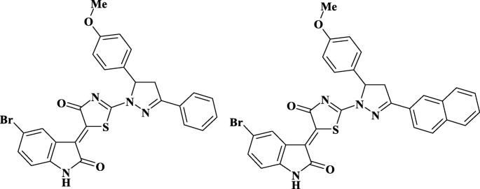
헤테로사이클릭 4-티아졸리디논 유도체(화합물 Les-3288 및 Les-3833, 그림 1)는 이전에 설명한 대로 우크라이나 Danylo Halytsky Lviv 국립 의과대학 제약, 유기 및 생물유기화학과에서 합성되었습니다[25].
<그림>
조사된 화합물의 구조 - Les-3288 및 Les-3833
세포 배양에 사용하기 전에 이들 화합물을 디메틸 설폭사이드(DMSO, Arterium, Lviv, Ukraine)에 용해시켰다. 용액을 끓는 수조에서 5분간 추가로 유지하고 작업 농도에 도달하도록 증류수로 희석하였다. 배양 배지에서 DMSO의 최종 농도는 0.1% 미만이었다. Dox는 우크라이나의 Pfizer(이탈리아) 대리점에서 현지 약국에서 구입했습니다.
고분자 나노캐리어
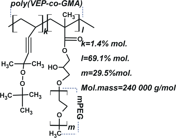
약물 전달을 위한 PNC는 앞서 설명한 방법론을 사용하여 우크라이나 Lviv Polytechnic National University의 유기 화학과에서 합성되었습니다[26, 27]. 폴리(VEP-co 합성 -GMA)-그래프트-PEG는 다음과 같은 후속 단계를 통해 수행하였다. 폴리(VEP-co -GMA)는 5-tert의 라디칼 공중합에 의해 합성되었습니다. 부틸퍼옥시-5-메틸-1-헥센-3-인(VEP, 0.41g, 0.5mol)(기술된 방법 [28]에 의해 합성된 퍼옥사이드 단량체) 및 글리시딜 메타크릴레이트(GMA, 7.72g, 12.2mol)(Sigma-Aldrich , USA) 에틸 아세테이트(7.9mL)(Merck, Darmstadt, Germany)에서 아조이소부티로니트릴(AIBN, 0.129g, 0.05mol)(Merck, Darmstadt, Germany)을 라디칼 개시제로 사용합니다. 중합은 65%의 최대 전환율에 도달할 때까지 343K에서 수행되었습니다. 폴리(VEP-co -GMA)는 폴리(에틸렌 글리콜) 메틸 에테르(mPEG)와 사이드 에폭사이드 그룹의 부가 반응을 통해 사이드 PEG 사슬을 부착하기 위한 백본으로 사용되었습니다. 삼불화붕소 에테레이트(0.027mL, 0.031mol)(Sigma-Aldrich, St. Louis, MO, USA)를 313K에서 디옥산(20mL; Merck, Darmstadt, Germany) 중 공중합체(1.0g)의 용액에 첨가하고 3시간 동안 교반하였다. mPEG(2.5mg, 3.35mmol)(Sigma-Aldrich, USA)를 디옥산(15mL)에 용해하고 용액을 백본 폴리머 용액과 혼합하고 313K에서 6시간 동안 교반했습니다. 미반응 mPEG는 투석 백을 사용하여 제거했습니다. 6-8 kDa의 분자량 컷오프(MWCO) 기공 크기(Sigma-Aldrich, USA). 결과 빗 모양 폴리(VEP-co -GMA)-그래프트-PEG를 일정한 중량이 될 때까지 진공 하에 실온에서 건조시켰다. PNC의 구조 및 일부 특성은 그림 2에 나와 있으며 이전에 설명되었습니다[29].
<그림>
PNC의 도식 구조 - poly(VEP-co- GMA)-이식 - 2-tert의 불포화 과산화물의 공중합체 염기에 주쇄가 있는 PEG -부틸퍼옥시-2-메틸-5-헥센-3-인(VEP, "k"로 표시), 측쇄 글리시딜 메타크릴레이트(GMA, "l"로 표시) 및 폴리에틸렌 글리콜(PEG, "m"으로 표시) )
수성 PNC 용액을 제조하기 위해 0.09g의 PNC를 0.9mL DMSO에 용해시켰다. 이 PNC 용액을 0.9% NaCl 용액 8.0mL에 첨가하였다. 그런 다음, 용액을 0.5시간 동안 교반하고 10초 동안 초음파 처리하였다. 복합체의 수분산액은 4-티아졸리디논과 PNC의 DMSO 용액을 다음 순서로 물에 떨어뜨림으로써 얻었다:PNC 0.045g을 DMSO(Sigma-Aldrich, USA) 0.15mL에 용해시킨 후, Les 0.0015g을 용해시켰다. -3288(또는 Les-3833)을 0.10mL의 DMSO에 용해시켰다. PNC 및 4-티아졸리디논 용액을 혼합하고 4.25mL의 1% NaCl 수용액에 첨가하고 10초 동안 초음파 처리했습니다.
이 표면 활성 PNC는 소수성 백본에 이식된 친수성 PEG 사슬을 포함합니다(그림 2a). 수용액에서 양친매성 PNC는 수불용성 화합물(예:약물)을 가용화할 수 있는 미셀과 같은 구조를 형성하거나 이러한 화합물이 나노입자의 표면에 흡착되어 생체 적합성을 증가시킬 수 있습니다. 4-thiazolidinone 기반 화학치료제의 매우 안정적인 수분산액을 얻기 위해 개발된 기술은 DMSO에 희석된 용액이 고분자 계면활성제 PNC를 포함하는 침전 식염수 용액으로 옮겨질 때 발생하는 나노 스케일 약물 입자의 핵형성으로 구성되어 있습니다. 나노 입자 표면. 그 결과, 응집 및 침강으로 인해 복합체가 안정화되고 분산이 방지되었다. 또한, 4-티아졸리디논 유도체의 나노규모 복합체는 주변의 생물학적 미세환경과의 가능한 상호작용으로부터 보호됩니다. 이러한 보호를 통해 안정성을 잃지 않고 체내에 더 오래 남아 있고 부정적인 부작용을 일으키지 않고 표적 조직이나 기관에 축적됩니다. 따라서 이러한 변형은 항암 화합물 나노입자의 수분산액의 응집 및 침강 안정성을 향상시킨다.
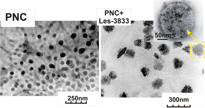
PNC의 폴리머 미셀 및 헤테로사이클릭 4-티아졸리디논과 PNC의 복합체의 크기는 Zetasizer Nano ZS(Malvern Instruments GmbH, Stuttgart, Germany) 및 DynaPro NanoStar(Wyatt Technology, Santa Barbara, CA, USA) 및 25°C에서 비침습적 후방 산란(NIBS) 기술을 사용하는 광자 상관 스펙트럼에 의한 것입니다. DLS 측정을 위한 시료는 위와 같이 준비하였으며, 필요한 경우 pH 6.5~7.0의 이중증류수로 희석하였다. 모든 샘플에 대해 3-5번의 측정이 이루어졌습니다(각 측정은 5주기로 구성되었고 측정 사이의 범위는 5분이었습니다). 그림 3은 DLS로 분석한 PNC의 미셀 구조와 헤테로사이클릭 4-티아졸리디논(Les-3833 및 Les-3288)과 PNC 복합체의 나노입자 크기 분포를 보여줍니다. PNC 미셀과 PNC로 코팅된 4-티아졸리디논 나노입자의 크기와 형태는 200kV의 가속 전압에서 투과 전자 현미경 JEM-200A(JEOL, Japan)를 사용하여 연구되었다[30, 31]. 상기와 같이 시료를 준비하고, 필요에 따라 이중증류수로 희석하였다. 시료는 초음파 분산제 UZDN-1A(Ukrrospribor Ltd., Ukraine)를 사용하여 기판에 시험 용액을 분사하여 준비했으며, 이는 기판에 균일한 코팅을 용이하게 합니다. 구리 그리드에 증착된 얇은 비정질 탄소막을 기판으로 사용했습니다. DLS 및 TEM 방법으로 연구한 PNC 미셀의 크기와 형태 및 헤테로고리 4-티아졸리디논(Les-3288 및 Les-3833)과의 복합체가 그림 3에 나와 있습니다.
<그림>
PNC(좌)와 PNC + Les-3833 복합체(우)의 투과전자현미경(TEM) 이미지
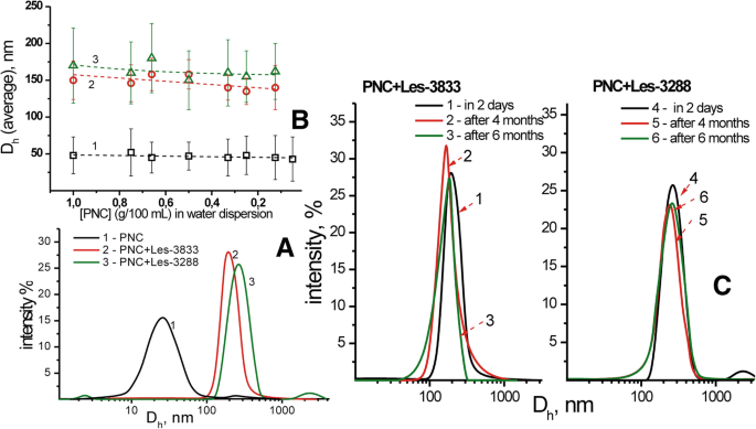
DLS 및 투과 전자 현미경(TEM) 연구는 PNC 미셀의 크기가 50 ± 25 nm이고 PNC와 헤테로고리 4-티아졸리디논 Les-3833 및 Les-3288의 복합체에 의해 형성된 나노입자가 140 ± 25 nm 및 155임을 입증했습니다. 각각 ± 30 nm. PNC와 4-티아졸리디논 유도체의 복합체 분산액은 매우 안정적이며 티아졸리디논 나노입자 표면에 흡착된 PNC 쉘에 의해 응집 및 침강으로부터 보호됩니다. 그림 4에서 볼 수 있듯이, 수계에 분산된 나노입자의 크기 변화는 물로 여러 번 희석했을 때와 4-티아졸리디논과 PNC 복합체의 수계 노화 6개월 후에도 무시할 수 있을 정도입니다.
<그림>
PNC 및 복합체의 유체역학적 직경에 대한 DLS 연구(a ) 및 분산 희석에 대한 PNC-약물 복합체의 평균 크기 의존성(b ):PNC(1)의 수분산, Les-3833+PNC(2) 및 Les-3288+PNC(3)의 착물, 뿐만 아니라 Les-3833+PNC(1-3) 및 Les-의 유체역학적 크기 3288+PNC(4–6) 2일(1.4), 4개월(2.5), 6개월(3.6) 보관 후 분산액(c )
세포 배양
C6 계통의 쥐 신경교종 세포는 우크라이나 국립 과학 아카데미(우크라이나 키예프)의 분자 생물학 및 유전학 연구소의 세포 배양 컬렉션에서 입수했습니다. 세포는 10% 태아 소 혈청(Sigma, USA)이 보충된 Dulbecco의 변형된 Eagle 배지(DMEM, Sigma, USA)에서 배양되었습니다. 세포는 CO2에서 성장했습니다. -37°C, 5% CO2에서 배양기 , 그리고 95% 습도. 세포재파종은 2~3일에 한번 1:5 비율로 진행하였습니다.
연구 물질의 세포독성 작용 평가
세포를 하나의 웰에 100,000개 세포/mL의 농도로 24웰 플레이트에 접종했습니다. (미국 노스캐롤라이나주 먼로 소재의 Greiner Bio-One North America, Inc.). 연구 중인 물질은 세포 파종 후 시작되는 24시간 적응 기간 후에 0.1, 0.5 및 1.0μM 농도로 추가되었습니다. 세포 현탁액에 첨가한 지 2분 후, 0.01% 최종 농도에서 Trypan Blue 염료(DV-T10282, Invitrogen, Thermo Fisher Scientific Corp., Waltham, MA, USA)를 사용하여 혈구 계수 챔버에서 일정한 간격으로 세포 계수를 수행했습니다. . 죽은 세포는 원형질막의 손상으로 인해 이 염료를 흡수했습니다.
유세포분석
세포 순환 및 세포 사멸을 연구하기 위해 C6 세포를 Les-3288, Les-3833 및 PNC와의 복합체 및 유리 형태의 PNC로 처리하고 인산염 완충 식염수(PBS, pH 7.4)로 세척하고 원심분리에 의해 펠렛화했습니다. 4°C에서 5분 동안 1000rpm에서 그리고 차가운 PBS(PBS 1mL당 2백만 세포)에 재현탁되었습니다. 그런 다음, - 20°C로 냉각된 무수 에탄올 4mL의 분취량을 첨가하고 부드럽게 혼합하여 세포를 고정했습니다. 고정된 샘플은 사용할 때까지 - 20°C에서 보관되었습니다(1주 이내). 형광 활성화 세포 분류(FACS) 분석을 위해, 세포 샘플을 4°C에서 5분 동안 1000rpm에서 원심분리하고, 상청액을 버리고 세포 펠렛을 1mL의 PBS에 재현탁했습니다. 100 마이크로리터의 DNase-free RNase(Sigma, USA)를 해당 현탁액에 첨가하고 샘플을 37°C에서 30분 동안 인큐베이션했습니다. 그 다음, 각 시료에 propidium iodide(PI)(Sigma, USA, 1 mg/mL) 100 μl를 보충하고 상온에서 10분간 배양하였다. 마지막으로 샘플을 플라스틱 팔콘 튜브로 옮기고 FACSCalibur 유세포 분석기(BD Biosciences, Mountain View, CA, USA) 및 Summit v3.1 소프트웨어(Cytomation, Inc., Fort Collins, CO, USA) 세포 순환 및 세포 사멸의 매개변수 측정을 위해.
Annexin V-양성(아폽토시스) 세포의 측정은 FACS 분석을 사용하여 수행되었습니다. 랫트 신경교종 C6 세포를 0.1 mg/mL, 0.5 mg/mL 및 1 mg/mL 농도의 PNC, Les-3288, Les-3833 및 그림 아래 표시된 이들의 고분자 접합체로 처리했습니다. 실험이 끝나면 Trypsin-EDTA 용액으로 접시 바닥에서 세포를 분리하고 PBS로 두 번 세척하고 Apoptosis Detection Kit (BD Biosciences, San Jose, CA, USA)를 사용하여 Annexin V-FITC로 염색했습니다. , 제조업체의 지침에 따라. 세척된 세포를 1/50 부피의 FITC-접합된 Annexin V 용액을 포함하는 Annexin V 결합 완충액(BD Pharmingen)에서 15분 동안 배양했습니다. 그런 다음, 샘플을 적절한 부피의 Annexin V 결합 완충액으로 2회 희석하고 유세포 분석기(Becton Dickinson, USA)의 FL1/FL2(FITC-PI) 채널에서 즉시 측정하였다. Annexin V 단일 양성 세포는 apoptotic으로 분류되었습니다.
DNA 혜성 분석
DNA 혜성 분석은 알칼리성 조건에서 수행되었습니다. 샘플당 10,000개의 세포를 저융점 아가로스(0.5%) 75μl에 혼합했습니다. 혼합 후, 샘플을 미리 1.5% 정상 융점 아가로스로 덮인 슬라이드에 피펫으로 옮겼습니다. 샘플을 용해 용액(2.5M NaCl, 100mM EDTA, 10mM Tris-base, 10% DMSO, 1% Triton X-100)에서 18시간 동안 4°C에서 인큐베이션했습니다. DNA의 풀림과 알칼리 불안정 손상의 발현을 허용하기 위해 슬라이드를 실온의 알칼리 용액에서 20분 동안 인큐베이션했습니다(암소에서 수행). 전기영동(30분 동안 ~74V/cm)을 위해 슬라이드를 수평 챔버로 옮기고 1x 전기영동 완충액(0.3M NaOH, 1mM EDTA, pH> 13)을 추가했습니다. 슬라이드를 얼음처럼 차가운 100% 메탄올에 고정하고 건조했습니다. 슬라이드를 80 μL 1x ethidium bromide(EtBr)로 5분 동안 염색한 다음 차가운 증류수에 담가 과도한 얼룩을 제거했습니다. DNA 혜성은 현미경(Carl Zeiss, Germany)을 사용하여 시각화되었고 이미지는 소프트웨어 'CASP'(Casplab-1.2.3b2 소프트웨어, CASPlab, Wroclaw, Poland)를 사용하여 분석되었습니다. 샘플당 100개의 혜성이 계산되었습니다. DNA 손상은 혜성 꼬리 크기에 따라 0(0-5% 손상), 1(5-25% 손상), 2(25-45% 손상), 3(45-70% 손상)의 5가지 유전독성 수준으로 분류되었습니다. ), 4(70% 이상의 데미지) [32]. 손상 지수(DI)는 앞서 설명한 대로 계산되었습니다[33].
메틸 그린 염료를 사용한 DNA 삽입 분석
화합물 Les-3288 및 Les-3833은 메틸 그린 분석을 사용하여 DNA 분자에 삽입되는 능력에 대해 조사되었습니다[34]. 연어 정자 DNA(10 mg/mL)를 15 μL의 메틸 그린 용액(H2 중 1 mg/mL)과 함께 37°C에서 1시간 동안 인큐베이션했습니다. 영형). 화합물을 1㎍/mL로 첨가하고 2시간 동안 암실에서 37℃에서 인큐베이션하였다. 샘플의 총 최종 부피는 1mL였습니다. 메틸 그린의 흡수는 BioTek 76 883 다중 채널 마이크로 광도계(BioTek, Winooski, VT, USA)를 사용하여 37°C에서 2시간 동안 샘플을 배양한 후 630nm에서 측정되었습니다. EtBr은 양성 대조군으로 사용되었습니다.
데이터 분석 및 통계
모든 실험은 각 변형에서 3개의 평행선으로 3번 반복되었습니다. 분산 분석(ANOVA)을 그룹 비교에 사용했습니다. 세포 주기의 여러 단계에서 C6 쥐 신경아교종 세포의 분포 데이터는 일원 ANOVA로 분석한 후 Tukey의 사후 다중 비교 테스트를 수행했습니다. 모든 데이터는 평균 ± SD로 표시됩니다. p <0.05의 값은 통계적으로 유의한 것으로 간주되었습니다.
<섹션 데이터-제목="결과">
결과
자유 형태 및 고분자 나노운반체와 복합적으로 사용되는 4-티아졸리디논 유도체의 세포독성 효과(트리판 블루 배제 테스트)
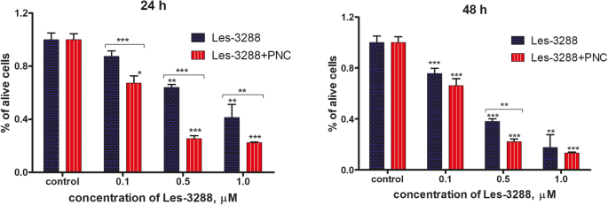
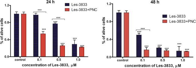
Trypan blue 배제 테스트의 결과는 PNC와 함께 Les-3288 및 Les-3833 복합체의 수성 형태가 이러한 화합물의 자유 형태(그림 5 및 6). 가장 높은 세포독성 효과는 PNC와의 복합체에서 Les-3288 및 Les-3833 화합물의 0.1 및 0.5 μM 용량에서 관찰된 반면, 이들 화합물의 최고 용량(1.0 μM)에서는 PNC와의 복합체화의 향상이 검출되었습니다. 24시간 동안 Les-3288의 경우에만 해당합니다(그림 5 및 6).
<그림>
다양한 농도(0.1, 0.5, 1.0μM)의 Les-3288 단독으로 24시간 및 48시간 동안 처리한 후 Trypan blue 배제 시험으로 측정한 살아있는 쥐 신경아교종 C6 세포의 수와 고분자 나노운반체 복합체의 작용 비교 PNC) *P <0.05; **P <0.01; ***P <0.001 ( 대조군과의 차이, 100%)
<그림>
다양한 농도(0.1, 0.5, 1.0μM)의 Les-3833 단독 처리 후 24시간 및 48시간 동안 트리판 블루 배제 시험으로 측정한 살아있는 쥐 신경아교종 C6 세포의 수와 고분자 나노운반체( PNC) *P <0.05; **P <0.01; ***P <0.001 (대조군과의 차이, 100%)
고분자 나노운반체를 포함하는 Les-3833 및 Les-3288 화합물의 복합체는 시험관 내에서 쥐 신경교종 C6 세포에 대한 세포자멸사 활성을 향상시킵니다.
선택된 4-티아졸리디논 유도체의 세포독성 가능성에 대한 PNC의 조절 효과를 평가하기 위해 두 가지 대안적 접근이 사용되었습니다. 먼저, 약물-PNC 복합체가 세포 주기에 미치는 영향을 조사하기 위해 세포 주기 분석을 수행했습니다. 또한 Annexin V/PI 이중 염색을 사용하여 이러한 나노복합체에 의해 유도된 세포 사멸의 정확한 유형을 구별했습니다.
저용량(0.1μg/mL 및 0.5μg/mL)의 Les-3288 및 Les-3833 화합물은 그림 7 및 표 1에서 볼 수 있듯이 세포 주기 진행이나 세포 사멸 유도에 가시적인 효과를 나타내지 않았습니다. PNC가 있는 두 화합물은 pre-G1에서 세포 수를 유의하게 증가시켰습니다. 단계(Les-3288의 경우 4배, Les-3833의 경우 6배)로, 이는 세포자멸사 촉진 가능성이 크게 향상되었음을 나타냅니다. 자유 형태의 PNC는 세포 주기 또는 세포자멸사 유도에 영향을 미치지 않는다는 점을 강조해야 합니다. 따라서 티아졸리디논-나노캐리어 복합체로 치료 중인 많은 pre-G1 세포 집단의 관찰된 출현은 PNC와 실험 약물의 단순한 상승 효과로 설명될 수 없습니다.
<그림>
4-티아졸리디논 유도체 Les-3288 및 Les-3833의 자유 형태 및 C6 쥐 신경교종 세포에서 고분자 나노운반체(PNC)와 복합체의 세포 주기 진행에 미치는 영향. 프로피디움 요오드화물(PI) 염색, 유세포 분석. R2, G1 이전; R3, G1; R4, S; R5, G2 단계. FL2-H, 유세포 분석기의 두 번째 채널(585/40 필터)의 피크 방출 값
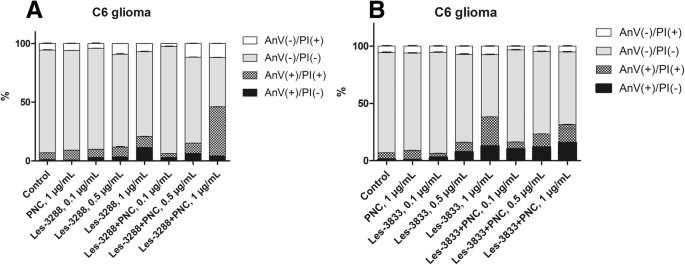
관찰된 sub-G1 피크가 apoptotic cell임을 확인하기 위해 Annexin V/PI 염색 결과를 분석하였다. 우리는 낮은 용량(0.1 및 0.5μg/mL) 및 높은 용량의 Les-3288(1.0μg/mL)을 사용한 C6 신경교종 치료 동안 포스파티딜세린(초기 세포자멸사의 주요 마커, FITC-접합 Annexin V에 의해 감지됨)의 유의미한 외부화가 관찰되지 않았습니다. , 이 화합물이 apoptosis의 약한 유도제임을 시사합니다. 대신에 괴사 세포 집단의 증가(대조군에서 1.2%에서 Les-3288의 경우 11.25%, 1.0μg/mL)를 발견했으며, 이는 Les-3288이 적어도 부분적으로 괴사 세포 사멸을 유발한다는 것을 나타낼 수 있습니다. 그러나 Les-3288과 PNC의 복합화는 이 약물의 작용 방식을 완전히 바꿨습니다. 초기 세포사멸 세포(Annexin V(+)/PI(-))와 후기 세포사멸 세포(Annexin V(+)/PI(+))의 수가 크게 증가한 반면 순수 괴사 세포의 전체 양은 대조군(미처리) 세포의 수준(그림 8a). 따라서 Les-3288을 PNC와 결합하면 신경교종 세포에 대한 세포자멸사 활성을 강력하게 향상시킬 뿐만 아니라 잠재적인 괴사성 세포 사멸 메커니즘을 감소시킵니다. 이러한 발견은 환자에게 불편한 염증의 대규모 유도로 인해 pro-necrotic 약물의 사용이 권장되지 않기 때문에 임상 실습에서 매우 중요할 수 있습니다.
<그림>
Les-3288의 pro-apoptotic 활동 비교(a ) 및 Les-3833(b ) 화합물 및 고분자 나노운반체(PNC)와의 복합체는 쥐 신경아교종 C6 세포를 향합니다. Annexin V/PI 이중 염색, 유세포 분석. PI, 요오드화 프로피듐
놀랍게도 우리는 Les-3833+PNC 복합체에 대해 동일한 효과를 관찰하지 못했습니다. 사실, Les-3833+PNC 복합체의 pro-apoptotic 활성은 모든 테스트 농도에서 유리 형태의 Les-3288에 비해 낮았습니다(그림 8b). 두 화합물의 이러한 큰 차이는 화학 구조의 특이성으로 설명될 수 있으며 Les-3833의 경우 폴리머 구조를 최적화하여 더 나은 결과를 얻을 수 있다고 추측합니다.
자유 형태의 4-티아졸리디논 유도체에 의해 유발되고 PNC와 복합된 쥐 신경교종 C6 세포의 DNA 손상에 대한 혜성 분석
알칼리성 혜성 분석을 통해 알칼리성 불안정 DNA 부위에서 단일 가닥 DNA 파손을 감지할 수 있습니다. 구한 결과는 OTM(Olive tail moment)의 평균값을 이용하여 추정하였다. 우리의 데이터는 3시간 동안 Les-3288, Les-3833 및 PNC(농도 0.5μg/mL)로 С6 쥐 신경교종 세포를 처리한 결과(그림 9 및 10) 심각한 DNA 손상을 일으키지 않았음을 보여주었습니다(OTM =0.7299 ± 0.1276, OTM =1.466 ± 0.3086, OTM =0.5846 ± 0.1078), 대조군에서 처리되지 않은 세포와 비교(OTM =0.4541 ± 0.07273). 그러나 Les-3833 + PNC 복합체(OTM =2.3880 ± 0.2212)를 사용한 세포 처리는 Les-3833의 유리 형태를 사용한 세포 처리보다 더 심각한 DNA 손상을 야기했습니다. 이러한 손상 증가는 Les-3288+PNC 복합체의 작용에서 관찰되지 않았습니다(그림 9 및 10).
<그림>
DNA 손상은 0.5μg/mL 농도에서 사용된 실험적 항종양 화합물로 3시간 동안 처리된 쥐 신경아교종 C6 세포에서 올리브 꼬리 모멘트를 사용하여 평가되었습니다. *피 ≤ 0.05; **피 ≤ 0.01; ***피 ≤ 0.001. PNC, 고분자 나노캐리어; Dox, 독소루비신(양성대조군)
<그림>
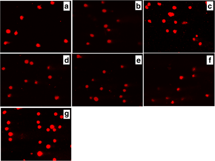
(a로 3시간 동안 처리한 후 쥐 신경아교종 С6 세포의 혜성 꼬리에 DNA 존재 ) 대조군(미처리 세포), Les-3288(b ), Les-3288+PNC(c ), Les-3833(d ) Les-3833 + PNC(e ), PNC(f ) 및 독소루비신(g ), 0.5μg/mL 농도에서 사용
An increase of cell incubation time to 6 h (Figs. 11 and 12) caused greater DNA damage in all experimental groups:Les-3288, Les-3288+PNC, Les-3833, Les-3833+PNC, and Dox (positive control). However, the effects of the Les-3288+PNC and Les-3833+PNC complexes appeared to be lower than the effects of the free forms of Les-3288 and Les-3833. It should be noted that the DNA-damaging effect of the complexes of both derivatives with the PNC was less than such effect from these derivatives used in free form.
DNA damage was evaluated using the Olive tail moment in rat glioma C6 cells treated for 6 h with experimental antineoplastic compounds used at a 0.5 μg/mL concentration. *피 ≤ 0.05; **P ≤ 0.01; ***P ≤ 0.001. PNC, polymeric nanocarrier; Dox, doxorubicin (positive control)
Presence of DNA in comet tail of rat glioma С6 cells after treatment for 6 h with (a ) control (untreated cells), Les-3288 (b ), Les-3288 + PNC (c ), Les-3833 (d ) Les-3833 + PNC (e ), PNC (f ), and doxorubicin (g ), used at a 0.5 μg/mL concentration
The results were evaluated using the mean value of the TailDNA%. The obtained data show that incubation of С6 rat glioblastoma cells with substance Les-3288, Les-3288+PNC, Les-3833, and PNC (all at concentration of 0.5 μg/mL) during 3 h (Fig. 13) does not lead to significant DNA damage (TailDNA% of 4.157 ± 0.682; TailDNA% of 3.530 ± 1,012, 8.807 ± 0.878; 3.298 ± 0.514, respectively) compared with control (TailDNA% =3.638 ± 0.406), but cell treatment with the Les-3833+PNC complex (TailDNA% =19.53 ± 1.48) causes more significant damage of DNA than the free form of Les-3833. An increase in the incubation time up to 6 h (Fig. 14) in all cases leads to greater damage to the DNA. Again, the level of TailDNA% detected from the action of complexes of both derivatives with the PNC was less than the level of that indicator for the action of these derivatives used in free form.
DNA damage evaluated using the % of DNA in comet tail of C6 rat glioma cells treated with experimental antineoplastic compounds during 3 h at a concentration of 0.5 μg/mL. *피 ≤ 0.05; **P ≤ 0.01; ***P ≤ 0.001. PNC, polymeric nanocarrier; Dox, doxorubicin (positive control)
DNA damage evaluated using the % of DNA in comet tail of C6 rat glioma cells treated with experimental antineoplastic compounds during 6 h at a concentration of 0.5 μg/mL. *피 ≤ 0.05; **P ≤ 0.01; ***P ≤ 0.001. PNC, polymeric nanocarrier; Dox, doxorubicin (positive control)
We also used an alternative, five-category classification system to classify DNA migration and calculated the DI. Table 2 summarizes the percentages of rat glioma C6 cells for each level of genotoxicity during 3 h of treatment. In the control (untreated cells), higher percentages of cells in levels 0 (81.3%) and 1 (18.6%) were detected compared to these levels in Les-3288 and PNC-treated cells. Complexation of Les-3288 with the PNC did not lead to a big change in the distribution pattern of the DNA comets. In contrast, Les-3833-treated cells showed a lower percentage of cells in 0 level (38.6%) and a higher percentage of cells in level 1 (60.0%), while Les-3833 + PNC-treated cells demonstrated a significantly lower percentage of cells in level 0 (8.0%) and much higher percentages of cells with levels 1 (76.0%), 2 (13.3%), and 3 (2.6%). The integrative indicator of DNA damage (DI) in the case of application of both Les-3288 and its complex with the PNC did not differ significantly from that for control or using free PNC, while the DI from using both Les-3833 and its complex with the PNC was even higher than the DI from using Dox.
During this time interval, Les-3833 + PNC-treated cells demonstrated a higher percentage of cells with level 4 (5.3%) DNA damage than cells treated with Les-3288 and the other experimental compounds, but the percentages of cells in levels 2 and 3 were lower (16.0% and 12.0%, respectively) than those from treatment with other compounds (Table 2). Table 3 summarizes the percentages of rat glioma C6 cells with DNA damage after 6 h of treatment. In general, a tendency for the appearance of DNA comets of higher classes was detected when complexes of Les-3833 and Les-3288 were used, compared with a spectrum of DNA comet classes induced by free forms of these agents (Table 3). However, the integrative indicator of DNA damage (DI) calculated for the action of complexes of Les-3833 and Les-3288 was found to be lower than such an indicator for the action of free forms of these agents. A possible explanation could be a very high DI level at 6 h action of studied agents that is even higher than such indicator found for the action of Dox (Table 3). Thus, we observed time dependence of DNA damage caused by complexes of Les-3833 and Les-3288:at 3 h of treatment, there was an increase of the DI in the case of action of Les-3833 complexes, compared to the action of the free form of Les-3833; however, at 6 h of treatment, there was a decrease in DNA damage for the action of complexes of both Les-3833 and Les-3288, compared to such damage observed for the action of the free forms of these agents. There was no significant time dependence for the action of the PNC, while a significant time-dependent (from 3 to 6 h) increase in the DI was observed for the action of Dox (from 41.2 + 0.40 to 185.2 + 0.23).
DNA Intercalation by 4-Thiazolidinone Derivatives—Les-3288 and Les-3833
Les-3288 and Les-3833 compounds demonstrated a certain capability for intercalating DNA molecules with approximately 15% and 10%, respectively, of replacement of methyl green dye. EtBr, a well-known DNA intercalating agent, was used as a positive control, and it was shown to replace the methyl green dye much more efficiently (70%) (Fig. 15). Thus, Les-3288 and Les-3833 are not capable of effectively intercalating in between two complementary base pairs in double-stranded DNA.
Replacement of methyl green dye intercalated into the DNA of salmon sperm by Les-3288 and Les-3833 compounds (1.0 μg/mL) and ethidium bromide (EtBr) used as a positive control
Discussion
There are two major problems that impede increasing the efficiency of treatment for cancer patients:(1) non-addressed actions of many anticancer drugs that lead to general toxicity and severe negative side effects due to damage of normal tissues and organs; (2) rapid (6–12 months) development of resistance of tumor cells to applied anticancer drugs that leads to a loss of efficacy of drug action. In addition, there is a third, technical problem of poor water solubility of many anticancer drugs. However, binding these drugs with specific carriers of the nanoscale size can help to solve the solubility problem.
In this study, we addressed the first and third of the above-mentioned problems by (a) selecting novel synthetic substances (4-thiazolidinone derivatives:Les-3288 and Les-3833) that possess both high anticancer activity and less general toxicity in tumor-bearing animals [7, 8, 23] and (b) applying a novel drug delivery platform that provides stable water-based forms of the complexes of the water-insoluble 4-thiazolidinone derivatives with a PEGylated polymer.
The 4-thiazolidinone derivatives—Les-3288 and Les-3833—are heterocyclic compounds with molecular mass of 400–600 Da. These and other related derivatives were synthesized at Lviv National Medical University (Ukraine), and most of them have been tested under the Developmental Therapeutic Program at the National Cancer Institute in Bethesda, MA, (USA) [35,36,37,38]. Based on these evaluations of their antineoplastic activity, the most promising substances were selected for further study of the mechanisms of their cytotoxic action and anticancer effects [25]. Les-3833 demonstrated high toxicity towards B16F10, WM793, and SK-Mel-28 melanoma cells, and against human lung A549, breast MCF-7, colon HCT116, and ovarian SKOV3 cancer cells and leukemia cells (L1210, Jurkat, HL-60 lines) [23, 25, 39]. Les-3288 and Les-3833 showed high toxicity against mammalian glioma cells (C6, U251, U373 lines) [23, 24].
Earlier, we found that Les-3288 and Les-3833 were the most toxic among the other studied 4-thiazolidinone derivatives towards rat glioma C6 cells and human glioblastoma U251 cells [23, 24], although their application for cell treatment was very complicated due to their absolute insolubility in water. Only using DMSO with additional heating was effective for preparing a soluble form of the derivatives (see the “Materials and Methods” section). Thus, it was reasonable to search for a way of improving the delivery of these derivatives to target cells, and we used a synthetic PNC to accomplish this task. The complexation of the studied derivatives of 4-thiazolidinone with the applied PNC also enhanced the effectiveness of their antineoplastic action. The mechanisms of the pro-apoptotic action of these compounds were demonstrated using western blot and FACS analyses [23, 24]. It should be noted that in healthy experimental animals, Les-3288 and Les-3833 produce much fewer negative side effects such as cardiotoxicity [40], hepatotoxicity [8], and nephrotoxicity [7] than the widely used anticancer chemotherapeutic agent Dox.
In a previous study, we showed that Les-3288 did not induce ROS production during its pro-apoptotic action in vitro and effects in experimental laboratory animals [23], while the action of Les-3833 led to such production; however, it was less than that of Dox. ROS are considered to be the main effectors of negative side effects of anticancer drugs, particularly Dox [41, 42]. Thus, the Les-3288 and Les-3833 compounds could be prospective anticancer drugs because they possess antitumor activity, but do not demonstrate general toxicity at the level characteristic for effective anticancer medicines such as Dox.
A great impediment to promoting Les-3288 and Les-3833 as anticancer drugs is their poor water solubility. This problem also exists for other anticancer medicines, such as taxols [2, 4]. For paclitaxel, this problem was solved by using a special oil for preparing the medicinal formulation [2, 4, 43]. However, specific delivery platforms are considered to be more promising for creating “smart” drugs that possess effective action in the organism [3, 13]. Specific functional polymer surfactants with block, comb-like, and block/branched architectures, including PEG-containing ones, as well as derived water forms have been developed and proposed for application as carriers for the delivery of Dox [26, 27] and the metal chemotherapeutic agent KP-1019 [10] at the Department of Organic Chemistry of Lviv Polytechnic National University.
Since the 4-thiazolidinone derivatives are water-insoluble compounds, DMSO was used to prepare their water-soluble forms. To avoid the negative consequences of using a toxic solvent like DMSO, we complexed these derivatives with the above-noted PNC. Highly dispersed and stable nanoscale water dispersions of these substances were obtained and applied for treatment of rat glioma C6 cells.
The use of complexes of 4-thiazolidinone derivatives with the PNC led to reduced general toxicity of these compounds in experimental animals. Earlier, we found that when experimental antitumor agents (Les-3288, Les-3833, Les-3882) were applied in a complex with a synthetic polymeric carrier, their action was accompanied by much smaller changes in the biochemical parameters in blood serum that are characteristic for cardiotoxicity [40], hepatotoxicity [8], and nephrotoxicity [7], compared to those for Dox. Thus, complexation of these antitumor agents with the PNC and their application in the form of water-soluble stable delivery systems reduces their toxic effects in experimental animals, compared with the action of these substances in a free form [44]. Several systems for paclitaxel delivery have been proposed including polymeric nanoparticles, lipid-based formulations, polymer conjugates, inorganic nanoparticles, carbon nanotubes, nanocrystals, and cyclodextrin nanoparticles [43].
Thus, complexation of Les-3288 with the PNC significantly increased the pro-apoptotic activity of this experimental drug, thereby allowing us to advance it to pre-clinical studies on animal tumor models. Complexation of Les-3833 with the same type of nanocarrier was less efficient, and thus another type of polymer should be used.
The developed water-based forms of the complexes, as well as the free derivatives, were applied for treatment of rat glioma C6 cells. It was found that these water-based forms of the complexes of the PNC with the 4-thiazolidinone derivatives—Les-3288 and Les-3833—enhanced the pro-apoptotic action of these compounds. In another study, we demonstrated that the induction of apoptosis by Les-3288 was not accompanied by ROS induction, opposite to the action of Dox and Les-3833 [23]. Thus, high antitumor activity of Les-3288 together with its low general toxicity when targeting the malignancy in NK/Ly lymphoma-bearing mice suggest great potential for this experimental anticancer compound. The presented data also predict the potential usefulness of Les-3288 and Les-3833 compounds complexed with the PNC for glioma treatment.
Cell division, differentiation, and death are principal physiological processes that regulate tissue homeostasis in multicellular organisms. A disruption of genomic integrity and impaired regulation of cell death may both lead to uncontrolled cell growth. A compromised cell death process can also promote genomic instability. It is becoming clear that dysregulation of the cell cycle and cell death processes plays a pivotal role in the development of major disorders such as cancer, cardiovascular disease, infection, inflammation, and neurodegenerative diseases [45].
We have found that some of the 4-thiazolidinone derivatives, namely, Les-3288 and Les-3833, were even more effective than Dox in the induction of apoptosis in rat C6 glioma and human U251 glioblastoma cells in vitro [23, 24]. It should be noted that brain tumors belong to malignancies that are the most resistant to applied chemotherapies, and survival rates in patients with these tumors are extremely low [44].
It has been shown that the complexation with this PNC enhanced the pro-apoptotic action of 4-thiazolidinone derivatives (Les-3288 and Les-3833) [44]. In another study [23], we demonstrated that the induction of apoptosis by Les-3288 was not accompanied by ROS induction, opposite to the action of Dox and partially opposite to that of Les-3833. These data could explain the lower general toxicity of Les-3288 compared with that of Dox [23, 24].
The results of FACS analysis are in agreement with the suggestion that an enhancement of the pro-apoptotic action of the studied 4-thiazolidinone derivatives is due to their complexation with a novel PNC. Such complexation of Les-3288 and Les-3833 increased the ratio of pre-G1 cells in the population of treated rat glioma C6 cells, decreased the ratio of G1 cells, and increased the action of G2/M cells. These data suggest an enhancement of both cytotoxic action (apoptosis) and cytostatic action (block in G2/M phase of cell cycle) of the studied derivatives complexed with the PNC. In general, the apoptosis mechanism of glioma cell killing by the Les-3288 + PNC complex and the Les-3833 + PNC complex was demonstrated by the results of FACS analysis of the appearance of Annexin V single-positive rat glioma C6 cells.
Involvement of the apoptosis mechanisms in the action of the studied 4-thiazolidinone derivatives was also confirmed by the results of the DNA comet assay. It was found that the complexation of Les-3833 with the PNC led to a decrease (compared to free Les-3833) in the ratio of rat glioma C6 cells in “0” DNA comets and sіmultaneous increase in the ratio of “1,” “2,” and “3” comets (3-h treatment of cells). At 6 h of treatment by the Les-3833+PNC complex, there was a large increase (compared to free Les-3833) in the ratio of “1” comets, an increase in the ratio of cells with the most expressed DNA damage (“4” comets), and a decrease in the ratios of “2” and “3” comets. It should be noted that the DNA damaging effects of Les-3288 and Les-3833 measured at 6 h was higher than such effects of complexes of these derivatives with the PNC (see Table 3). A possible explanation here could be a very high level of the DI at 6 h action of studied agents that is even higher than the level of that indicator found for the action of Dox (Table 3). There was no time dependence in the action of the PNC, while a significant time-dependent (from 3 to 6 h) increase (from 41.2 + 0.40 to 185.2 + 0.23) in the DI was detected for the action of Dox.
Thus, a complexation of 4-thiazolidinone derivatives—Les-3288 and Les-3833—with a PNC enhanced the cytotoxic effect of these compounds towards rat glioma C6 cells, and that effect was achieved through the apoptosis mechanisms including DNA damage observed as DNA comets in the electrophoresed cells.
We used the DNA intercalation test to investigate the direct targeting of DNA by the 4-thiazolidinone derivatives and revealed only weak replacement of methyl green dye (15% and 10% replacement for Les-3288 and Les-3833, respectively) that intercalated into the DNA of salmon sperm, compared to the replacement by EtBr, used as a positive control (70%).
Thus, conducting the cytotoxicity experiments in vitro with the 4-thiazolidinone derivatives—Les-3288 and Les-3833—was more convenient due to using the water-based forms of the 4-thiazolidinone derivatives—Les-3288 and Les-3833—complexed with the novel PNC. Besides, we have demonstrated that the novel synthetic 4-thiazolidinone derivatives (Les-3833 and Les-3288) possessed treatment effects i n vivo towards NK/Ly lymphoma grafted to BALB/C mice, and those effects were comparable to such effects of Dox. At the same time, the action of these derivatives led to much fewer negative side effects measured as changes in the number of erythrocytes, neutrophils, and lymphocytes in the animals’ blood and the activity of aspartate and alanine aminotransferases in their blood serum, compared with the action of Dox.
결론
The complexation of Les-3288 and Les-3833 with the PEG-containing PNC enhanced their cytotoxic action towards rat glioma C6 cells. The cytotoxic action was realized by apoptotic mechanisms and confirmed by FACS analysis as the presence of the pre-G1 fraction in the rat glioma C6 cells and of Annexin V-single-positive cells. DNA comet analysis revealed single-strand brakes in the nuclear DNA of treated glioma C6 cells that probably was not caused by the intercalation of the studied compounds into the DNA molecule. The Les-3288 and Les-3833 stabilized by the amphiphilic PEG-containing PNC resulted in the water-based forms and provided enhancement of their antitumor effect in vitro in comparison with the free form of the drugs.