전기화학적으로 자가 조직화된 Titania 나노튜브 어레이에 대한 검토:합성, 변형 및 생물의학 응용
초록
양극 산화에 의해 성장된 티타니아 나노튜브는 많은 독특하고 잠재적인 특성으로 재료 과학계의 관심을 끌었으며 기술의 합성은 성숙 단계에 접어들고 있습니다. 현재 검토는 TiO2에 초점을 맞출 것입니다. 자기 조직화 티타니아 나노튜브 층의 합성과 양극 산화를 조정하여 크기, 모양, 질서도 및 결정화 단계에 영향을 미치는 수단을 비판적으로 강조하는 Ti 금속 기판으로부터 자기 조직화된 전기화학적 양극산화에 의해 성장된 나노튜브 매개변수 및 후속 열 어닐링. 양극 TiO2의 치수와 속성 간의 관계 나노튜브 어레이가 제시될 것입니다. 양극성 TiO2 형성 메커니즘 연구의 최근 진행 상황과 의의 나노튜브에 대해 간략히 설명합니다. 또한, 최근에 보고된 생물 의학적 방향과 도핑, 표면 개질 및 열 어닐링에 의해 수행된 개질에서 양극으로 형성된 TiO2의 특성을 개선하기 위해 수행된 가장 유망한 응용 프로그램을 보여줍니다. 나노튜브. 마지막으로 이 분야의 해결되지 않은 몇 가지 문제와 가능한 미래 방향이 제시됩니다.
소개
20세기 초부터 이산화티타늄(TiO2 )은 자외선 차단제, 페인트, 센서, 광촉매, 태양 전지, 전기 변색 장치, 약물 전달 등의 상업적 생산으로 사용되었습니다[1,2,3,4,5,6,7]. TiO2 현상 조명 조사 하에서 광 생성된 전자-정공 쌍을 생성할 수 있으며 물을 산소와 수소로 분해하는 데 도움이 될 수 있으며, 가장 잠재적인 연료로서 미래의 에너지 위기를 해결하는 데 도움이 됩니다. Fujishima와 그의 동료들은 TiO2에서 광촉매 물 분할을 처음 보고했습니다. 자외선(UV) 광선 아래 전극[8,9,10], 그 이후로 이산화티타늄은 재료 과학에서 가장 많이 연구된 화합물 중 하나가 되었습니다. 모든 전이금속 산화물 중에서 화학적 불활성, 내식성, 안정성과 같은 광범위한 기능적 특성, 특히 생체적합성 향상[11], 전기적 광학적 특성[1]을 나타낸다. Iijima는 1991년 [12] 탄소나노튜브를 발견하여 형상과 기능의 독특한 조합을 보여주며 형상에 직접적으로 영향을 받을 수 있는 특성을 나타냄으로써 기본적으로 화학, 물리, 생물의학 분야의 나노기술 분야에서 엄청난 노력을 기울여 왔습니다. 재료 과학.
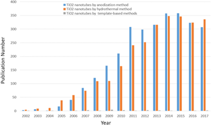
지금까지 가장 많이 연구된 나노물질은 여전히 탄소이지만, 일반적으로 전이금속 산화물을 기반으로 하는 또 다른 종류의 나노튜브 물질은 지난 20년 동안 상당한 관심을 끌었다. 양극산화된 티타니아 나노튜브를 형성하기 위한 첫 번째 시도는 알칼리 과산화물 처리를 사용한 후 크롬산을 함유한 전해질에서 전기화학적 양극산화를 사용한 Assefpour-Dezfuly[13]에 의해 이루어졌습니다. 그리고 Zwilling et al. 그들이 1999년에 불소 이온을 함유한 크롬산 전해질에서 전기화학적 양극산화에 의해 Ti 기판 상에 자기 조직화된 나노튜브 층을 처음으로 생성했다고 보고한 이후, 그 분야는 엄청나게 빠르게 확장되었다[14]. 지난 10년 동안 "티타니아 나노튜브"를 키워드로 33,800편 이상의 논문이 발표되었습니다. 그림 1은 TiO2 분야에서 연간 분류된 총 출판물을 보여줍니다. 나노튜브 및 2002-2017 기간의 다양한 합성 방법을 비교하여 기하급수적인 성장 추세를 보일 뿐만 아니라 자기 조직화된 양극 TiO2 나노튜브 어레이는 큰 잠재력과 장점으로 많은 주목을 받고 있습니다. 최근 Lee et al. 다른 합성 접근법에 대한 간략한 설명과 함께 성장, 변형, 특성 및 응용을 포함한 거의 모든 측면을 다루는 양극성 티타니아 나노튜브 분야에서 포괄적이고 최신 견해를 제시했습니다[15]. 수력/용매열 [16,17,18] 및 템플릿 보조 방법 [19, 20]과 같은 다른 제조 방법과 비교할 때 직접 산화는 크기 조정을 통해 원하는 제어 가능한 나노 구조, 인가된 전위, 시간, 온도, pH 및 전해질 조성과 같은 산화 매개변수를 최적화함으로써 형태 및 질서 정도를 성장시킬 수 있다[15]. 특정한 기하학적 구조로 인해 고도로 조직화된 구조와 표면적 비율을 갖는 자기정렬 산화물 나노튜브층은 매우 높은 기계적 강도, 큰 비표면적과 같은 독특한 특성을 나타내며, 심지어 높은 전자와 같은 전자적 특성을 제공합니다. 이동 속도 또는 양자 구속 효과[15, 21]. 또한 전기화학적 양극산화는 저비용 공정이며 티타늄에 국한되지 않고 다른 전이 금속 Hf[22], Zr[23], Nb[24], Ta[25], V[26] 또는 합금에도 적합할 수 있습니다. TiAl[27], TiZr[28]. 현재 검토는 여전히 TiO2에 중점을 둡니다. Ti 금속 기판으로부터 자가 조직화된 전기화학적 양극산화에 의해 성장된 나노튜브. 게다가, 우리는 이러한 유형의 자가 조직화된 티타니아 나노튜브 층의 합성과 4가지 다른 세대를 포함하는 양극 산화 매개변수 조정 및 후속 열 어닐링을 통해 크기, 모양, 질서도 및 결정화 단계에 영향을 미치는 수단을 강조할 것입니다. 전해질 종 및 정의된 2단계 양극 산화와 다름. 양극 TiO2의 치수와 특성 간의 관계 나노튜브 어레이가 제시될 것입니다. 양극성 TiO2 형성 메커니즘 연구의 최근 진행 상황과 의의 나노튜브에 대해 간략히 설명합니다. 우리는 최근에 보고된 생물의학적 방향과 도핑, 표면 개질 및 열 어닐링을 통해 양극으로 형성된 TiO2의 특성을 개선하는 개질에 대해 보고된 가장 유망한 응용 분야를 보여줍니다. 나노튜브. 우리는 또한 이 분야의 미해결 문제와 가능한 미래 방향을 고려합니다. 주요 단락 텍스트는 여기에서 바로 이어집니다.
<그림>
연구 동향. TiO2와 관련된 연간 분류된 논문 수 2002년부터 2017년까지 다양한 합성 방법으로 차별화된 나노튜브. (데이터는 티타니아 나노튜브를 사용하여 확장된 Science Citation Index Expanded에서 수집되었으며, anodization 또는 hydrothermal 방법 또는 템플릿 기반 방법을 키워드로 사용)
TiO의 합성2 전기화학적 양극산화에 의한 나노튜브 어레이
최근 몇 년 동안 나노로드, 나노입자, 나노와이어, 나노튜브 등 다양한 형태의 나노구조 이산화티타늄이 성공적으로 개발되었지만[29,30,31], 나노튜브는 고유한 자기조립 구조로 인해 기술 응용에 대한 관심이 증가하고 있습니다. 더 나은 후보로 표면적 종속 응용 프로그램에 적용할 수 있는 큰 계면 영역과 크기 및 모양의 편리한 제어. TiO2의 기능을 다루기 위한 여러 우수한 리뷰[1, 2, 15, 32,33,34]가 있습니다. 다양한 합성 방법으로 분류되는 나노 물질. 전기화학적 양극산화는 비교적 간단한 자동화 기술로 티타니아 나노튜브를 얻는 가장 효과적인 방법 중 하나로 입증됐다. 양극 TiO2를 제작하기 위한 주요 기술을 지정할 것입니다. 아래는 나노튜브입니다.
자체 조직 양극 TiO2 나노튜브 어레이
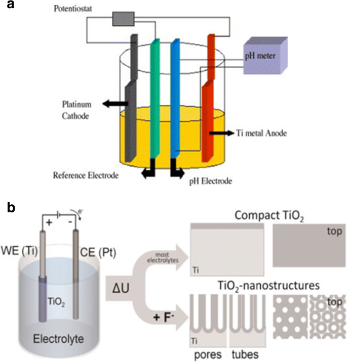
광범위하게 연구된 바와 같이, 티타니아 나노튜브 층은 특정 설정 환경 조건에서 형성될 수 있습니다. 산화 장치는 세 부분으로 구성됩니다. (I) 아세톤, 에탄올 및 탈이온수에서 순차적으로 초음파 처리하여 탈지되는 작업 전극으로 준비된 Ti 호일이 있는 3전극 시스템, 상대 전극으로 백금 및 일반적으로 Ag/AgCl 참조 전극으로 사용(그림 2a), pH 전극은 때때로 F
-
의 최종 농도를 얻기 위해 추가됩니다. 및 HF[35] 또는 양극으로 Ti 호일과 음극으로 불활성 금속 전극으로 구성된 또 다른 간단한 2전극 시스템(그림 2b)[36]; (II) 일반적으로 불화물 이온, 염화물 이온, 크롬 이온, 브롬화물 이온 또는 과염소산염 함유 전해질; 및 (III) DC 전원 공급 장치. 티타니아 나노튜브의 유망한 응용 분야에 영향을 미치는 양극 산화 형성 조건에 의해 영향을 받는 두 가지 주요 특징이 있습니다. 생물의학. 즉, 전기화학적 양극산화 파라미터(인가전위, 양극산화 지속시간, 불소이온 농도를 포함하는 전해질 시스템, 전해질 내 물, 전해질 온도, 전해질 pH 등)를 제어함으로써 더 자세히 논의될 것이다. "Synthesis of TiO2 Nanotube Arrays by Electrochemical Anodization" 섹션), 평평한 콤팩트 산화물[1], 다공성 층[1, 36], 무질서한 TiO2와 같은 다양한 티타니아 나노구조를 제조할 수 있습니다. 다발로 성장하는 나노튜브 층[37], 또는 최종적으로 고도로 조직화된 규칙적인 TiO2 나노튜브 또는 고급 나노튜브 층:가지 모양의 튜브[38], 대나무 모양[38, 39], 이중벽[40], 나노레이스[38] 또는 이중층[39] 구조로 특성이 다르게 발견될 수 있습니다. 그림 3 및 4는 이러한 TiO2의 전형적인 예의 전계 방출 주사 전자 현미경(FE-SEM) 이미지를 표시합니다. 나노튜브 형태.
<그림>
도식 설정. 아 준비된 Ti 호일을 작업 전극으로, 백금을 상대 전극으로, 일반적으로 Ag/AgCl을 기준 전극으로, pH 전극을 pH 측정기로 사용하는 3전극 시스템의 예시도. ref에서 재생산. [35]. ㄴ Ti 호일을 양극으로, 불활성 금속 전극을 음극으로 사용하는 단순한 2전극 시스템의 예시도. 양극 산화는 다른 조건에서 다른 양극 산화 산화물 층으로 이어집니다. 대부분의 중성 및 산성 전해질에서는 조밀한 티타니아가 형성될 수 있습니다. 그러나 묽은 불소 전해질을 사용하면 나노튜브/나노다공성 산화물 층이 금속 표면에 직접 부착됩니다. ref에서 재생산. [36]
<그림>
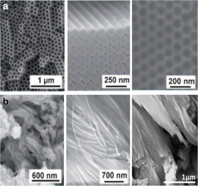
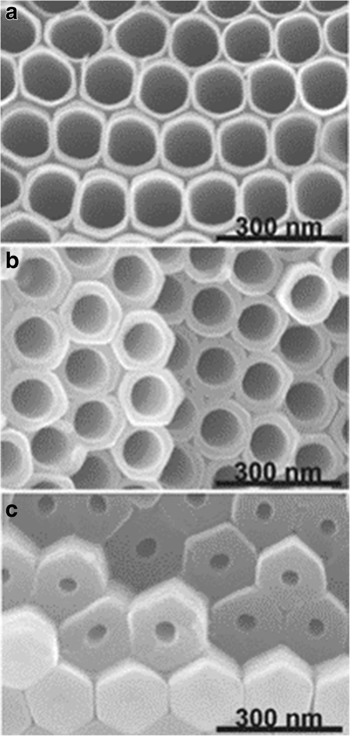
양극산화된 TiO2의 SEM 이미지 Ti의 다양한 양극 산화 공정에 의한 나노튜브 층. 아 고도로 정렬된 TiO2 나노튜브(상단 및 측면 보기)는 유기 전해질 시스템에서 얻어지며, 튜브 층이 제거될 때 실제로 금속 표면인 자체 정렬된 표면 딤플(오른쪽)이 있습니다. ref에서 재생산. [1]. ㄴ 무질서한 TiO2 나노튜브는 표면적의 패치에서 성장하고 급속 분해 양극 산화(RBA)로 알려진 초고속 양극 산화 기술에 의해 전해질을 함유한 염화물 번들로 함께 융합됩니다. ref에서 재생산. [1] 및 [37]
<그림>
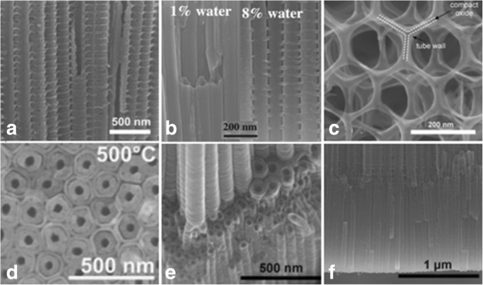
고급 TiO2의 SEM 이미지 나노튜브 형태. 아 대나무형 강화 TiO2 나노튜브는 0.2 mol/L HF로 구성된 에틸렌 글리콜의 특정 교류 전압(AV) 조건에서 120V에서 1분, 40V에서 5분의 시퀀스로 제작됩니다. [38]. ㄴ 매끄러운에서 대나무와 같은 TiO로의 전환2 나노튜브는 0.135 M NH4에 물을 조절하여 첨가(수분 함량:1 ~ 8%)하여 양극 산화 처리하여 유도할 수 있습니다. F/에틸렌 글리콜 전해질
참조에서 재생산. [39]. ㄷ 2D 나노레이스 구조는 120V에서 50초 및 0V에서 600초의 시퀀스로 불화물 함유 전해질에서 장기간 수행된 전압 사이클링 하에서 얻어집니다. [38]. d 이중벽 TiO2 나노튜브는 1°C s
−1
의 가열 속도로 500°C에서 어닐링한 후 120V에서 에틸렌 글리콜 전해질을 함유한 불화물에서 Ti의 양극 산화에 의해 성장됩니다. . ref에서 재생산. [40]. 이 분지된 나노튜브는 먼저 120V(6시간)에서 다음으로 40V(2시간)에서 전압 스테핑으로 관찰할 수 있습니다. ref에서 재생산. [38]. 에 같거나 두 개의 다른 튜브 직경을 가진 이중층 나노튜브를 볼 수 있습니다. ref에서 재생산. [38]
(현재 TiO2 10 ~ 500 nm 범위의 튜브 직경, 수백 나노미터 ~ 1000 μm 범위의 층 두께, 2 ~ 80 nm 범위의 벽 두께를 갖는 나노튜브 어레이를 얻을 수 있습니다[15, 41].)
Masuda와 Fukuda가 양극산화 조건을 최적으로 조정하여 고순도 다공성 알루미나를 처음 보고한 것은 20년 전이었습니다[42]. 나중에 연구원들은 TiO2에 대해서도 유사하게 조직화된 구조에 노력을 기울였습니다. 나노튜브 층. 그리고 양극 TiO2의 질서도에 영향을 미치는 세 가지 중요한 요소가 있습니다. 나노튜브 어레이(층의 폴리곤 및 튜브 직경의 표준 편차에 따라):Ti 기판, 인가 전압 및 반복적인 양극 산화 [33, 43]. 절연 파괴[33]와 이상적으로는 육각형 자기 정렬 TiO2 아래의 가능한 가장 높은 전압에서 고순도 재료에 대해 배열의 결함이 더 적게 얻어질 수 있다는 것은 분명합니다. 그림 5와 같은 나노튜브는 2차 튜브 성장에 의해 크게 향상될 수 있습니다[43]. Sopha et al. 불순물은 두 번째 양극산화 이후에 생성되는 나노튜브의 치수와 배열에 큰 영향을 미친다는 것을 보여주었습니다[44]. 또한, Ti 기질 입자의 결정학적 방향은 TiO2의 성장 특성에 결정적인 영향을 미치는 것으로 밝혀졌습니다. 전자 후방 산란 회절(EBSD)에 의한 나노튜브 어레이. Leonardi et al. 나노튜브는 밸브 금속 산화물이 결정립에 형성되어 1M(NH4 )H2 PO3 +0.5 중량% NH4 F는 전해질로 사용되었다[45]. 이와 유사하게 Macak과 동료들은 최근 문헌[46]에서 알려진 바와 같이 수성 전해질을 사용한 경우에 비해 널리 사용되는 에틸렌글리콜계 전해질에서 결정립 상의 나노튜브 성장이 지연되지 않는다고 보고하였다[46]. 연마된 Ti 시트에서 [0 0 0 1] 방향 또는 이에 가까운 방향의 결정립이 이상적인 결정립으로 판명되었으며 이상적인 방향의 단결정 Ti를 사용하는 것은 가장 균일한 나노튜브 어레이를 얻는 데 큰 발전이 될 것입니다[46] .
<그림>
TiO2의 SEM 이미지 나노튜브. 나노튜브는 0.27 M NH4를 포함하는 에틸렌 글리콜 전해질에서 형성됩니다. F는 Ti의 반복적인 양극 산화에 의한 것입니다. 단면은 레이어의 상단, 중간 및 하단에서 가져옵니다. ref에서 재생산. [43]
그럼에도 불구하고 여전히 질서의 정도에 영향을 미치는 몇 가지 결함이 있습니다. 최근에는 Ti를 균일하게 나노임프린팅하여 더욱 확장되었습니다. Kondo et al. 이상적으로 정렬된 양극 TiO2의 처리량 제작을 알아냈습니다. 정렬된 볼록부가 있는 Ni 몰드를 사용하여 상부에 Al 층이 있고 하부에 Ti 층이 있는 Ti 표면 또는 2층 시편을 나노임프린팅함으로써. 그리고 TiO2 NH4의 후속 양극산화에 의해 사전 텍스처링된 패턴의 얕은 오목부가 개시 부위로 작용하는 더 질서 있는 방식으로 층을 생성할 수 있습니다. F 에틸렌 글리콜 용액[47, 48]. 밀접하게 따라 Sopha et al. 먼저 원자층 증착(ALD)에 의해 준비된 Ti 기판에 TiN 보호층을 덮은 후 집속 이온 빔(FIB)으로 사전 텍스처링을 수행한 다음 에틸렌 글리콜 전해질을 사용하여 양극 산화 처리하여 두께를 가진 육각형으로 완벽하게 배열된 나노튜브 층을 생성합니다. 나노튜브가 주어진 개시 부위에서만 성장하도록 제한하고 결함 없이 양극산화 시간을 연장할 수 있는 2 μm의 [49].
양극 TiO의 형성 메커니즘2 나노튜브
양극 산화 기술 및 양극 TiO2 형성 메커니즘 연구 나노튜브는 다양한 분야에서 오랫동안 폭넓은 관심을 받아왔습니다. 1969년 Diggle이 조밀한 양극 산화물과 다공성 양극 산화물의 필름에 대해 보고한 메커니즘 연구[50]는 현재까지도 매우 중요한 안내 역할을 하고 있습니다. 상당한 양의 최근 연구에서 구멍에서 튜브로의 전환이 점진적인 특성을 갖는다는 것을 보여줍니다[1, 27, 36]. 그러나 완전한 이론적 모델과 추론은 제공되지 않았습니다.
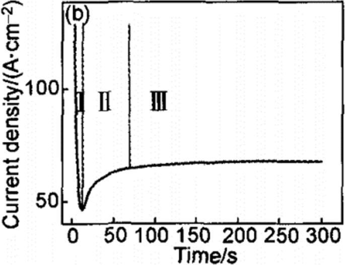
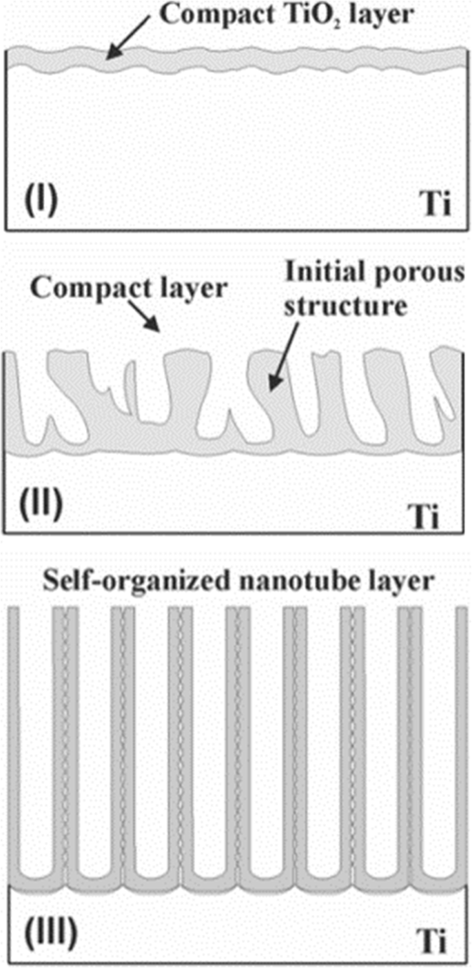
기존의 현장 보조 용해(FAD)가 가장 수용 가능한 이론입니다[1, 33, 51]. 바로 전기화학적 양극산화(TiO)2 과정 중입니다. 나노튜브 어레이는 상대적으로 독립적인 세 가지 절차로 인해 티타니아의 자가 조직화에 의해 형성됩니다. Ti의 TiO2로의 전기화학적 산화 , TiO2의 전기장 유도 용해 , TiO2의 불소 이온 유도 화학적 용해 , 섬세한 균형에 도달합니다. 나노튜브 형성을 유도하는 불화물을 포함하는 전해질에 대한 그림 6의 특성 전류 시간 곡선[51]과 그림 7의 형성 과정을 개략적으로 설명하는 데 도움이 될 수 있는 전형적인 이미지[33]로, 과도 현상은 다음과 같이 나눌 수 있습니다. (I) 첫 번째 부분에서는 새로 형성된 차단 산화물에 의해 발생하는 전류 감쇠가 있습니다. 두 가지 주요 과정인 O
2-
금속/산화물 계면을 향한 이온 및 Ti
4+
의 외부 이동 이온이 산화물/전해질 계면으로 이동하여 균형을 이룹니다. (II) 두 번째 부분에서는 양극의 표면적 증가로 인한 시차와 함께 전류가 다시 상승하기 시작합니다. 지연 시간이 짧을수록 불소 농도는 형성된 TiO2의 불소 유도 용해로 인해 더 높아집니다. , 그리고 기공은 무작위로 제작되기 시작하여 TiO2의 초기 형성으로 판명되었습니다. 나노튜브. (III) 그 후, 금속 산화물 계면에서의 기공 성장 속도와 형성된 TiO2의 유도 용해 속도가 되면 전류는 정상 상태에 도달한다. 외부 인터페이스에서 평형 상황에 도달합니다. 따라서 최종 튜브는 점점 더 V자 모양이 됩니다. 즉, 튜브의 상단은 튜브가 밀폐 포장된 바닥보다 훨씬 더 얇은 벽을 갖습니다. 그림 5에서 튜브 벽 두께의 구배는 튜브를 따라 전해질에 대한 노출 시간과 농도가 다르기 때문일 수 있습니다[43].
<그림>
불소를 함유한 전해질의 정전압에서의 일반적인 전류 시간 곡선. 과도 현상은 세 가지 별개의 영역으로 나눌 수 있습니다(I –Ⅲ ). (나 ) 첫 번째 부분에는 급격한 전류 감쇠가 있습니다. (II ) 두 번째 부분에서 시간 지연과 함께 전류가 다시 상승하기 시작합니다. (Ⅲ ) 세 번째 부분에서 전류는 ref에서 재현된 정상 상태에 도달합니다. [51]
<그림>
TiO2의 형성 과정 나노튜브 어레이. TiO2의 형성 나노튜브 어레이는 세 가지 형태학적 단계로 나눌 수 있습니다(I –Ⅲ ). (나 ) 차단 산화물이 형성됩니다. (II ) 표면이 국부적으로 활성화되고 모공이 무작위로 자라기 시작합니다. (Ⅲ ) 자체 조직화된 나노튜브 층이 형성되어 ref. [33]
그러나 이 이론은 나노다공성 구조와 달리 튜브로 분리되는 현상을 아직 명확하게 설명할 수 없으며 Fahim et al. 적절한 전압에서 플루오르 이온이 없는 황산 용액에서 티타니아 나노튜브를 얻는 것이 가능하며, 이 경우 I-t 곡선은 위에서 논의한 것과 유사하다는 것을 관찰했습니다[52]. Houser와 Hebert가 지적했듯이, 티타니아 다공성막의 과정과 I-t 곡선 사이의 정량적 관계를 설명하기 위한 성장 메커니즘은 아직 개발되지 않았습니다[53]. 해석이 충분히 설득력이 없기 때문에 점성유동모형과 두 해류의 성장모형과 같은 메커니즘에 대한 새로운 지적이 최근 등장하고 있다. 이러한 메커니즘과 관련하여 리뷰 [51]는 기존의 장 보조 용해 이론에 대한 많은 한계를 보여주고 점성 흐름 모델 및 두 흐름의 성장 모델에 대한 연구의 최신 진행 상황과 의의에 대해 몇 가지 설명합니다.
기하학적 특성 및 특성에 영향을 미치는 양극 산화 조건의 영향
전해질의 조성과 농도는 나노튜브 어레이 형성에 상당한 영향을 미친다. 우리가 사용하는 전해질의 차이에 따라 현상은 기본적으로 세 단계로 나뉩니다. 표 1은 결과 TiO2의 양극 산화 조건과 치수를 요약한 것입니다. 현재까지 다른 연구 그룹에서 조사한 3세대 나노튜브 어레이.
1세대:불산(HF) 기반 수성 전해질
이정표는 Gong et al. 처음으로 HF 기반 수성 전해질에서 Ti의 양극 산화에 의한 균일한 티타니아 나노튜브 어레이를 제시했습니다[54]. pH가 상대적으로 낮은 HF 수용액 전해질에서 높은 농도의 수소 이온을 의미하는 TiO2의 화학적 용해 불소 이온에 의해 유도되는 것은 양극 산화 공정에서 지배적인 위치를 차지합니다[55]. 티타니아 나노튜브를 형성하는 과정에서 짧은 시간에 동적 평형이 이루어지므로 최대 나노튜브 길이를 약 0.5μm로 제한하였다[54,55,56].
2세대:완충 전해질
후속 연구에서 화학적 용해를 줄이기 위해 튜브를 길게 하기 위해 Cai et al. KF 또는 NaF와 같은 더 약한 산을 완충 용액에 첨가하고 황산 또는 수산화나트륨을 사용하여 pH를 약산성(pH =4.5)으로 조정함으로써 길이가 약 4.4μm인 나노튜브가 달성되었음을 입증했습니다[57]. PH 값은 전기화학적 에칭 및 화학적 용해를 방해하는 것으로 판명된 티타늄 이온의 가수분해에 영향을 미칩니다. Cai et al. 또한 낮은 pH 값은 더 짧지만 깨끗한 나노튜브를 생성하고 더 높은 pH 값은 더 긴 나노튜브를 생성하지만 원치 않는 파편을 생성한다고 지적했습니다[57]. pH 값이 올라감에 따라 가수분해 속도가 증가하여 화학적 용해가 느려져 더 긴 나노튜브가 생성되는 반면 알칼리성 용액은 나노튜브 성장에 적합하지 않습니다[57, 58]. 적절한 전압의 중성 NaF 전해질에서 산성 용액보다 훨씬 더 긴 나노튜브를 얻을 수 있음이 Macak et al.에 의해 입증되었습니다. [58]. 불소 함유 전해질의 특정 전압이 주어지면 pH 구배를 조정하여 필요한 종횡비와 층 두께를 얻을 수 있습니다[59].
3세대:극성 유기 전해질
글리세롤[59], 디메틸 설폭사이드[60], 포름아미드 또는 디에틸렌 글리콜[61, 62], 에틸렌 글리콜[41, 63]과 같은 전해질, NH4와 같은 불화물 종 함유 F, NaF, KF가 서서히 나타납니다. Macak과 동료들은 점성 글리세롤 전해질을 사용하여 두께가 약 7μm이고 평균 튜브 직경이 40nm인 티타니아 나노튜브 어레이를 제작하는 데 앞장섰습니다[59]. 더 높은 종횡비 TiO2 나노튜브는 전해질 pH의 적절한 제어로 인해 이러한 극성 유기 전해질에서 성장될 수 있으며, 이는 티타니아의 화학적 용해를 감소시킨다[64]. Paulose et al. 0.25wt% NH4를 함유하는 에틸렌 글리콜을 사용하여 제조된 약 134μm 길이의 나노튜브 형성 17시간 동안 60V의 양극 산화 전위에서 F [60]. 곧이어 250μm 두께 이상의 TiO2 나노튜브 어레이는 Albu[65]에 의해 보고되었습니다. 게다가 수분 함량은 이 과정에서 이중 역할을 합니다. 물은 티타니아 형성에 필수적이지만 화학적 용해를 가속화하기도 합니다[63]. 따라서 TiO2의 두께와 차수를 높이려면 수분 함량의 영향을 최소화하는 방법이 필요합니다. 나노튜브 어레이. 일반적으로 수분 함량을 5% 미만으로 제한하는 것이 매우 긴 나노튜브를 성공적으로 달성하는 열쇠이며[60], 잘 조직화된 티타니아 나노튜브를 형성하기 위해서는 최소한의 수분 함량(0.18 중량%)이 필요합니다[66]. 물을 첨가하면 기록된 전류 밀도가 감소하여 무수 에틸렌 글리콜 용액에서 가장 높은 것으로 보고되었다[66]. Paulose et al. 0.6wt% NH4를 함유한 에틸렌 글리콜에서 216시간 동안 60V에서 길이가 약 1000μm인 자가 조직화된 육각형 티타니아 나노튜브 어레이의 형성이 처음 보고되었습니다. F 및 3.5% 물 [41]. 또 다른 눈에 띄는 현상은 매끄러운 관 벽이 낮은 수분 함량에서 성장하는 반면 측벽의 잔물결은 그림 4b와 같이 높은 함량에서 형성된다는 것입니다[59, 67]. 가장 많이 사용되는 전해질 유형으로서, 물과 불소 이온을 함유하는 에틸렌 글리콜은 항상 이중벽 나노튜브 구조로 이어집니다(그림 4d)[40, 68,69,70]. 적절한 어닐링 처리 후 간단한 화학적 에칭 공정. 내부 쉘을 제거한 후 확장된 튜브는 TiCl4 기반의 반복적인 접근 방식을 사용하여 나노 입자로 레이어별로 장식할 수 있습니다. -가수분해 [71]. 단일 벽 튜브는 전체 튜브의 두께가 기본적으로 동일하고 내부 쉘이 더 이상 나타나지 않는 염료 감응 태양 전지(DSSC)[71, 72]에서 상당히 향상된 전도도 및 전자 수송 시간을 보인 반면, Mirabolghasemi et al. 이중벽 및 단일벽 튜브를 비교하고 1.5 M H2를 포함하는 전해질에 디메틸 설폭사이드(DMSO)를 첨가하여 원하는 단일벽 튜브를 제시했습니다. O 및 0.1M NH4 여 [72].
최근에 비불소계 전해질이 TiO2를 성장시키는 것으로 보고되었습니다. 염산, 과산화수소, 과염소산 용액 및 이들의 혼합물을 포함하는 4세대 합성으로 간주될 수 있는 나노튜브 어레이[73, 74]. Allama와 Grimes는 길이가 300nm, 내부 직경이 15nm, 외부 직경이 25nm인 잘 발달된 나노튜브 어레이가 10~13V의 산화 전압에서 3M 염산(HCl) 수용액에서 얻어졌다고 설명했습니다. 그러나 낮은 농도의 H3 추가 PO4 나노튜브에서 막대로의 변화를 가져왔습니다. 그들은 또한 3M보다 낮거나 높은 농도에서 HCl 함유 전해질에서 자체 조직화된 나노튜브 어레이를 달성할 수 없다고 제안했습니다[73]. Allama는 염산을 포함하는 수용액에 과산화수소를 첨가하는 것이 더 두꺼운 산화물 층에 따라 강한 산화 특성을 갖는 티타니아 나노튜브를 늘리는 가능한 방법이 될 수 있다는 것을 발견했으며, 이는 불소 이온이 성장에서 염화 이온으로 성공적으로 대체될 수 있음을 보여줍니다. 나노튜브 어레이 [74]. 게다가, 유리 불화물 종을 첨가하지 않은 이온성 액체는 최근 몇 년 동안 티타니아 나노튜브를 위한 또 다른 유형의 용매 시스템으로 취급되어 왔다[75, 76].
표준 매개변수 외에도 생성된 나노튜브의 기하학적 구조는 전해질의 반복적인 사용("사용된 용액 효과")에 따라 달라집니다. 새로운 용액으로 얻은 튜브와 비교하여 한 번 사용한 용액을 사용하면 나노튜브 길이가 증가하고 달성된 나노튜브 성장 속도가 60V 이상에서 한 번 사용한 용액에 대해 지속적으로 더 높은 품질을 나타냈습니다[77]. 약간 다르지만 구별 가능한 전류 과도 동작이 주목될 수 있습니다[66]. 또한 2회 사용한 용액에서는 F
-
의 결핍으로 인해 나노관 구조가 아닌 산화막이 얻어졌다. 종 [78]. 그러나 Sopha et al. TiO2의 형태에 대한 에틸렌 글리콜 기반 전해질의 다양한 연령 조사 오래된 전해질에서 어레이가 더 낮은 종횡비를 나타낸다는 것을 보여주는 나노튜브[79].
적용 가능성
양극 산화 전압은 튜브 직경을 제어하는 중요한 요소입니다[80, 81]. 나노튜브 어레이의 크기는 전극을 가로질러 전위창이라고 하는 적절한 전압 범위를 적용함으로써 간단히 예측할 수 있습니다[67]. 낮은 전압에서 전기장 용해가 적어 TiO2를 형성합니다. 더 작은 직경의 나노튜브. 전압이 너무 낮으면 TiO2 층이 조밀해 지지만 나노튜브 구조는 관찰되지 않는다. 반대로 전압이 너무 높으면 스폰지 같은 다공성 구조가 나타납니다. 제어 가능한 전압에서 나노튜브의 직경은 전압에 비례합니다[81]. Furthermore, studies show that the range of voltage forming nanotubes is also related to the electrolyte system. In aqueous electrolytes, the potential window should be controlled from 10 to 25 V, which in organic electrolytes is much wider between several volts and some hundred volts. Wang and Lin found out the fact that in aqueous electrolytes, the anodization potential exhibits significant influence on the growth of TiO2 nanotube arrays, which exhibited slight influence in non-aqueous electrolytes in this regard [82]. The voltage dependence has a significant reduction in non-aqueous electrolytes which is attributed to a large extent to the low conductivity of organic electrolytes [83, 84].
The Duration on Anodization
The duration of anodization affects the nanotubes mainly in two aspects:(I) the formation of the tubes or not and (II) the length of the tubes. That is, in the early stage of the anodization, a compact TiO2 film is formed. If the duration is too short to reach an equilibrium in reaction, the regular nanotube array cannot be achieved instead of a disordered porous layer [67]. With increasing the anodization time, porous structure gradually grows deeper and converts into the TiO2 nanotubular array [1, 33, 51]. If other electrochemical parameters are kept unchanged, increase in the nanotube length is observed over time while no significant effect on diameter and tube wall thickness until a steady-state situation occurs [67, 85, 86]. However, due to the decrease of the F
−
concentration in the electrolyte, where the ion transport rate decreased, the growth rate of nanotubes is reduced. After reaching a stable condition between tube growth at the bottom and chemical/electrochemical dissolution at the top, we will find no further increase in length of the nanotubes [87]. As time continues to go, pipe orifice becomes an irregular polygon resulting in TiO2 spikes and coverings which can be seen on the surface of the TiO2 nanotube arrays [36]. It is worth mentioning that enlightened by the success of aluminum-repeated anodization for self-organized porous alumina [88], the two-step anodization of titanium for such a highly ordered hexagonally packed nanostructure of titania has appeared [43, 77, 89,90,91]. After the first-step anodization, the first nanotube layer from the Ti foil should be removed ultrasonically or by using an adhesion tape which leads to a surface where the remaining Ti is covered by comparably ordered dimples. Researches have shown that the former treatment helps to avoid potential mechanical damage to the Ti surface and also improve the structural uniformity of the TiO2 nanotubes to a great extent [77, 90, 91]. In the second anodization step, the pretreated Ti foil would be used as anode again with or without changes in parameters of oxidation conditions. It is subsequently found that the highly ordered and vertically oriented titania nanotubes, have greater potential in such fields as photocatalysis [77], photoelectriochemical activity [92, 93], and biological interaction with cells [94] than the disordered nanotubular titania.
Electrolyte Temperature
Temperature restricts the growth and quality of titania nanotube arrays, directly affecting the rate of oxide growth, length, and wall thickness of the structure [64, 95]. Wang and Lin first reported the effect of electrolyte temperature in both aqueous and non-aqueous electrolyte on anodic oxidation of titanium [82]. In aqueous electrolyte, with the temperature increasing, a slight diminish in the internal diameters was observed while the external diameters remained the same [68]. The reason may be the dissolution induced by electrical field and fluoride ions are similar while the oxide formation rate is higher than that at lower temperature. In non-aqueous electrolyte containing fluoride ions, the outer nanotube diameter was found to be largely increased by the increasing electrolyte temperature [82]. This may be because at lower temperature, the ion mobility of fluorine in some viscous electrolyte is further inhibited, leading to much slower dissolution of newly formed titania, which subsequently lead to a smaller nanotube diameter. As chemical dissolution rate increases, surface of TiO2 nanotubes arrays can easily produce excessive corrosion, resulting in lodging nanotubes and agglomeration. Therefore, the appropriate bath temperature for stable TiO2 nanotube arrays is at room temperature [82, 95, 96].
Modification of Nanotubes Properties
Increasing applications of TiO2 nanotubes as a novel semiconductor are closely related to its photoelectriochemical (PEC) performance; however, they are sometimes prevented by two fundamental drawbacks:(I) the wide band gap (3.0 eV for the rutile phase and 3.2 eV for the anatase phase) can only absorb ultraviolet light, which accounts for less than 10% of the sunlight [97], resulting in low average utilization ratio of solar energy and (II) the low electrical conductivity cannot efficiently transfer photogenerated carries. At the same time, the photoelectrons and vacancies can be easily recombined, thus making low electron mobility rate or quantum confinement effects [98]. Hence, post-treatment of TiO2 nanotubes is the key to improve the performance of its materials and related devices successfully. Considerable researches have been reported on modified methods to reduce the recombination of photogenerated electron-hole pair rate, speed up the electron transfer rate, and enhance the photoelectriochemical activity of TiO2 nanotubes. The research of the methods for the improvement of the photoelectriochemical properties of TiO2 nanotubes will be reviewed, including thermal annealing, doping, and surface modification. As for promising modification in biomedical fields, we will present in the application section.
Thermal Annealing
The crystallinity of the nanotube arrays and their conductivity, lifetime of charge carrier, and photoresponse depend mainly on the thermal annealing temperature and atmosphere [99, 100]. The as-prepared TiO2 nanotubes above are amorphous in nature but can be annealed to anatase or rutile phase, or mixtures of both phases relying on the specific temperature [1, 3, 40, 92, 100]. It is demonstrated that amorphous nanotube layers grown in a glycerol-based electrolyte containing fluoride ions have low photocurrents and an incident photon-to-electron conversion efficiency (IPCE) below 5% due to lots of structural defects while anatase phase nanotubes exhibit an IPCE value up to 60% thus attracting more interest to applications such as dye-sensitized or perovskite solar cells [93]. As well in mixed water-glycerol electrolyte with F
−
, Das et al. stated their points that if the self-organized TiO2 nanotube arrays with thickness about 1 μm were annealed around 300–500 °C, the anatase phase of TiO2 as the most preferred crystalline structure could be observed. The single anatase structure of nanotubes with the best photoelectriochemical properties and the lowest resistivity could be fabricated when annealed at 400 °C. At temperature higher than 600 °C, a track of typical rutile appeared and with a further increase in annealing temperature the percentage and quality of the rutile phase increased [92]. It should be noted that in Jaroenworaluc’s work, rutile phase was detected in anodic nanotube layers grown in aqueous NaF/Na2 SO4 with thickness of approximately 1.5 μm at 500 °C heat treatment and became the dominant phase at 600 °C. Whereas at 550 °C, partial nanotubes began to break down [101]. It begins to cause the collapse of the entire nanostructure formed in aqueous NaF/Na2 SO4 with the continuous increase of temperature (800–900 °C) or the extended annealing time [3]. While for extended temperature, the crystalline structure of the nanotubes completely converts to rutile phase at above 900 °C [3]. Some researchers demonstrated a loss of the typical single-walled nanotube layers morphology when the annealed temperature rose above 580 °C [102]. Besides the whole annealing process especially the heating rate controls, the morphological structures of the entire nanotube arrays [40]. The double-walled nanotube layers prepared from ethylene glycol (containing less than 0.2 wt% H2 O), with the addition of HF and H2 O2 , have such a high stability that can keep their structure intact until temperature is higher than 900 °C with a heating rate of 1 °C s
−1
. However, the double-walled nanotubes begin to collapse as soon as the temperature reaches 500 °C when the heating rate is 25 °C s
−1
. Most extraordinarily, with the high speed of 50 °C s
−1
the entire separated nanotubes fuse into a highly ordered porous membrane [40]. Xiao et al. obtained crystallized titania nanotubes arrays with calcination in different gases like dry nitrogen, air, and argon indicating nanotubes in dry nitrogen appeared to have enhanced electrochemical and photoelectrical properties who also found out that with the increasing temperature internal diameter decreased while wall thickness increased at the expense of nanotubes length [103].
As shown in Fig. 8, the conductivity along the TiO2 nanotubes with three different thickness is strongly affected by annealing temperature. Smallest resistance is observed at about 350–450 °C when the amorphous nanotube arrays are totally converted into anatase layers [99]. And it is evident to see that specific resistivity increases with thicker nanotube arrays which can be shown more clearly in the inset in Fig. 8. Furthermore, calcination temperature is responsible for the decrease in the length of the anatase TiO2 nanotubes. As shown in Fig. 9a, increment of temperature between 300 and 500 °C causes the as-prepared nanotube arrays slightly changing in thickness from 13.6 to 12.6 μm. When annealing temperature continuously increases to 600 °C, the average length of the nanotubes decrease dramatically to 6.6 μm. Figure 9b shows conversion from anatase TiO2 to rutile phase TiO2 occuring at 500 °C when the rutile barrier layer is formed on the bottom of the TiO2 nanotube arrays along the anatase nanotubes by consuming the bottom layer if the annealed temperature is further increased. This leads to a length decrease and corresponding photocatalytic activity decline [104].
Electrical resistance as a function of the annealing temperature for the different nanotube layer thicknesses. The curve shows electrical resistance measurement for different titania nanotube arrays grown in ethylene glycol based electrolyte containing HF and water at different temperature and the influence of thickness on resistance. The inset shows more details about the relationship between the thickness of the nanotube arrays annealed at 250 °C and their specific resistivity. Reproduced from ref. [99]
Evolution of titania nanotube arrays at different calcination temperatures. The electrolyte was ethylene glycol containing 0.3 wt% ammonium fluoride and 5 vol% distilled water. 아 The decrease in the thickness of titania nanotube arrays at different annealing temperature from 300 to 600 °C. The insets are corresponding SEM images and the scale bar is 5 μm. ㄴ The schematic of crystallization process of anodic titania nanotubes annealed at (1) 450 °C, (2) 500 °C, and (3) 600 °C in air. Reproduced from ref. [104]
Doping
Doping ions or atoms into titania lattice, a substitution within the lattice either at Ti
4+
또는 O
2−
sites, on the one hand, changes the lattice constants and bond energy. On the other hand, it is beneficial to the separation between photogenerated electron and hole pair, which in turn adjusts the band gap and improves the photoelectrochemical performance of nanotubes [15]. The impurity doping has been commonly applied to extend the light absorption onset of TiO2 nanotubes by either introducing subbandgap states or adjusting its bandgap width [105]. Lately, co-doping approach has been proposed as a more efficient way to reduce the band gap and adjust energy band level in favor of photoelectriochemical reactions [106, 107]. There are various kinds of doped-elements and preparation methods, and Table 2 summarizes some methods and the doping effects of doped titania nanotubes.
The most typical doped TiO2 nanotubes are as follows:
i.
Metal-doped TiO2 nanotubes such as Nb [107], Fe [108], Cu [109], Cr [110], Zr [111], Zn [112], and V [113]
ii.
Non-metal-doped TiO2 nanotubes such as N [105], F [114], B [115], C [116], S [117], and I [118]
iii.
Co-doped TiO2 nanotubes such as N–Ta [105], N–Nb [107], and C–N–Ni [119]
Choiet systematically studied the photoreactivities of 21 metal ion-doped quantum-sized TiO2 doping with Fe, Mo, Ru, Os, Re, V, and Rh significantly increases quantum efficiency, while Co and Al doping decreases the photoreactivity [120]. Momeni et al. recently obtained Fe-TiO2 nanotube (Fe-TNT) composites using different amounts of irons to decorate anodically formed TiO2 nanotubes with potassium ferricyanide as the iron source, indicating that Fe doping efficiently accelerates the photocatalytic performance for water splitting [108]. Not limited to transition metals, other elements including N [105], F [114], B [115], C [116], S [117], and I [118] are successfully explored. Nitrogen-doped TiO2 nanotubes turns out to be a promising path to narrow the band gap energy with enhanced photocurrent response in the visible light and the tube length influences the magnitude of conversion efficiency [121, 122]. Kim and co-workers proved that TaOxNy layer-decorated N-TNT (N-doped TiO2 nanotubes) as dual modified TNTs have significantly improved both visible (3.6 times) and UV (1.8 times) activities for water splitting [105]. At present, more researches are aimed at co-doping which exhibits remarkable synergistic effect causing a significant improvement on photoelectriochemical properties. Chai et al. grew Gd–La co-doped TiO2 nanotubes by an ultrasonic hydrothermal method, enhancing visible light photocatalysts [123]. Cottineau et al. modified titania nanotubes with nitrogen and niobium to achieve co-doped nanotubes with noticeably enhanced photoelectriochemical conversion efficiency in the visible light range [107]. Nevertheless, the mechanism for increasing photoconductivity and synergistic effect of various elements on co-doping remains a further study.
Surface Modification
Surface modification means decoration on surface of TiO2 nanotube arrays with nanoparticles (metal, semiconductors, and organic dyes). Nanowire arrays can also be fabricated by electrodeposition into titanium oxide nanotubes [124]. TiO2 nanotube is a semiconductor with a wide band gap, which can only absorb ultraviolet light [97, 125]. Any other nanomaterials which possess a narrow band gap or can absorb the visible light can be used as a sensitizer for titania nanotubes. Silver nanoparticles can be decorated on the tube wall by soaking the titania nanotube arrays in AgNO3 solutions and photocatalytically reducing Ag
+
on a TiO2 surface by UV illumination [126]. Ag/TiO2 nanotubes show a significantly higher photocatalytic activity and good biological performance compared with neat TiO2 nanotubes [126, 127]. Some compositions such as graphene oxide GO [128], CdS [129], CdSe [130], and ZnFe2 O4 [131]. can be modified on TiO2 nanotube arrays. Lately, GO have attracted much scientific interest in nanoscale devices and sensors which is easy to combine with nanostructure materials to compose some compounds. Titania nanotubes fabricated by anodization in water-ethylene glycol electrolyte consisting of 0.5 wt% ammonium fluoride (NH4 F) can be incorporated with GO by cyclic voltammetric method, which achieve higher photocatalytic activity and more effective conversion efficiency (GO-modified vs pure nanotubes =26.55%:7.3%) of solar cell than unmodified TiO2 nanotubes [128]. Semiconductor composite is a method improving the performance of titania nanotubes via, in some specific way, combining two kinds of semiconductors with different band gap [132]. Yang et al. decorated CdSe nanoparticles on the surface of TiO2 nanotubes by applying an external electric field to accelerate CdSe nanoparticles in nanochannels resulting in a material with more stable and higher photoresponse to visible light. Furthermore, the degeneration rate of anthracene-9-carbonxylic acid when exposed to the green light irradiation indicating that CdSe dominates the photocatalytic process under visible light [130].
Besides, other oxide nanoparticle deposition such as WO3 [133] or TiO2 [134] onto TiO2 nanotubes by the hydrolysis of a chloride precursor also turns out to augment the surface area and improve the solar cell efficiency. Another very effective approach is to consider organic dyes as sensitizers for TiO2 nanotubes to improve its optical properties [135]. Lately, atomic layer deposition (ALD) becomes an established procedure to modify TiO2 nanotube layers. ALD appears to be a very uniform and precisely controllable deposition process to functionalize nanotubes in conformably coating the surface of the nanotube layers with one atomic layer after another of a secondary material, such as Pd [136], ZnO [137], Al2 O3 [138], CdS [139], or TiO2 [140].
Biomedical Applications
Historically, the mentioned milestones were reported on the fabrication of titania nanotube arrays contributing to widen the promising applications over the past 20 years in the areas ranging from anticorrosion, self-cleaning coatings, and paints to sensors [141,142,143], dye-sensitized and solid-state bulk heterojunction solar cells [144,145,146], photocatalysis [147, 148], eletrocatalysis, and water photoelectrolysis [149, 150]. They also outperform in biomedical directions as biocompatible materials, toward biomedical coatings with enhanced osseointegration, drug delivery systems, and advanced tissue engineering [15, 135, 141, 142, 151]. In the following section, we will give an overview of current efforts toward TiO2 nanotubes biomedical applications. Titania nanotubes possess good biocompatibility as they show some antibacterial property, low cytotoxicity, good stability, and cytocompatibility including promoting adhesion, proliferation, and differentiation of osteoblast and mesenchymal stem cells (MSCs) with a high surface area-to-volume ratio and controllable dimensions [152,153,154,155].
However, Ti products have inadequate antibacterial ability and efforts have been made to improve their antibacterial properties such as modifications on titania nanotubes for biomedical applications like bioimplant [126, 156].
Biological Coatings And Interactions with Cells
A number of in vitro and in vivo studies have demonstrated that MSCs, osteoblasts and osteoclasts show size-selective response which means the effect of size holds an important position in cell interaction where the optimized size for cell adhesion, proliferation, growth, and differentiation is ranging from 15 to 100 nm [153, 157, 158]. Particularly, it was demonstrated that the TiO2 nanotubes with a diameter of 70 nm was the optimal nanoscale geometry for the osteogenic differentiation of human adipose-derived stem cells (hASCs) [159]. Smith et al. reported increased dermal fibroblasts and decreased epidermal keratinocyte adhesion, proliferation, and differentiation on TiO2 nanotube arrays (diameter 70–90 nm, length 1–1.5 μm) [160]. As shown in Fig. 10, Peng et al. found that nanotubular surface preferentially promoted proliferation and function in endothelial cells (EC) while decreased in vascular smooth muscle cell (VSMC) by measuring EdU, a thymidine analog which is incorporated by proliferating cells [161]. Furthermore, it is pointed out that surface wettability of the TiO2 nanotube layers is recognized as a critical factor for cell behavior which can be adjusted by changing the diameter of the nanotubes. That is to say, water contact angles can be altered without changing the surface chemistry [158]. To get further understanding of the effect of TiO2 nanotube layers to bone-forming cells as well as stem cells response, Park et al. seeded green fluorescent protein-labeled rat MSCs on TiO2 nanotube layers with six different diameters (15, 20, 30, 50, 70, and 100 nm), resulting in cell activity that is sensitive to nanoscale surface topography with a maximum in cell activity obtained for tube diameters of approximately 15–30 nm. Such lateral spacing exactly corresponds to the predicted lateral spacing of integrin receptors in focal contacts on the extracellular matrix, forcing clustering of integrins into the closest packing, resulting in optimal integrin activation. While tube diameters larger than 50 nm, severely impaired cell spreading, adhesion, and spacing of 100 nm may lead to the cell apoptosis [94]. Besides adjusting the size of the nanotubes, surface modification loaded with bioactive factors should be highlighted, in which case biomedical properties can be further optimized. In the case of bone implants, hydroxyapatite (HA) formation is important for osseointegration. Recent works have shown hydroxyapatite nanocrystalline coating onto the nanotubular TiO2 results in further enhanced osseointegration with strong adhesion and bond strength, and a drastic enhancement of deposition rate is observed [162, 163]. Nanotubular TiO2 surface can greatly enhance the natural apatite growth rate in simulated body fluid (SBF) compared with flat surfaces [10, 164]. The alkaline-treated TiO2 nanotubes with NaOH solutions are more bioactive in SBF, where sodium titanate can significantly accelerate nucleation and the growth of HA formation presenting a well-adhered bioactive surface layer on Ti due to its larger surface area and promoted mechanical interlocking between HA and TiO2 nanotubes [165, 166]. Electrodeposited with hydroxyapatite, higher adhesion of TiO2 nanotubes has been described in the literature by means of adhesive tape test and the live/dead cell staining study which is essential for early bone formation [166]. The results also showed that at the length of 560 nm the highest adhesion of HA surface on the nanotubes is observed. Also the nanotube surface can indeed strengthen Collagen type I expression in vivo experiment which is considered to be a basic initial bone matrix protein in bone formation [167]. Moreover, annealing of the amorphous nanotubes to anatase or a mixture of anatase and rutile was found to be an important factor in the apatite formation process [164].
Ratio of EdU positive a ECs and b VSMCs on flat or nanotube substrate. It is normalized by the average proportion of positive cells on flat surfaces on day 1 and 3. Data is presented as average ± standard deviation. *p < 0.05, **p < 0.01 versus same day flat control, n = 6 reproduced from ref. [161]
Drug Delivery and Antibacterial Ability
Furthermore, the tubular nature of TiO2 in biomedical devices may be exploited as gene and drug delivery carriers with living matter due to its high surface area, controllable pore, and self-ordered structure [1, 15]. When the orthopedic bioimplant is placed into the bone defect, persistent and chronic infection is one of the most common and serious complications associated with biomedical implantation [16, 168]. Certain dimension and crystallinity may be useful to prevent bacteria adhesion and promote bone formation. The thermal annealing has decreased the number of bacteria adhering to the Ti surface. It could be in part because heat treatment removes the fluorine content which has a tendency to attract bacteria. The research also indicates that nanotubes with 60 or 80 nm in diameter decrease the number of live bacteria as compared to lower diameter (20 or 40 nm) nanotubes [169, 170].
Bauer et al. loaded epidermal growth factor (EGF) and bone morphogenetic protein-2(BMP-2) onto the TiO2 nanotubes surface by covalent attachment. They observed positive influence on the behavior of MSCs on 100-nm nanotube arrays where cell count was at much higher levels compared to the untreated one [171]. Lately, titania nanotubes loaded with antibiotics contribute to suppressing bacterial infections. As gentamicin sulphate (GS) is mostly widely used with highly water solubility, Feng et al. loaded titania nanotubes with GS through physical adsorption and cyclic loading which can treat many types of bacterial infections [172]. Zhang et al. fabricated titania nanotubes loaded with vancomycin to investigate the increasing biocompatibility and obvious antibacterial effect on Staphylococcus aureus [173]. However, systemic antibiotics in clinical will bring many side effects. The release of antibiotics from the nanotubes is too fast to maintain the long-term antibacterial ability, and the use of antibiotics may develop resistant strains [126, 168, 174]. Ensuring a constant release rate becomes a crucial but difficult part in the field of drug delivery. In strategies like surface modification, controlling the dimension of nanotube arrays, biodegradable polymer coating have been employed to solve the issue [21]. Drug release of several drugs such as antibiotics or growth factors from titania nanotube arrays can be adjusted by varying their diameters and lengths [152, 175, 176]. Feng et al. covered a thin film comprising a mixture of GS and chitosan on GS-loaded titania nanotubes and showed a controlled release of the drug providing sustained release effects to a certain extent [172]. Titania nanotube arrays as drug nanoreservoirs on Ti surface for loading of BMP-2 were fabricated by Hu et al. and then further covered with gelatin/chitosan multilayers to control the release of the functional molecule meanwhile maintain the bioactivity for over 120 h via a spin-assisted layer-by-layer assembly technique which is mainly based on electrostatic interactions between polyanions and polycations as well as promote osteoblastic differentiation of MSCs [177]. Lai et al. successfully fabricated Chi/Gel multilayer on melatonin-loaded TiO2 nanotube arrays to control the sustained release of melatonin and promote the osteogenic differentiation of mesenchymal stem cells [178]. Karan et al. synthesized titania nanotubes loaded with the water-insoluble anti-inflammatory drug indomethacin and modified lactic-co-glycolic acid on surface as a polymer film in order to extend the drug release time of titania nanotubes and produce favorable bone cell adhesion properties, with reduced burst release (from 77 to> 20%) and extended overall release from 4 days to more than 30 days [152]. As previous study reported that surface treatment of implants with N -acetyl cysteine (NAC) may reduce implant-induced inflammation and promote faster bone regeneration [179], Lee et al. examined the feasibility of N -acetyl cysteine-loaded titania nanotubes as a potential drug delivery system onto an implant surface, and the data indicates the enhanced osseointegration and the value of the small animal model in assessing diverse biological responses to dental implants. Besides, TiO2 nanotube arrays are suitable for loading inorganic agents like Ag, Sr, and Zn to obtain long-term antibacterial ability and osseointegration [126, 180,181,182]. Ag nanoparticles have been incorporated into TiO2 nanotube arrays previously with satisfactory small possibility to develop resistant strains, a broad-spectrum antibacterial property, low cytotoxicity, and good stability by immersion in a silver nitrate solution followed by ultraviolet light radiation [126]. Zhang et al. demonstrated that a series of porous TiO2 coatings with different concentrations of silver had significant inhibition effect on Escherichia coli 및 황색포도상구균 . Besides, only with the optimum amount of silver can the coatings retain the antibacterial effect but without any measurable cytotoxicity to cells [183]. Due to cytotoxicity observed by the excessive release of Ag
+
subsequently, titania nanotube arrays with Ag2 O nanoparticles embedded in the wall are prepared on Ti by TiAg magnetron sputtering and anodization in order to get slower and more controllable silver ion release [184]. That is because the TiO2 barrier is surrounded thereby minimizing the cytotoxicity induced by burst or large Ag
+
release.
Similar to Ag, Zn possesses antibacterial and anti-inflammation properties, and osteogenesis induction [185,186,187]. Huo et al. produced anodic TiO2 nanotube arrays at 10 V and 40 V (NT10 and NT40) incorporated with Zn by hydrothermal treatment at 200 °C for 1 and 3 h (NT10-Zn1, NT10-Zn3, NT40-Zn1, and NT40-Zn3) in Zn containing solutions, followed by annealing at 450 °C for 3 h in air. NT40-Zn3 has the largest Zn loading capacity and releases more Zn compared with other samples. The amounts of Zn released diminish gradually with time and nearly no Zn can be detected 1 month later except sample NT40-Zn3 (Fig. 11). The NT-Zn samples present different antibacterial ability. It is evident that NT40-Zn3 and NT10-Zn3 effectively kill more adherent bacteria as well as surrounding planktonic bacteria in the early stage. Figure 12a describes a synergistic effect of both released and surface incorporated Zn while Fig. 12b explains the effect of the released Zn [181].
아 Total amounts of Zn incorporated into the NT-Zn samples for the 1 cm
2
coatings and b non-cumulative Zn release profiles from NT-Zn into PBS. Reproduced from ref. [181]
아 Antibacterial rates versus adherent bacteria on the specimen (Ra) and b antibacterial rates against planktonic bacteria in the medium (Rp) *, **p < 0.05 and 0.01 vs NT10;
#
,
##피 < 0.05 and 0.01 vs NT40;
★
,
★★피 < 0.05 and 0.01 vs NT10-Zn1;
%
,
%%피 < 0.05 and 0.01 vs NT10-Zn3;
$
,
$$피 < 0.05 and 0.01 vs NT40-Zn1. Reproduced from ref. [181]
결론
This review presents the historical developments and traditional formation mechanism of titania nanotube arrays grown by electrochemical anodization as well as the approaches to influence and modify morphology in order to improve their performances. We also focus on current efforts toward TiO2 nanotubes applications in biomedical directions. Those steady progresses have demonstrated that TiO2 nanotubes are playing and will continue to play an important role in material science, but there are still some aspects needed to be further improved.
<리> 1.
The synthesis of TiO2 nanotube arrays is already comparatively mature so far in fact, but how to simplify the technology for the purpose of large-scale production in industry with extending practical operability and how to precisely control nanotube geometry efficiently by varying the anodic parameters so as to obtain optimized properties have yet to be further investigated.
<리> 2.
The formation mechanisms of anodic TiO2 nanotubes have gradually become a hotspot of research due to their unique structure and excellent performances but the exact mechanism remains controversial. Conventional FAD explains the growth process and the porous structure of TiO2 nanotubes, but the combination of viscous flow model and growth model of two currents can give a comprehensive explanation to the growth process. Notably, the validity of oxygen evolution resulting from electronic current has much room for investigation.
<리> 3.
Modification is key for improving performances of titania nanotube arrays. Thus, we need to explore more methods for modification and take full advantage of the self-organized nanostructure. Through self-assembling inorganic, organic, metallic, and magnetic nanoparticles into or onto the tubes as nanocomposites with broad spectral response to visible light, high quantum efficiency, and stabilizing properties, applications could be widened. Currently, ALD appears to be an option to coat the titania nanotube layers homogenously and precisely from the bottom to the tube mouth, resulting in many advanced functionalities of the newly prepared nanotube layers. Nevertheless, further optimization of the ALD process toward coatings and inner fillings is demanded.
<리> 4.
TiO2 nanotube researches in biomedical directions are still in their infancy and have a long distance to go in clinical use. The biological reaction between cells and titania nanotubes has to develop from cellular level to molecular level and from morphological changes to molecular alterations. It has been shown that adhesion, spreading, and growth of osteoblast and mesenchymal stem cells strongly depends on nanotube diameter, so the regularity and principle of this phenomenon as well as other factors affecting cells’ behaviors need to be further explored.