오늘날 상업 제품에서 나노 입자(NP)의 사용이 증가하고 있지만 잠재적인 유해성에 대한 포괄적인 이해와 일치하지 않습니다. NP의 물리화학적 특성이 세포 내에서의 삼킴과 세포 내 인신매매, 운명 및 독성을 어떻게 안내하는지를 다루기 위해서는 더 많은 시험관 내 조사가 필요합니다. 이러한 나노-바이오 상호작용은 특히 기계적 관점에서 아직 광범위하게 다루어지지 않았습니다. 세포 역학은 세포 이동, 조직 무결성 및 세포골격 재배열을 통한 분화와 같은 과정을 조절하기 때문에 세포 건강의 중요한 지표입니다. 여기에서 우리는 SiO2에 의해 유도된 Young's modulus modifier 측면에서 Caco-2 및 A549 세포주의 탄성 섭동을 시험관 내에서 조사했습니다. NPS 및 TiO2 NPS . TiO2 NP는 SiO2에 비해 세포 탄성에 더 강한 영향을 나타냄 NP는 액틴 네트워크에서 상당한 형태학적 및 형태학적 변화를 유도했습니다. TiO2 NPS A549 세포에서 반대 효과가 관찰된 반면 Caco-2 세포의 탄성이 증가했습니다. 이러한 결과는 NP의 물리화학적 특성과 테스트된 특정 세포에 따라 달라지는 세포 탄성의 변경과 NP 독성 사이의 상관 관계가 있음을 보여줍니다.
<섹션 데이터-제목="배경">
배경
상업 제품에서 공학 나노입자(ENP)의 대규모 사용은 인간과 환경에 대한 잠재적 독성에 대한 인식을 높이고 있습니다[1]. 독성의 분자 메커니즘을 밝히기 위한 목적으로 지금까지 많은 in vitro 및 in vivo 연구가 수행되었습니다[2, 3]. 그러나 나노입자(NPs)와 살아있는 유기체 사이의 상호작용을 이해하는 것은 표준화된 운영 절차가 없기 때문에 다소 어렵습니다. 나노입자의 역효과는 그들의 물리화학적 특성과 시험된 특정 세포 또는 유기체에 엄격하게 의존한다는 것이 확립되어 있습니다[6]. 이러한 이유로 NP의 특성화는 신뢰할 수 있는 데이터를 얻는 데 기본이 됩니다[7]. 금속 산화물 나노입자는 상용 제품에 광범위하게 사용됩니다[8]. 이 중 비정질 SiO2 NP 및 결정질 TiO2 나노입자는 의약품, 화장품 첨가제, 건강관리 제품, 프린터 토너, 도료, 식품 포장재, 식품 첨가물 등 다양한 산업 분야에서 사용되고 있다[9, 10]. 따라서 이러한 나노입자는 다양한 경로(섭취, 흡입 및 피부 침투)를 통해 살아있는 유기체에 접근할 수 있을 가능성이 있습니다[11]. 예는 TiO2 기반 식품이지만 이에 국한되지 않습니다. NP(상업 라벨에 E171로 표시됨) 및 SiO2 NP(상업 라벨의 E551, E554, E556)는 엄청난 성장을 이뤘습니다[12,13,14]. SiO2에 대한 현재 연구 NP 및 TiO2 NP는 중요한 세포 메커니즘을 적극적으로 방해한다고 제안합니다. 예를 들어, 그들은 장내 미세융모를 손상시키고[18, 19], ROS 생성을 유도하고[20], ATP 합성을 억제하고[21], 유전독성 [22,23,24,25,26]. 그러나 이러한 NP가 세포 역학과 상호 작용하는지 여부를 조사한 연구는 거의 없었으며 [27], 추가 조사가 필요한 주제입니다. 세포 접착 및 세포골격 재배열은 실제로 세포 항상성을 유지하는 데 중요합니다[28]. 세포골격 구조의 모든 변화는 세포 역학을 교란시키고 세포 탄력성과 이동 역학에 영향을 줄 수 있습니다[29]. 이 연구에서 우리는 20 nm SiO2의 생체역학적 효과를 주의 깊게 평가했습니다. NP 및 TiO2 NP에 노출된 조직과 가장 유사한 모델인 Caco-2 및 A549 세포의 NP. 우리는 사전에 그들의 진입 메커니즘을 조사했을 뿐만 아니라 슈퍼옥사이드 디스뮤타제(SOD) 및 말론디알데히드(MDA) 활성화와 함께 세포 생존력, 막 손상 및 ROS 생성을 평가했습니다. 그런 다음 원자력 현미경(AFM)으로 NP 배양 시 세포 탄성(영률)의 변화를 특성화하는 데 중점을 두었습니다. 우리의 결과는 영률의 변화에 의해 확인된 바와 같이 NP가 피질 액틴의 상당한 재구성을 유도할 수 있음을 보여줍니다. 특히 SiO2의 주요 생체적합성 TiO2의 만성 독성에 대한 NP NP가 관찰되었습니다. 세포독성 조사를 생체역학적 특성화와 결합하는 우리의 접근 방식은 NP-독성 평가에서 프로토콜을 표준화하기 위한 새로운 잠재적인 방법을 나타냅니다.
방법
무정형 SiO2의 합성2 NP
Malvindi et al. [25]. 요약하면, Triton X-100 880 μL, 사이클로헥산 3.75 mL, 물 170 mL, TEOS(98%, Sigma-Aldrich) 50 μL를 혼합하고 30분 동안 교반하였다. 나중에 NH4 30 μL OH(28.0–30.0%, Sigma-Aldrich)를 마이크로에멀젼에 첨가했습니다. 24시간 후, 현탁액을 원심분리(4500 rpm)로 분리한 후 에탄올(98%, Sigma-Aldrich) 및 milliQ 물로 5회 세척했습니다. 그런 다음 나노 입자를 물에 분산시켰다.
TiO의 합성2 NP
TiO2 NP는 Leena et al.에 의해 기술된 졸-겔 방법에 따라 제조되었습니다. [30] 약간의 수정이 있습니다. 간단히 말해서, 티타늄(IV) 이소프로폭사이드(TTIP, 99.9% Sigma-Aldrich)를 산성 조건(pH 3)에서 교반하면서 에탄올과 milliQ 물(5:1:1)의 용액에 떨어뜨렸습니다. 나노입자를 먼저 30 °C에서 5 시간 동안 배양한 다음 430 °C에서 3시간 동안 배양하여 백색 나노 분말을 얻었다.
TEM 특성화
투과 전자 현미경(TEM) 특성화는 100Kv의 가속 전압에서 작동하는 JEOL Jem 1011 현미경으로 수행되었습니다(JEOL USA, Inc.). TEM 샘플은 탄소 코팅된 구리 그리드(Formvar/Carbon 300 Mesh Cu)에 물에 희석된 NP 용액을 떨어뜨려 준비했습니다.
DLS 및 ζ-잠재적 측정
SiO2의 평균 유체역학적 크기 및 제타 전위 NP 및 TiO2 NP는 633nm에서 작동하는 4.0mW HeNe 레이저와 애벌랜치 광다이오드 검출기가 장착된 Zetasizer Nano-ZS(모델 ZEN3600, Malvern Instruments Ltd., Malvern, 영국). 측정은 수용액 및 10% 및 20% pH 7에서 FBS(Sigma-Aldrich)가 보충된 세포 배양 배지(DMEM, 고 포도당, Sigma-Aldrich)에서 25°C에서 이루어졌습니다. DLS 측정값과 ζ 전위의 평균값을 얻기 위해 두 개의 독립적인 기술 복제를 사용하여 각 샘플을 세 번 실행했습니다.
XRD 특성화
TiO2의 결정상 분석을 위한 분말 X선 회절(XRD) NP는 필터링된 Cu-Ka 방사선을 사용하여 Bragg-Brentano 반사 기하학의 Rigaku 회절계에서 수행되었습니다. XRD 패턴은 0.02의 2Q 증분과 2 s/step의 고정 카운팅 시간을 사용하여 단계적 스캐닝에 의해 2Q =20–80 범위에서 기록되었습니다.
세포 배양
Caco-2(ATCC® HTB-37™) 및 A549(ATCC® CCL-185™)는 100 U/mL 페니실린 및 100 mg/mL 스트렙토마이신이 보충된 50 μM 글루타민과 함께 DMEM에서 유지되었습니다. FBS의 백분율은 A549의 경우 10%, Caco-2 세포의 경우 20%였습니다. 세포는 공기/CO2의 비율이 95~5%인 가습 제어된 대기에서 배양되었습니다. , 37 °C에서.
SiO2의 세포내 흡수 측정2 NPS 및 TiO2 NP
10
5
Caco-2 및 A549 세포를 6웰 플레이트의 1 mL 배지에 접종했습니다. 37 °C에서 24시간 동안 배양한 후, 배지는 SiO2를 포함하는 새로운 배지로 교체되었습니다. NP 및 TiO2 15 μg/ml 및 45 μg/ml 농도의 NP. 37℃에서 48시간, 72시간, 96시간 배양 후 DMEM을 제거하고 PBS(pH 7.4)로 4회 세척하여 세포막에 결합할 수 있는 나노입자를 제거하였다. 세포를 트립신 처리하고 자동 세포 계수 챔버를 사용하여 계수하였다. 360,000개의 세포를 200 μL의 milliQ에 현탁시키고 HCl/HNO3로 처리했습니다. 3:1(v /v ) 및 5 mL로 희석:생성된 용액을 분석하여 Si 및 Ti 함량을 평가하였다. 원소 분석은 Varian Vista AX 분광기를 사용하여 유도 결합 플라즈마 원자 방출 분광법(ICP-AES)으로 수행했습니다.
WST-8 분석
Caco-2 및 A549 세포를 96웰 마이크로플레이트에 5 × 10
3
농도로 시딩했습니다. 24 h 안정화 후 세포/웰. NP 스톡 솔루션(SiO2 NP 및 TiO2 NPs)를 15 μg/ml 및 45 μg/ml로 세포 배지에 첨가하였다. 세포를 24 h, 48 h, 72 h 및 96 h 동안 배양했습니다. 종점에서 표준 WST-8 분석(Sigma-Aldrich)을 사용하여 세포 생존력을 결정했습니다. De Matteis et al.에서 이전에 설명한 절차에 따라 분석을 수행했습니다. [31]. 데이터는 평균 ± SD로 표시되었습니다.
LDH 분석
Caco-2 및 A549 세포는 SiO2로 처리되었습니다. NP 및 TiO2 WST-8 분석에 대해 보고된 절차에 따른 NP. 젖산 탈수소효소(LDH) 분석은 제조업체의 지침에 따라 CytoTox-ONE Homogeneous Membrane Integrity Assay 시약(Promega)을 적용하여 마이크로플레이트에서 수행되었습니다. 배양액을 채취하고 Bio-Rad 마이크로플레이트 분광광도계를 이용하여 490 nm에서 흡광도를 판독하여 LDH 수준을 측정하였다. 데이터는 평균 ± SD로 표시되었습니다.
DCF-DA 분석
Caco-2 및 A549 세포를 96웰 마이크로플레이트에 접종하고 SiO2로 처리했습니다. NP 및 TiO2 15 μg/ml 및 45 μg/ml의 최종 농도에서 NP. 24 h, 48 h, 72 h 및 96 h 세포-NP 상호작용 후, DCF-DA(Sigma) 분석은 De Matteis et al. [32] 데이터는 평균 ± SD로 표시되었습니다.
SOD 분석
Caco-2 및 A549(24 h, 48 h, 72 h 및 96 h 동안 15 μg/ml, 45 μg/ml와 함께 배양) 세포 추출물은 [33]에 설명된 프로토콜에 따라 준비되었습니다. 3가지 유형의 SOD(Cu/ZnSOD, MnSOD 및 FeSOD)를 모두 측정하는 SOD 분석(Cayman Chemical Company, Michigan, OH, USA)을 적용하여 마이크로플레이트에서 분석을 수행했습니다. 분석은 크산틴 산화효소 및 하이포크산틴에 의해 생성된 슈퍼옥사이드 라디칼의 검출을 위해 테트라졸륨 염을 사용했습니다. SOD의 1단위는 슈퍼옥사이드 라디칼의 50% 불균일화를 나타내는 데 필요한 효소의 양으로 정의됩니다. SOD 활성은 Bio-Rad 마이크로플레이트 분광광도계를 사용하여 440–460 nm에서 흡광도를 판독하여 측정되었습니다.
MDA 분석
Caco-2 및 A549(24 h, 48 h, 72 h 및 96 h 동안 15 μg/ml, 45 μg/ml와 함께 배양) 세포 추출물은 이전에 설명된 절차에 따라 준비되었습니다[33]. 분석은 지질 과산화(MDA) 분석 키트(Abcam)를 적용하여 마이크로플레이트에서 수행되었습니다. 샘플의 MDA는 티오바르비투르산(TBA)과 반응하여 MDA-TBA 부가물을 생성했습니다. 이 경로에는 MDA-TBA 부가물이 형성되는 동안 생성된 적색의 분광광도 측정이 포함되었으며, 이는 Bio-Rad 마이크로플레이트 분광광도계를 사용하여 532 nm에서 흡광도를 판독하여 정량화할 수 있습니다(nmol/mg 단백질 측면에서).
CLSM 분석
세포를 24웰 플레이트에 10
5
농도로 시딩했습니다. 세포/웰 및 SiO2와 함께 연속적으로 인큐베이션 NP 및 TiO2 24 h, 48 h, 72 h 및 96 h 동안 15 μg/ml 및 45 μg/ml 농도의 NP. 처리 후, 각 시점마다 나노입자가 포함된 배지를 제거하고 세포를 PBS로 3회 세척하고 0.25% glutaraldehyde(v /v PBS, Sigma-Aldrich)에서 20분 동안 처리하고 마지막으로 0.1% Triton(v /v PBS, Sigma-Aldrich)에서 5분 액틴 염색을 위해 Phalloidin-ATTO 488(Sigma-Aldrich)을 1 μg/ml의 농도로 30분 동안 사용했습니다. 핵은 7분 동안 1 μg/ml의 농도에서 DAPI(Sigma-Aldrich)에 의해 표시되었습니다. Axio Observer Z1(Zeiss) 도립현미경이 장착된 Zeiss LSM700(Zeiss) 공초점 현미경에서 레이저 주사 공초점 현미경 검사를 이미징용 × 100, 1.46 개구수 오일 침지 렌즈를 사용하여 수행했습니다. ZEN2010 소프트웨어(Zeiss)를 사용하여 공초점 데이터 파일을 처리하고 ImageJ 1.47 분석 소프트웨어를 사용하여 15개 세포에서 형태계측 정량화(F-액틴의 일관성 및 통합 밀도)를 수행했습니다. OrientationJ 플러그인은 로컬 이웃의 구조 텐서 측정을 기반으로 공초점 획득에서 특정 ROI 시퀀스를 선택하여 일관성 매개변수를 정량화하는 데 사용되었습니다. 동시에 소프트웨어는 액틴 섬유가 배향된 정도를 나타내는 배향 및 일관성 값을 계산했습니다. 더 무질서한 섬유는 0에 가까운 값을 갖는 반면 완벽하게 정렬된 섬유는 약 1의 일관성 값을 나타냅니다[34]. 통합 밀도는 또한 세포의 액틴 섬유 양을 정량화하기 위해 공초점 획득에 대한 ROI의 픽셀 값의 합으로 계산되었습니다.
AFM 분석
Caco-2 및 A549 세포를 플라스틱 페트리 접시(Corning)에 10
5
농도로 시딩했습니다. 세포/웰에서 70-80% 합류될 때까지 성장합니다. 그런 다음 세포를 45 μg/ml의 TiO2로 처리했습니다. NPS 및 SiO2 72 h 동안 DMEM의 NP. 이어서, NP를 제거하고 세포를 PBS로 세척하였다. 세포를 글루타르알데히드 0.25%로 20분 동안 고정한 후 PBS로 세척하였다. 측정은 도립 광학 현미경(Zeiss Observer Z1, Zeiss GERMANY)에 장착된 고급 주사 프로브 현미경(Bioscope Catalyst, Bruker Inc., USA)으로 수행되었습니다. 전체 시스템은 환경적 기계적 진동과 관련하여 절연체 역할을 하는 베이스에 배치됩니다. AFM 실험은 공칭 스프링 상수가 0.01 N/m인 고감도 질화규소 캔틸레버인 V형 Bruker의 Sharp Microlever(MSNL, tip C)를 사용하여 힘-체적 모드에서 수행되었습니다. 이 값은 AFM 획득을 수행하기 전에 열 튜닝 방법[35]에 의해 정확하게 추정되었습니다. 사용된 매개변수는 다음과 같습니다:스캔 영역 50 μm, 램프 속도 3 Hz, FV 스캔 속도 0.03 Hz, 트리거 임계값 100 nm, 샘플 수 128, 라인당 샘플 64 및 라인 64. 영률(E)은 다음에서 결정되었습니다. 20개의 세포에서 핵 영역에 해당하는 25개의 힘-거리 곡선과 세포질 영역의 25개 곡선이 추출되었습니다. 추출된 곡선에서 파생된 접근 데이터(접점에서 최대 힘 값까지) 세트는 수정된 Sneddon 모델에 적합했습니다.
여기서 z 및 δc 실험 하중 데이터(각각 높이 및 캔틸레버 편향), α 팁의 반각, kㄷ 캔틸레버의 탄성 상수 값이고, ν 포아송 비율(생물학적 시료의 경우 0.5로 가정)입니다. 맞춤 알고리즘에서는 접점을 맞춤 변수로 처리하고 접착력을 고려하여 20개의 셀에 대해 획득했습니다.
통계 분석
데이터는 평균값 및 관련 표준 편차로 표현되었습니다. 서로 다른 평균값 간의 차이는 학생 tp로 테스트 값 ˂ 0.05(<0.05*, <0.01** 및 <0.005***).
<섹션 데이터-제목="결과">
결과
SiO의 특성2 NP 및 TiO2 NP
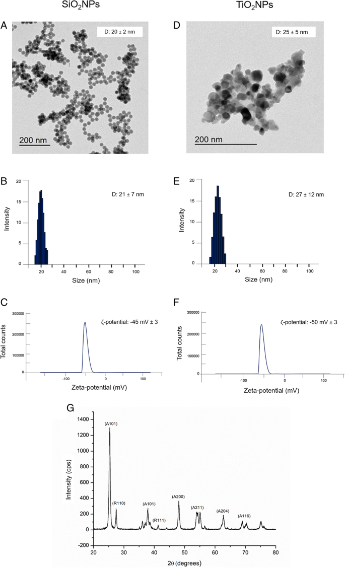
SiO2 NP 및 TiO2 NP는 좁고 제어된 크기 분포를 갖는 NP를 얻기 위해 다양하고 재현 가능한 합성 경로로 합성되었습니다("방법" 섹션 참조). 그런 다음, NP는 물과 다양한 농도의 단백질 공급원(FBS)이 있는 세포 배양 배지(DMEM) 모두에서 TEM, DLS, ζ-전위 및 XRD를 통해 깊이 특성화되었습니다. 이것은 미디어 단백질이 나노입자 표면을 덮을 수 있으므로 물리화학적 특성과 생물학적 효과를 변화시킬 수 있기 때문에 중요합니다[36]. TEM 분석은 SiO2 NP는 평균 직경이 20 ± 2 nm인 구형입니다(그림 1a). TiO2 NP는 크기가 비슷하지만(25 ± 5 nm) 형태가 다릅니다(그림 1). 96 h에서 수중에서 수행된 DLS 측정은 SiO2에 대해 21 ± 7 nm 및 27 ± 12 nm의 유체역학적 반경을 확인했습니다. NP 및 TiO2 NPs(그림 1b 및 그림 1e). 예상대로 이러한 데이터는 TEM 관찰과 잘 일치합니다. ζ-전위 분석은 또한 SiO2에 대해 - 45 ± 3 mV의 물에서 표면 전하 값을 확인했습니다. TiO2의 경우 NP 및 − 50 ± 3 mV NP(그림 1c, f). 예상대로, NP의 물리화학적 특성은 세포 배양 배지 내에서 접종 시 변하였다. DLS는 특히 20%의 FBS가 보충된 DMEM의 존재에서 NP 크기의 상당한 증가를 확인했습니다(표 1). 특히, SiO2 NP는 29 ± 9 nm의 크기를 보인 반면, TiO2 NP는 96 h 후에 41 ± 14nm까지 증가했습니다. DMEM 측정에서 관찰된 DLS 피크의 확대(FBS 유무에 관계없이)는 NP 응집의 징후이며, 이는 매체의 이온 강도에 의해 촉진될 수 있습니다(데이터는 표시되지 않음). 또한 ζ 전위 측정은 두 NP의 표면 전하가 더 음의 값으로 이동했음을 보여주었습니다. 이 큰 시간 의존적 현상은 NP의 표면에 흡착된 세포 배양 배지의 혈청 단백질의 존재에 의해 유도된 매우 안정적인 단백질 코로나 형성 [37, 38] 때문이었습니다. NP의 크기와 전하가 함수로 변합니다 FBS 농도의.
<그림>
SiO2의 특성 NP 및 TiO2 물 속의 NP. 아 –d 대표적인 TEM 이미지. ㄴ –이 동적 광산란(DLS) 및 c –f ζ-전위 측정. 지 TiO2의 X선 회절 분석(XRD) 패턴 NP
TiO2의 XRD 패턴 NPS , 430 °C에서 하소된 아나타제와 금홍석 결정상의 혼합물을 보여주었습니다(그림 1g) . 2θ =25.4°(101), 48.1°(200), 54.1°(211), 62.4°(204) 및 68.8°(116)에서 지배적인 피크는 표준 JCPDS 데이터( 카드 번호:21–1272). 루틸상은 27.5°(110), 36.2°(101) 및 41.2°(111)에서 회절 피크로 표시되었습니다.
Caco-2 및 A549 세포에서 NP의 흡수
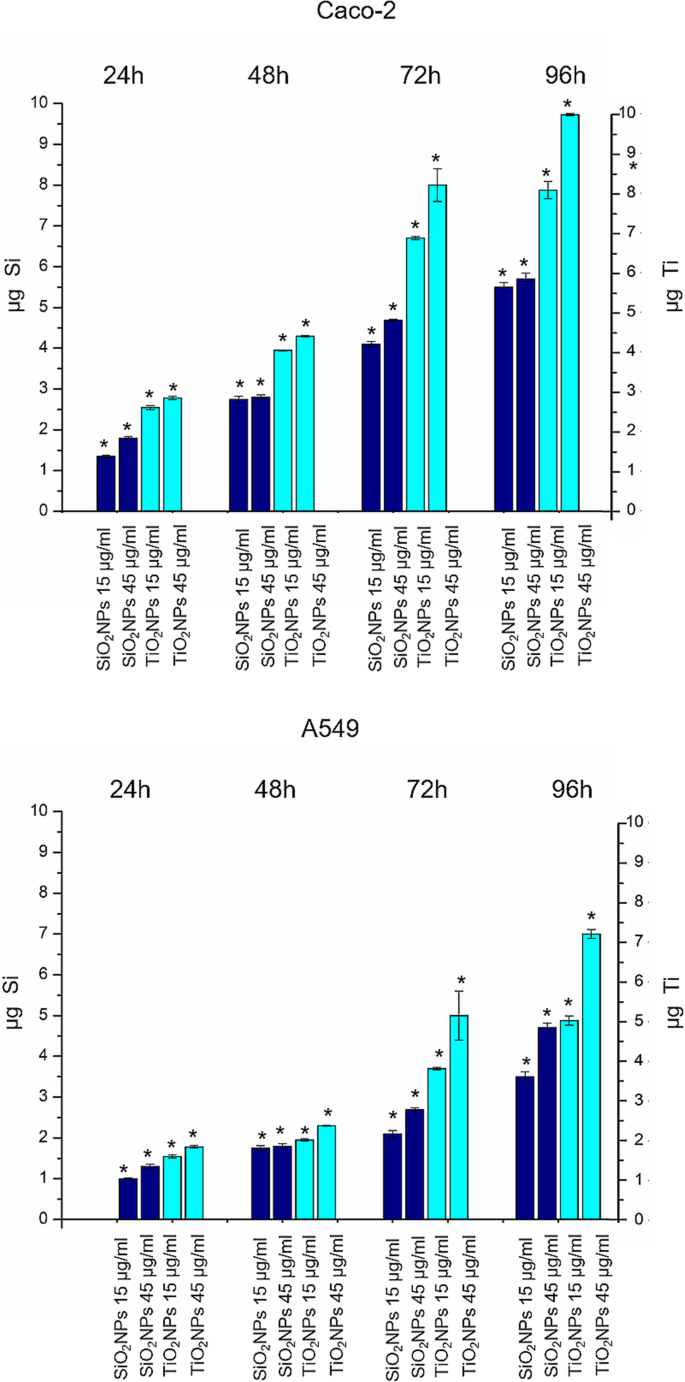
SiO2의 양을 정량화하기 위해 NP 및 TiO2 NPS 세포에 의해 흡수, 예비 조사로 용해된 세포에 대해 ICP-AES 원소 분석을 수행했습니다. 세포를 15 μg/ml 및 45 μg/ml의 NP로 처리하였다. 실험 데이터는 SiO2의 존재를 확인했습니다. NP 및 TiO2 두 세포주의 NP, 시간 의존적 내재화 효율(그림 2a). TiO2 NP는 SiO2와 관련하여 더 큰 흡수를 보였습니다. NP. 이것은 Ti 함량이 72 h 및 96 h 후에 각각 8.2 ± 0.4μg 및 9.7 ± 0.031μg의 세포내 농도에 도달한 Caco-2에서 특히 분명했습니다. A549에서 검출된 Ti의 양은 72 h 후에 5 ± 0.599μg, 96 h의 배양 시간 후에 7.12 ± 0.11μg으로 더 낮았습니다. SiO2 NP는 TiO2에 비해 세포에 덜 흡수되었습니다. 내재화가 Caco-2에서 더 뚜렷하더라도 NPs. 이 경우에도 실제로 내재화된 SiO2의 양이 Caco-2 세포의 NP는 72 h 후 4.69 ± 0.031μg, 96 h 배양 후 5.78 ± 0.045μg였습니다. 값은 A549에서 감소했으며, 여기서 72 h 후 2.58 ± 0.045μg 및 96 h 후 4.7 ± 0.04μg를 정량화했습니다.
<그림>
TiO2 NP 및 SiO2 15 μg/ml 및 45 μg/ml의 TiO2에 노출된 Caco-2 및 A549 세포주에서 NP 축적 NP 및 SiO2 24 h, 48 h, 72 h 및 96 h에 대한 NP. 그런 다음 세포를 수확하고 살아있는 세포를 계수하고 360,000개 세포(μg Ti 및 μg Si)에서 Ti 및 Si 함량을 측정했습니다. 3개의 독립적인 실험에서 평균 ± SD로 보고된 데이터; p에 대한 노출된 세포 대 대조군 세포의 통계적 유의성 값 <0.05(<0.05*, <0.01** 및 <0.005***)
CaCo-2 및 A549에 대한 NP의 영향:세포 생존, 막 손상 및 ROS 생성
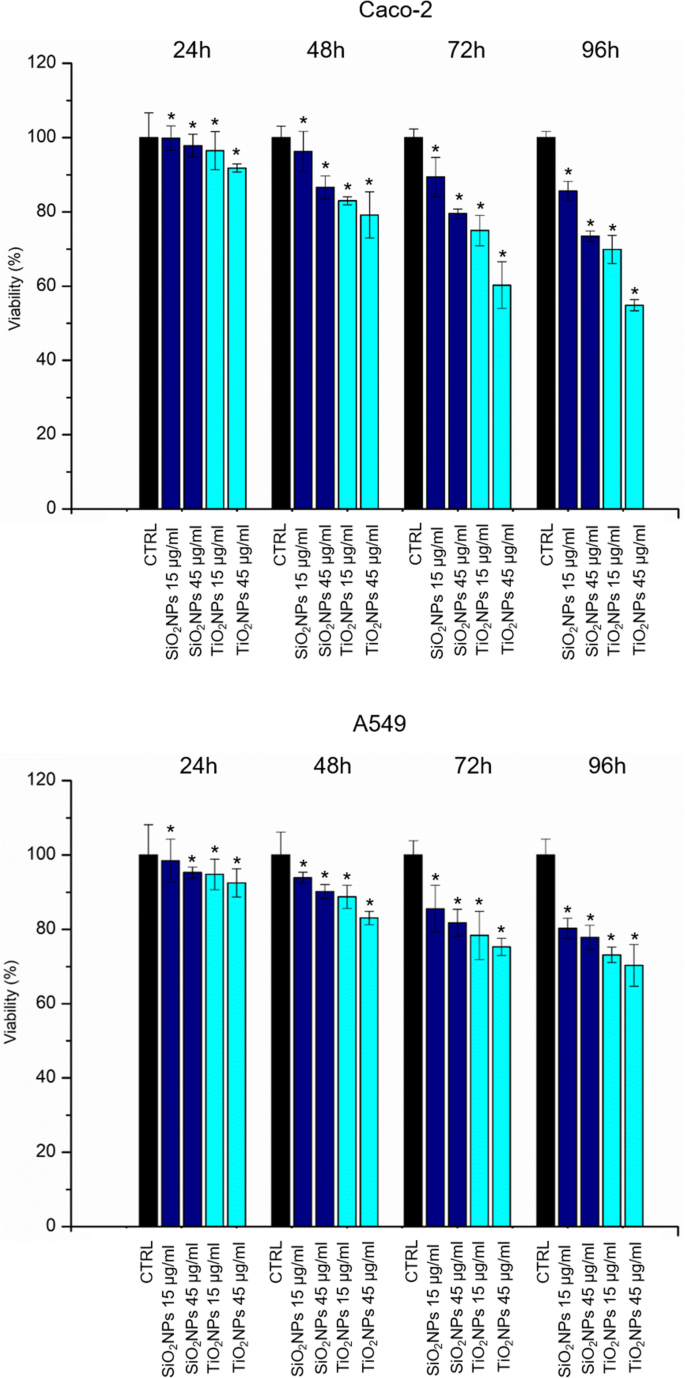
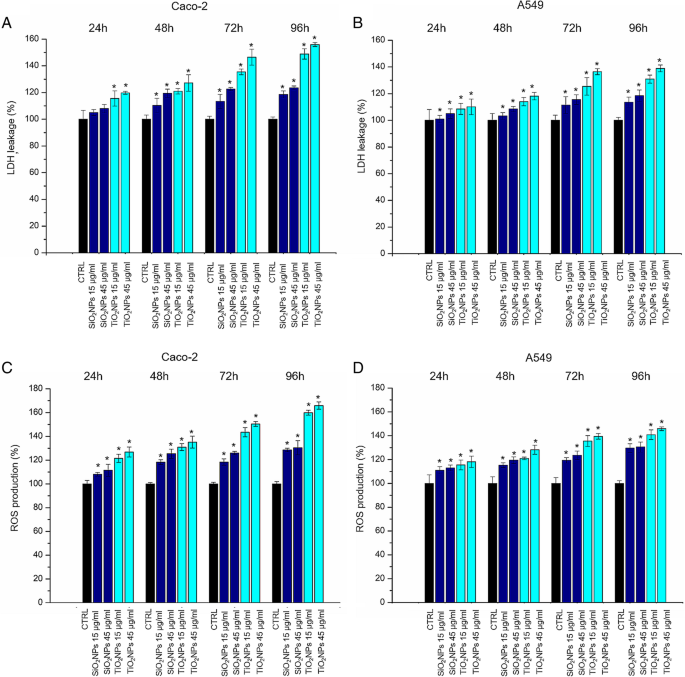
Caco-2 및 A549 세포 생존력은 WST-8 분석으로 평가되었습니다. SiO2로 처리 NP 및 TiO2 NP는 테스트된 두 세포주에서 약간의 용량 의존적 생존력 감소를 유도했습니다(그림 3). TiO2 NP는 SiO2와 관련하여 강화된 세포독성을 유도했습니다. TiO2 처리 시 NP 및 CaCo-2 세포의 세포 생존율은 A549보다 더 큰 영향을 받았습니다. NP. 특히, 우리는 45 μg/ml의 TiO2로 처리된 Caco-2에서 약 40%의 생존율 감소를 관찰했습니다. 72 h 동안의 NP. 이 감소는 96 h 이후에 최대 50%까지 감소한 반면, A549 세포주에서는 TiO2 NP는 96시간의 처리 후에만 생존력의 30% 감소를 유도했습니다. LDH 방출 및 ROS 생성은 TiO2 노출 시 Caco-2 및 A549 세포에서 평가되었습니다. NP 및 SiO2 NP. 그림 4a, b에서 볼 수 있듯이 NP는 생존 결과와 밀접하게 일치하는 세포막 천공(및 실제로 LDH 방출)을 유도했습니다. 그 효과는 특히 TiO2에서 A549와 관련하여 Caco-2에서 더 분명했습니다. 가장 높은 시점(72 및 96 h)에서 NP 처리. LDH 방출 비율은 96시간 노출 후 처리되지 않은(대조군) 세포에 비해 약 160% 증가했습니다. ROS 생성은 NP에 의해 유도되는 주요 효과 중 하나이기 때문에 열성적으로 연구되었습니다[39]. 이 현상은 생물학적 항산화 방어 반응을 방해하지만[40], 실제 작용 메커니즘이 아직 조사 중이라는 점을 언급하는 것이 중요합니다. 잠재적인 NP-유도 산화 스트레스는 DCFH-DA 분석에 의해 추정되었다. 예상대로 NP와 세포 간의 상호 작용은 TiO2에 대한 Caco-2의 강력한 영향과 함께 용량 의존적 방식으로 ROS 생성을 자극했습니다. NP 처리(그림 4c, d). 방출 비율은 테스트된 최고 농도에서 대조군 세포에 대해 165%의 값에 도달했습니다.
<그림>
Caco-2(a)의 생존력 분석(WST-8) ) 및 A549(b ) 두 가지 용량(15 μg/ml 및 45 μg/ml)의 TiO2에 24시간, 48시간, 72시간, 96시간 노출 후 세포 NP 및 SiO2 NP. NP 처리된 세포의 생존력은 처리되지 않은 대조군 세포로 정규화되었습니다. 양성 대조군(P)으로서, 세포를 5% DMSO와 함께 인큐베이션하였다(데이터는 나타내지 않음). 3개의 독립적인 실험에서 평균 ± SD로 보고된 데이터는 대조군(n =8) p의 경우 값 <0.05(<0.05*, <0.01** 및 <0.005***)
<그림>
LDH(a –b ) 및 ROS(c –d ) Caco-2 및 A549 세포에 대한 분석. 세포를 15 μg/ml 및 45 μg/ml의 TiO2와 함께 배양했습니다. NP 및 SiO2 24 h, 48 h, 72 h 및 96 h에 대한 NP. 나노입자 처리된 세포의 LDH 누출 백분율은 처리되지 않은 대조군 세포와 비교하여 표시됩니다. 양성 대조군(P)은 0.9% Triton X-100으로 세포를 처리하여 ca. 500% LDH 증가(데이터는 표시되지 않음). ROS 수준은 15 μg/ml 및 45 μg/ml의 TiO2로 Caco-2 및 A549 세포를 노출시키는 것으로 기록되었습니다. NP 및 SiO2 24 h, 48 h, 72 h 및 96h에 대한 NP는 100 μM DCFH-DA와 함께 배양되었습니다. 세포 형광을 측정하였다. 양성 대조군(P)으로 세포를 500 μM H2와 함께 배양했습니다. O2 ca를 보여줍니다. 300% DCFH-DA 증가(보이지 않음). 3개의 독립적인 실험에서 평균 ± SD로 보고된 데이터는 대조군(n =8) p의 경우 값 <0.05(<0.05*, <0.01** 및 <.005***)
Caco-2 및 A549 세포의 항산화 활성 및 지질 과산화에 대한 NP의 영향
SOD 효소는 산화 스트레스 유도 후 항산화 방어 시스템에 관여합니다. 이 효소는 반응성이 높은 슈퍼옥사이드(O2
•−
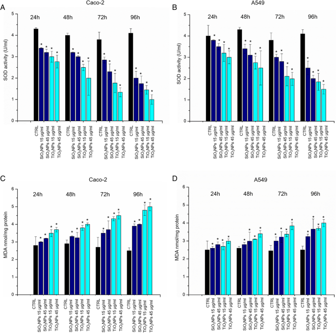
) 과산화물 H2로의 음이온 O2 [41]. 우리는 SiO2와 함께 배양한 후 Caco-2와 A549 모두에서 SOD 효소 활성의 용량 의존적 감소를 관찰했습니다. NP 및 TiO2 다른 시점(24~96 h)에서 NP(15 μg/ml, 45 μg/ml)(그림 5a, b). 세포 독성 평가와 밀접하게 일치하여 TiO2에 대한 Caco-2의 효과가 더 분명했습니다. NP 노출. 예를 들어, SOD 활성 수준은 45 μg/ml의 TiO2에 노출된 Caco-2 세포에서 대조군의 4.1 ± 0.2U/ml에서 1.03 ± 0.325U/ml로 감소했습니다. NP, 96 h 이후. 동일한 농도의 SiO2에 노출 NP는 SOD 활성을 1.45 ± 0.12U/ml로 줄였습니다. MDA 기반 분석은 잠재적인 ROS 매개 지질 과산화를 확인하는 데 사용되었으며, 이는 차례로 세포 산화 스트레스를 확인하는 또 다른 방법입니다. [42] MDA의 세포 수준은 Caco-2와 A549 모두에 대해 두 가지 유형의 NP에 노출된 후 증가했습니다(그림 5c, d). 예상대로 증가된 MDA 수치는 농도 및 노출 시간에 비례했습니다.
<그림>
아 –d Caco-2 및 A549 세포에 대한 SOD 및 MDA 분석. 세포를 15 μg/ml 및 45 μg/ml의 TiO2와 함께 배양했습니다. NP 및 SiO2 24 h, 48 h, 72 h, 96 h에 대한 NP. SOD 분석은 xanthine oxidase와 hypoxanthine에 의해 생성된 슈퍼옥사이드 라디칼의 검출을 위해 tetrazolium salt를 사용했습니다. 표준 곡선을 양성 대조군으로 사용했습니다(데이터는 표시되지 않음). MDA 수준은 MDA-TBA 부가물(OD =532 nm)의 정량화에 의해 검출되었습니다. 3개의 독립적인 실험에서 평균 ± SD로 보고된 데이터는 대조군(n =8) p의 경우 값 <0.05(<0.05*, <0.01** 및 <0.005***)
NP에 의해 유도된 형태역학적 효과
15 μg/ml 및 45 μg/ml의 SiO2와 함께 배양된 Caco-2 및 A549의 공초점 현미경 분석 NP 및 TiO2 24 h, 48 h, 72 h 및 96 h에 대한 NP는 그림 6 및 7. 대조군 Caco-2 세포는 정점 측에서 단단한 접합부 및 브러시 경계를 갖는 장내 장세포와 유사한 형태를 나타내었다[43]. NP로 처리하면 세포의 단단한 접합부가 무너지고 세포 패턴이 길어진 모양으로 분리되었습니다. 이러한 효과는 세포가 TiO2로 처리되었을 때 더 분명했습니다. 72 h의 배양 시간 동안 45 μg/ml의 NP는 세포 형태의 변화뿐만 아니라 액틴 패턴의 관련 변경을 보여줍니다. 처리되지 않은 A549 세포는 자갈과 같은 모양과 기능적인 세포-세포 접착을 나타냈지만[44], NP로 처리하면 세포-세포 접촉이 감소하고 세포 형태가 더 길쭉한 형태로 변형됩니다. 그림 8은 액틴 분포의 변화를 감지할 수 있는 확대된 공초점 그림을 보여줍니다. NP 노출 후 액틴 네트워크의 변경된 조직(45 μg/ml의 SiO2의 72 h NP 및 TiO2 NPs)는 ImageJ 소프트웨어를 사용하여 형광 밀도 및 일관성에 의해 정량적으로 분석되었습니다(그림 9). 통합 밀도를 통해 액틴의 양을 정량화할 수 있었고 일관성을 통해 주변 환경과 비교한 섬유 배향 정도에 대한 정보를 얻을 수 있었기 때문에 우리는 이 두 매개변수를 특별히 선택했습니다[45]. 처리되지 않은 Caco-2 세포는 129.4 ± 16의 밀도 값을 가졌으며 이는 NP 처리에 영향을 받지 않은 채로 남아 있었습니다. 값은 SiO2에 노출된 후 127.7 ± 20 및 128.5 ± 18이었습니다. NP 및 TiO2 NP, 각각 그림 9a). 유사하게, 액틴 염색 네트워크의 밀도도 처리 전후 A549에서 동일하게 유지되었습니다(음성 대조군의 경우 68.4 ± 14, 67.9 ± 15 및 67.7 ± 18, SiO2) NP 및 TiO2 NP, 각각 그림 9b). NP 처리는 액틴 양의 변경을 유도하지 않았지만 일관성 분석은 액틴 공간 재구성이 유사하지 않음을 시사했습니다. Caco-2에서 SiO2에 대한 처리된 세포의 일관성 값 NP(0.16 ± 0.04) 및 TiO2의 경우 NP (0.09 ± 0.02) treatment decreased with respect to that of the control (0.26 ± 0.03) (Fig. 9c). Even the A549 cells underwent a decrease of coherency after interacting with SiO2 NPs and TiO2 NPs (0.2 ± 0.07 and 0.158 ± 0.04) compared to the control cells (0.4 ± 0.03) (Fig. 9d). Hence, NPs induced a significant reorganization of actin network, which showed an actin isotropic orientation, but they did not change the overall quantity of actin expressed. In addition to cytoskeletal rearrangements, we also analyzed the area described by the nucleus/cytoplasm ratio. Values of N/C ratio were calculated as the ratio between nuclear area and the whole cellular area (measured performed on 15 cells). We observed opposite values following the treatment with 45 μg/ml of NPs for 72 h with significant statistical validity. In particular, the ratio was (0.40 ± 1.7) in untreated Caco-2 cells, and this increased up to 0.554 ± 0.09 and 0.62 ± 0.12 after SiO2 NP 및 TiO2 NP exposure (Fig. 9e). The trend was different in A549. The nuclear/cytoplasm ratio dropped down upon exposure to NPs from values of 0.550 ± 0.04 for control cells to 0.334 ± 0.06 for SiO2 NPs and 0.225 ± 0.09 for TiO2 NP. After the morphological investigations, we analyzed the elastic properties of cells after exposing them to 45 μg/ml of SiO2 NPs and TiO2 NPs for 72 h by AFM, in force–volume mode. We measured the different elasticity expressed by Young’s modulus values in the nuclear and cytoplasmic region. Caco-2 cells displayed Young’s modulus value of 105 ± 25 kPa for nuclear region and 47 ± 21 kPa for the cytoplasm. After SiO2 NP treatment, we observed a reduction of value to 42 ± 8 kPa for the nucleus and 19.59 ± 2 kPa for the cytoplasm. The effects were more evident after treatment with TiO2 NPs:the Young modulus for the nucleus was 27 ± 4 kPa and 18 ± 4 kPa for the cytoplasm (Fig. 10a). We found an opposite outcome concerning the elastic properties of A549 cells. In this case, Young’s modulus was 129 ± 24 kPa for the nuclear region and 147 ± 26 kPa for the cytoplasmic area. After SiO2 NP treatment, the values of elasticity increased to 152 ± 23 kPa for nucleus and 152 ± 25 kPa for cytoplasm. When cells were doped with TiO2 NPs, Young’s modulus values drastically increased to 372 ± 60 kPa for nucleus region and 549 ± 40 kPa for cytoplasmic region (Fig. 10b).
Effects of SiO2 NPS and TiO2 NPs on actin network of Caco-2 cells. Caco2 were treated with 15 μg/ml and 45 μg/ml of NPs for 24 h, 48 h, 72 h, and 96 h; cells were fixed and then stained with Phalloidin–ATTO 488 and DAPI. The 2D images of cortical actin were acquired by a Zeiss LSM700 (Zeiss) confocal microscope equipped with an Axio Observer Z1 (Zeiss) inverted microscope using a × 100, 1.46 numerical aperture oil immersion lens. All data were processed by ZEN software (Zeiss)
Effect of SiO2 NPS and TiO2 NPs on actin network on A549 cells. A549 were treated with 15 μg/ml and 45 μg/ml of NPs for 24 h, 48 h, 72 h, and 96 h; successively they were fixed and stained with Phalloidin–ATTO 488 and DAPI. The 2D images of cortical actin were acquired by a Zeiss LSM700 (Zeiss) confocal microscope equipped with an Axio Observer Z1 (Zeiss) inverted microscope using a × 100, 1.46 numerical aperture oil immersion lens. All data were processed by ZEN software (Zeiss)
Local enlargement of confocal acquisitions acquired in Figs. 6 and 7 showed (more in details) the effect of SiO2 NPS and TiO2 NPs on actin network of Caco-2 and A549 cells after the exposure of 45 μg/ml of NPs for 72 h and 96 h
Integrated density (a , b ) and coherency (c , d ) for Caco-2 and A549 cells treated with 45 μg/ml of SiO2 NPs and TiO2 NPs after 72 h. The integrated density and coherency values were expressed as a mean value and relative SD, calculated from confocal acquisitions by ImageJ (calculation on 15 cells). The mean values and their standard deviations were reported in the histograms. Results were statistically significant for p < 0.05 (< 0.05*, < 0.01**, and < 0.005***). 이 Analyses of nucleus/cytoplasm ratio on Caco-2 and A549 after exposure to 45 μg/ml of SiO2 NPs and TiO2 NPs for 72 h. The ratio was calculated on 15 cells by ImageJ. The mean values and the SD were reported in the histogram. Results were statistically significant for p < 0.05 (< 0.05*, < 0.01**, and < 0.005***)
Young’s modulus values expressed in kPa, calculated from the nuclear region and the cytoskeletal area of Caco-2 (a ) and A549 (b ) cell lines after a treatment to 45 μg/ml of SiO2 NPs and TiO2 NPs for 72 h
Discussion
The spread of different kind of ENPs in several fields raises awareness about the importance to assess their potential toxicity in living organisms and the environments as well, taking into account their potential application in biomedical field [46,47,48]. In vitro and in vivo investigations are crucial to enrich the scientific knowledge and to release reliable clinical and epidemiological data [49]. The toxicity tests performed on different cells are considered the golden standard to assess the safety of NPs. However, few studies have investigated the interactions between NPs and cells from a biomechanical point of view. Cell mechanic is an important factor that influences many cellular pathways, including apoptosis, differentiation, migration, cancer metastasis, and wound healing [50]. In our work, we have addressed this point and related cell viability with the changes in mechanical properties of cells treated with different NPs. Firstly, we synthetized amorphous SiO2 NPs and crystalline TiO2 NPs with a size of c.a. 20 nm. NPs were stable in water and DMEM up to 96 h, even upon incubation with 10% and 20% of FBS. This was found to induce an increase in NPs size due to the formation of protein corona, in perfect agreement with the literature data. [51]. Since the entry route of NPs often occurs through inhalation and ingestion, we opted to investigate the potential effects on Caco-2 and A549 cells, which are representative models for the intestinal tract and airways mimicking oral and inhalation uptake [52]. As primary investigation, we quantified the cellular internalization of SiO2 NPs and TiO2 NPs by elemental analysis. The most effective uptake was observed in Caco-2 cells, especially upon treatment with TiO2 NPs in a time-dependent manner. It has been reported that amorphous SiO2 NPs, with a small size range of 15–20 nm, can bind the plasma membrane and then passively pass across the lipid bilayer to get access into the cells [53]. As demonstrated in A549 [54] and Caco-2 [55], in fact, small SiO2 NPs can translocate in the cytoplasm with no apparent membrane encapsulation. The anatase crystalline form of TiO2 NPs is the more chemically reactive [56] showing a faster absorption with respect to rutile, as previously reported [32]. However, the uptake mechanisms of Caco2 are still unclear, despite that some hypothesis have been formulated, some of these include metal ion release upon NPs degradation in the intestinal barrier lumen or/and direct uptake by endocytosis. [57]. In A549 cells, TiO2 NPs were localized in cytoplasm and close the nucleus region [58]. We used WST-8 assay to assess the influence of different concentrations of SiO2 NPs and TiO2NPs on cell viability. We have observed a general decrease of viability, especially in Caco-2, with TiO2 NPs displaying the strongest toxicity. After assessing the viability, we monitored the extracellular release of the cytoplasmic enzyme LDH. We confirmed that the NPs induced an extensive membrane damage, which relates also to the increase of intracellular ROS levels, resulting in oxidative stress. In this context SOD, which acts as strong antioxidant against ROS [59], was significantly reduced most probably because of the unbalance of the redox repair systems. In addition, the oxidative stress increased the lipid peroxidation [60], as demonstrated by MDA measurements after NPs incubation. This is particularly evident in Caco-2 cells after TiO2 NP exposure. It is worth mentioning that this effect can decrease membrane fluidity, which can further explain the observed higher entry levels of the TiO2 NPs [61]. This was in significant accordance with the intracellular oxidative stress levels measured by SOD inhibition, as well as with the reactive oxygen species generation. After these assessments, we investigated the modulation of the cell cytoskeleton, as an increase of intracellular ROS could affect the F-actin organization [62]. The cytoskeleton is characterized by a set of filaments (actin microfilaments, microtubules, and intermediate filaments) organized in a network that affects the mechanical properties of cells, as well as their behavior [29]. In particular, actin filaments are crucial for cell mechanics, and any alterations may induce aberrations in cell morphology under sub-toxic conditions [63]. It has been demonstrated that actin was one of the most commonly bound protein by SiO2 NPs and TiO2 NPs in cellular extracts. This definitely suggests that the actin functions, as well as cell motility and organelles trafficking, can be strongly affected by the presence of these NPs [64, 65]. As a further proof, several in vivo studies have revealed the potential of NPs to induce alterations in the expression of genes related to the cytoskeleton [63]. In order to understand how NPs modulate the cytoskeleton, we performed qualitative and quantitative confocal analyses on Caco-2 and A549 cells, after SiO2 NP and TIO2 NP treatment. We focused on actin stress fibers and cortical actin because they allowed to maintain the physiological mechanical architecture of cells. As reported in Figs. 4 and 5, the treatment with NPs induced a significant reorganization of actin. This was more evident after 72 h of treatment with 45 μg/ml of NPs, and especially with the use of TiO2 NP. The adverse effects were stronger in intestinal cells, where we have observed the formation of protrusions and philopodia at the plasma membrane level, together with the disruption of tight junctions. Fluorescence coherency and fluorescence density have been used as quantitative parameters to assess alterations of actin distribution in the cytoskeleton. While coherency gives information about the organization of actin, density quantifies the levels of fluorescent actin. Caco-2 and A549 exposed to NPs showed a reduction of coherency compared to untreated cells, especially upon incubation with TiO2 NP. This was in good agreement with the qualitative confocal imaging analyses. The fluorescence density of actin was not altered by NP treatment in both the cell lines, even if untreated Caco-2 cells showed higher values with respect to untreated A549. These data could suggest a potential difference in the amount of actin, which is dependent on>the specific cell type. We also evaluated the nucleus-to-cytoplasm ratio as the relative area of the nucleus over the whole cell. We confirmed a reduction of values in A549 and an increase of the ratio in Caco-2 with respect to the control cells. This indicates changes in cell morphology after NP treatment:Caco-2 underwent an increase of nucleus area, whereas A549 became larger through cytoplasm extension. As a final point, we explored any potential change in cell elasticity upon NP treatment. Cell elasticity is commonly expressed by Young’s modulus (E), which is a ratio between the stress and the applied strain (with unit in Pascal) [66, 67]. Changes in cell elasticity due to cytoskeleton reorganization is often associated to disease progression [68], hence (E) can be a refined indicator of potential pathological states [67]. The deformability of cells was measured through indentation experiments by AFM [69]. Many studies showed the detrimental effects of NPs on the F-actin that induced an enhancement of cell elasticity. However, a clear relation between change in cell stiffness, actin rearrangement and cell viability has not been clearly established yet. Here, we have covered such topic and found that Caco-2 and A549 cells significantly change their (E) upon NP treatment, even though in two different ways. Caco-2 cells are softer as confirmed by the decreased Young’s modulus, which has been found to be a function of both the NPs type and the cell regions analyzed. In particular, TiO2 NPs induced a general enhancement of elasticity, and this effect is more evident in the nuclear regions rather than the cytoplasmic one. On the other side, A549 displayed a remarkable increase of Young’s modulus after TiO2 NP exposure in cytoplasm region, compared to control cells (594 ± 40 kPa versus 146 ± 26 kPa, respectively). These data indicated that TiO2 NPs induce dose-dependent changes in force–deformation profiles in both cell lines, whereas SiO2 NPs showed little effects. The decrease of Young’s Modulus, and consequently an increase of elasticity after NPs exposure, can potentially impact cell homeostasis and physiological pathways. The reorganization F-actin, together with a reduction of coherency, showed a strong modulation of mechanical cell properties. NPs have been demonstrated to make the nuclear region of Caco-2 cells softer than untreated cells. The increase of elasticity (corresponding to a reduction of Young’s modulus) is a critical factor in tumor progression, because it is an indicator of disruption of cell junctions, which promotes in turn cell migration and metastatization [70]. Therefore, the treatment with NPs on Caco-2 (and TiO2 NPS in particular) can potentially promote migration due to change of elastic properties and deformability of cells. Also, the larger and softer nucleus area can be associated to cancer progression [71].
Conclusion
In this paper, we careful assessed the adverse effects of SiO2 NPs and TiO2 NPs on two different cell lines (Caco-2 and A549), mimicking the typical tissue that are exposed to NPs (ingestion and inhalation). SiO2 NPs presented a low cytotoxicity in comparison with TiO2 NPS. We demonstrated how the cellular exposure to high doses of NPs induced morphostructural changes in term of actin reorganization, coherency, density and nucleus/cytoplasm ratio, which were more evident upon TiO2 NP treatment. Cell membrane deformability measurements showed different behavior in the two cells. In Caco-2, the cell elasticity increased after NP treatment, whereas A549 displayed an increase of stiffness. These results demonstrated that NPs induce modifications of cytoskeleton structures and, as consequence, a different Young’s Modulus values. Hence, the phenotype of cancer cells can turn into a more invasive profile, characterized by enhanced migration. On the other side, the increased stiffness observed in A549 might not promote the mobility of this specific cell indeed. We are sure that the analysis of cell mechanics upon NP exposure, combined with standard toxicological assays, will represent a golden standard to accurately assess the safety of NPs and to predict any potential possible diseases triggered by NPs.
약어
A549:
Human adenocarcinoma alveolar basal epithelial cells