약물 내성 박테리아에 대한 가장 유망한 방법 중 하나는 살생물성 나노입자 및 나노복합체로 표면 개질된 물질일 수 있습니다. 여기에서 우리는 새로운 다기능 항균 및 항진균 재료로서 산화 그래핀(GO) 표면에 은 나노 입자(Ag-NPs)가 있는 나노 복합재를 제시합니다. 초음파 기술은 폴리우레탄 호일을 코팅하는 효과적인 방법으로 사용되었습니다. 그람 음성 박테리아에 대한 독성(대장균 ), 그람 양성 박테리아(황색 포도상구균 및 표피 포도상구균 ) 및 병원성 효모(Candida albicans )은 세포 형태 분석, PrestoBlue 분석을 사용한 세포 생존력 평가, 젖산 탈수소효소 분석을 사용한 세포막 완전성 분석 및 활성 산소 종 생산에 의해 평가되었습니다. 항균제로 널리 사용되는 Ag-NPs 및 GO와 비교하여 당사의 나노복합체는 박테리아 및 효모 세포에 대해 훨씬 더 높은 항균 효율을 나타냅니다.
<섹션 데이터-제목="배경">
배경
항생제의 개발은 세균 감염의 수를 조절하는 데 중요한 역할을 했습니다. 그러나 항생제의 부적절한 사용과 남용으로 인해 많은 박테리아 종에서 다제 내성이 발생했습니다. 일부 균주는 베타-락탐, 테트라사이클린 및 아미노글리코사이드와 같은 일반적으로 사용 가능한 거의 모든 약제에 내성을 갖게 되었습니다[1]. 주요 내성 병원체는 메티실린 내성 황색 포도상구균입니다. , 반코마이신 내성 Enterococcus , 및 확장 스펙트럼 β-락타마제 생성 Klebsiella pneumoniae 및 대장균 [2, 3]. 매우 큰 개체군과 빠른 증식 시간을 가진 박테리아는 박테리아 개체군의 하위 집합이 항생제 치료에서 살아남을 때 항생제 내성 메커니즘을 빠르게 개발할 수 있습니다. 더욱이, 항생제 내성 박테리아는 다른 먼 관련 박테리아에 대한 내성 메커니즘을 암호화하는 DNA 사본을 전달할 수 있으며, 이는 내성 유전자를 다음 세대에 전달할 수 있습니다. 따라서 항생제 내성균의 출현은 새로운 항균제의 개발로 극복할 수 있는 심각한 문제이다. 항균제는 섬유 산업, 물 소독, 의약품 및 식품 포장에서 매우 중요합니다. 나노입자와 나노물질은 항생제의 대안으로 사용될 수 있다[4]. 나노 입자의 항균 활성 메커니즘은 나노 입자의 유형에 따라 다릅니다. 일부 제안된 메커니즘은 나노입자의 물리화학적 구조와 관련이 있는 반면, 다른 메커니즘은 나노입자 표면에서 항균 이온의 방출 증가와 관련이 있습니다. 미생물에 대한 다중 동시 작용 메커니즘은 내성 발달을 위해 동일한 미생물 세포에서 다양한 동기 DNA 돌연변이를 필요로 합니다. 따라서 박테리아 세포가 나노 입자 및 나노 물질에 내성을 갖기가 어렵습니다. 은, 구리, 풀러렌 및 단일벽 탄소 나노튜브와 같은 항균성 나노물질은 고유한 물리화학적 특성과 높은 표면적 때문에 여러 이점을 제공할 수 있습니다[5,6,7,8]. 다양한 박테리아에 대한 나노입자(NP) 독성의 정확한 메커니즘은 완전히 이해되지 않았습니다. 현재 연구에 따르면 나노입자의 항균 효과의 기본이 되는 주요 과정은 박테리아 세포막 파괴, 금속 이온 방출, ROS 생성, 박테리아 세포막 침투, DNA 및 DNA와의 상호 작용을 포함한 세포 내 항균 효과 유도입니다. 단백질 [9, 10]. NP는 정전기적 상호작용에 의해 세균막에 부착되어 세균막의 완전성을 파괴할 수 있습니다. NP 표면의 양전하는 접착에 필수적입니다. 양전하는 NP와 미생물의 음전하를 띤 세포막 사이에 정전기적 첨가를 가능하게 한다[11]. 세균 세포 표면에 존재하는 황 함유 단백질과 나노입자 사이의 정전기적 연결은 세포벽 구조의 비가역적 변화를 일으켜 세포벽과 막의 손상을 초래한다[12]. 세균막은 세포의 항상성 및 대사 에너지 변환을 위한 선택적 투과성을 제공하기 때문에 세포의 대사 상태에 관계없이 중요합니다. NPs의 두 번째 항균 및 항진균 활성은 ROS 및 자유 라디칼 종을 생성하는 능력 때문입니다[13]. ROS의 증가된 수준은 지질, 단백질 및 DNA의 과산화를 유도합니다[14].
또한, 많은 유형의 나노입자의 구조는 항균제를 운반하는 데 적합합니다[15, 16]. 보균자는 표적 박테리아의 내성으로부터 약물을 보호하는 데 도움이 될 수 있습니다. 나노입자 기반 약물 전달 시스템은 항생제를 감염 부위에 표적화하여 전신 부작용을 최소화하는 데 도움이 될 수 있습니다. 기타 장점으로는 소수성 약물의 용해도 향상, 전신 순환 시간 및 약물 반감기 연장, 약물 방출 지속 등이 있습니다[4].
최근 탄소의 새로운 동소체인 그래핀이 항균 활성이 있음이 입증되었습니다. 그래핀은 탄소 원자가 육각형의 반복 패턴으로 결합되어 만들어진 물질입니다. 그래핀 플레이크의 독특한 특징은 표면에 대한 두께의 비율입니다. 그래핀의 표면은 전자 구름으로 덮여 있는데, 이는 아마도 이 물질이 전자 공여체가 되도록 하고 특별한 결합을 만드는 능력을 부여할 것입니다. 그래핀의 가장자리에는 다른 결합(다이아몬드 sp3형 결합의 특징)이 있으며 이러한 위치는 다른 물리화학적 특성을 가질 수 있습니다[17]. 이러한 특성은 그래핀이 박테리아 세포를 포함한 다양한 세포간 구조에 플라스틱 접착에 노출될 수 있음을 시사합니다[18,19,20]. 또한 그래핀은 두 개의 활성면(표면과 가장자리)이 있기 때문에 가장자리에 생물학적 분자를 부착하고 세포 표면에 부착할 수 있습니다. 산화된 형태의 그래핀인 산화 그래핀(GO)은 산소 작용기의 존재로 인해 물 및 기타 유기 용매에 쉽게 분산될 수 있습니다. 산소화된 그룹은 공유 및 비공유 상호작용을 통해 GO 시트의 직접적인 화학적 기능화를 가능하게 합니다. GO의 강력한 항균 활성이 보고되었습니다. GO의 항균 활성은 산화 그래핀 나노시트의 날카로운 모서리에 의해 유발된 막 응력에 할당되어 있으며, 이는 세포막에 물리적 손상을 일으켜 박테리아 막 무결성을 상실할 수 있습니다[21]. 최근 그래핀 기능이 있는 항균 나노입자가 유망한 항균 재료로 사용되고 있다[22, 23]. 나노복합체는 개별 구성요소의 한계를 극복할 수 있습니다. 예를 들어, 그래핀 기판에 부착된 항균 나노물질은 더 안정적이고 잘 분산됩니다[24]. 이러한 나노복합체에는 금속, 금속 산화물 및 중합체가 포함될 수 있습니다.
약물 내성 박테리아에 대한 가장 유망한 방법 중 하나는 살생물성 나노 입자로 표면 개질된 물질일 수 있습니다. 초음파 기술은 항균 및 살균 물질로 다양한 재료를 코팅하는 효과적인 방법으로 확인되었습니다 [25,26,27,28]. 많은 연구자들은 초음파 방법을 "녹색 기술"로 분류합니다[29, 30]. 이 방법은 액체 매질에서 공동화 기포의 형성, 성장 및 붕괴인 공동화 현상의 사용을 기반으로 합니다[31, 32]. 내파하는 거품은 짧은 시간 내에 최대 5000K의 미세 영역과 최대 2000atm의 압력에서 엄청난 양의 에너지를 생성합니다[33, 34]. 결과적으로 고체 표면을 향한 충격파와 소위 마이크로젯이 생성됩니다[35]. 액체 매질에 위치한 NP는 내파 효과에 의해 상승하고 고체 표면에서 고속(> 100m/s)의 제트 기류를 일으켜 층을 형성합니다[36]. 음향 캐비테이션은 초음파 처리된 물체의 물리적 특성을 변화시킬 수도 있습니다(예:GO 플레이크 크기 조정[37, 38].).
우리는 Salmonella enterica에 대한 이전 연구에서 유망한 결과를 얻었습니다. 및 리스테리아 모노사이토제네스 깨끗한 그래핀, GO 및 환원된 GO로 처리 [20]. 여러 종류의 그래핀 중에서 GO가 낮은 농도에서 가장 높은 항균 활성을 나타내는 것으로 나타났습니다. 박테리아 세포는 GO의 전체 표면에 분포되어 있습니다. 이 연구에서 우리는 은 나노입자(GO-Ag)로 장식된 GO가 베어 GO 또는 은 나노입자(Ag-NP)보다 미생물 세포에 더 강한 독성 영향을 미칠 것이라고 가정했습니다. 2개의 활성면(표면과 가장자리)이 있기 때문에 GO 산화물은 Ag-NP를 가장자리에 부착하고 세포 표면에 부착할 수 있습니다. 그래핀 기반 나노복합체의 항균 활성은 세포막의 파괴와 산화 스트레스 때문일 수 있다. 이 연구의 목적은 그람 음성 박테리아(Escherichia coli ), 그람 양성 박테리아(황색 포도상구균 및 표피 포도상구균 ) 및 병원성 효모(Candida albicans ) 시험관 내 모델을 사용합니다. 조사는 X선 회절, 라만 분광 투과, FT-IR, 전자현미경(TEM), 주사전자현미경(SEM) 및 원자력현미경(AFM)을 이용한 나노복합체의 구조 분석, 미생물 세포 형태 평가, PrestoBlue™ 분석에 의한 세포 생존력, 젖산 탈수소효소 분석(LDH)에 의한 세포막 무결성 조사, 활성 산소종(ROS) 생산 평가
방법
그래핀 산화물의 합성, 변형 및 특성화
이 연구에서는 상업적으로 이용 가능한 흑연 분말(Acros Organics, New Jersey, USA)을 변형된 Hummers 방법으로 산화시켰다[39]. 10g의 흑연 분말을 10°C 미만의 진한 황산(98%) 230mL와 혼합했습니다. 그 다음, 황산과 흑연 혼합물에 4.7g의 질산나트륨과 30g의 과망간산칼륨을 온도를 10℃ 이하로 유지하면서 서서히 첨가하였다. 그런 다음, 혼합물을 30°C로 가열하고 2시간 동안 교반했습니다. 다음 단계에서 100mL의 물을 첨가하고 혼합물 온도는 ~ 100°C에 도달했습니다. 마지막으로 혼합물을 10mL의 과산화수소로 처리했습니다. 정제를 위해 슬러리를 여과하고 여과액의 pH가 6.5에 도달할 때까지 탈이온수로 세척했습니다.
GO의 탄소, 수소, 질소 및 황 함량의 중량 분석은 Elementar Analysensysteme GmbH(Langenselbold, Germany)에서 생산한 Vario EL III 장치를 사용하여 수행되었습니다. GO의 화학적 분석 측정을 수행하기 전에 샘플을 50°C의 진공(0.05mbar)에서 탈착 스테이션(VcPrep 061, Micromeritics, Norcross, GA, USA)에서 24시간 탈착했습니다. 산소 함량은 100% 중량에서 결정된 탄소, 수소, 질소, 황 함량을 빼서 계산했습니다.
라만 분광법은 inVia Raman 현미경(Renishaw, UK)을 사용하여 수행되었습니다. 산화 그래핀은 초기 출력의 5%인 514nm 레이저 파장으로 분석되었습니다. 스펙트럼은 샘플의 5가지 다른 지점에서 수집되었습니다. 노출 시간은 10이며 2개의 스캔이 수집되었습니다.
FT-IR 측정은 다이아몬드 결정의 감쇠 전반사 모드에서 Nicolet iS10 분광계(Thermo Fisher Scientific, USA)를 사용하여 수행되었습니다. 5 마이크로리터의 산화 그래핀 수 현탁액을 다이아몬드 결정의 표면에 적하하고 건조되도록 두었다. 건조 후 스펙트럼은 400–4000cm
-1
범위에서 수집되었습니다. .
평균 입자 크기 및 제타 전위 측정은 각각 동적 광산란(DLS) 모드 및 레이저 도플러 전기영동을 사용하여 실온(23°)에서 Malvern Instruments Ltd.(Malvern, UK)에서 제조한 Zetasizer Nano-ZS ZEN 3600을 사용하여 수행했습니다. 다).
나노물질의 TEM/SEM/AFM 분석
분말 및 호일의 형태는 투과 전자 현미경(TEM; JEM-1220 JEOL, Tokyo, Japan, 가속 전압 80kV) 및 주사 전자 현미경(SEM; Zeiss, Ultra Plus, Oberkochen, Germany)을 사용하여 결정되었습니다. TEM 관찰을 위한 샘플은 TEM 그리드(영국 스탠스테드 소재 Agar Scientific의 3mm 200 Mesh Cu Grid의 Formvar)에 하이드로콜로이드 액적을 배치하여 준비했습니다. 액적을 공기 건조시킨 직후 격자를 현미경에 삽입했습니다.
SEM 분석을 위해 스퍼터 코팅기(SCD 005/CEA 035, BAL-TEC, Pfäffikon, Switzerland)를 사용하여 샘플을 얇은 탄소 층으로 코팅했습니다. 내부 실험실 측정 절차가 적용되었습니다(P5.10, 2015년 8월 26일 6판).
AFM(원자력 현미경) 이미징은 Asylum Research MFP3D Bio 소프트웨어(버전:Asylum Research MFP3D 15.106.09)를 사용하여 수행되었습니다. 표면 지형 이미징 및 테스트된 포일 표면에서 GO의 감지는 두 가지 이미징 모드, 즉 위상차 이미징을 위한 AC 모드와 GO가 마찰력을 감소시키기 때문에 GO 감지를 위한 횡력 현미경(LFM)을 사용하여 수행되었습니다[40].
GO 및 Ag-NP로 코팅된 폴리우레탄 포일의 준비
폴리우레탄 호일을 덮기 위해 Ag-NPs(HydroSilver1000, Amepox, Łódź, Poland) 및 GO의 현탁액이 사용되었습니다. GO, Ag-NPs 및 GO-Ag(GO(200μg/mL), Ag-NPs(100μg/mL), GO(200μg/mL) + Ag-NPs(100μg/mL)) 현탁액은 탈이온수(컨덕턴스 0.09μS/cm, 탈이온수:HLP 20UV, Hydrolab, Staszyn, Poland)에서 준비됨. 현탁액은 추가 정제 및 여과 없이 사용되었습니다.
폴리우레탄 호일(15 × 15 × 0.05 mm)의 초음파 코팅은 부피가 50 ml인 유리 플라스크에서 수행되었습니다. 포일 샘플을 스탠드(Teflon)에 고정한 후 준비된 현탁액에 담그십시오. 코팅 공정은 현탁액에 존재하는 호일 샘플에 직각으로 배치된 초음파 혼(Ti 혼, Ø13 mm, 60% 효율, 20kHz, Sonics &Materials, Inc., Newtown, CT, USA)을 사용하여 수행되었습니다. 공정 온도는 30 ± 1 °C였습니다. 덮인 샘플을 탈이온수로 씻어내고 층류 챔버에서 건조시킨 다음 멸균 패키지에 포장했습니다.
표면 자유 에너지
젖음성 테스트는 Data Physics OCA – 20 goniometer(DataPhysics Instruments GmbH, Filderstadt, Germany)를 사용하여 수행되었습니다. 표면 자유 에너지(SFE)는 탈이온수와 디오도메탄이라는 두 가지 테스트 액체를 사용하여 Owens, Wendt, Rabel, and Kaelble(OWRK) 방법을 사용하여 계산되었습니다[41].
박테리아 및 효모 배양 및 준비
황색포도상구균 (ATCC 25923) 및 표피 포도상구균 (ATCC 14990), 대장균 (ATCC 25922) 및 Candida albicans (90028)은 LGC Standards(폴란드 Lomianki)에서 입수했습니다. 균주는 20%(v /v ) - 20°C에서 글리세롤. 실험에 사용하기 전에 균주를 해동하고 세균 세포를 증류수로 세척하여 글리세롤을 제거했습니다. 박테리아와 효모는 다음 영양 배지에서 성장했습니다:S용 트립신 한천 . 구균 및 E . 대장균 , S용 뇌심장 한천 . 표피 , 그리고 C를 위한 Sabouraud의 한천 . 알비칸 (Merck Millipore, 다름슈타트, 독일). 한천 플레이트에서 성장한 박테리아와 효모는 멸균 증류 식염수로 플레이트를 부드럽게 세척하여 수확했습니다. 세포 현탁액의 박테리아 수를 계산하려면 600nm에서 현탁액의 광학 밀도(OD600 ) 분광광도계(Helios Epsilon, Unicam, Milwaukee, WI, USA)를 사용하여 측정하였다. 일련의 10배 희석(최대 10
- 5
)을 수행하여 각 미생물에 대한 검량선을 작성했습니다. ) 알려진 광학 밀도의 박테리아 및 효모 현탁액. 각 희석액 1 밀리리터를 영양 배지가 들어 있는 페트리 접시에 펼쳤습니다. 37°C에서 24시간 배양 후 페트리 접시에 형성된 집락의 수를 계산했습니다. 열거 결과(3회 수행)를 기반으로 원래 세균 현탁액의 밀도(CFU)/mL를 계산했습니다.
항균 분석
항균 분석을 위한 접종원은 활발하게 성장하는 유기체(대수 단계)에서 준비되었습니다. 모든 미생물의 접종원은 37°C의 Mueller-Hinton(MH) 브로쓰에서 호기적으로 성장한 밤새 배양에서 준비했습니다. 박테리아 및 효모 농도는 600nm(OD600 ). 간단히 말해서, 박테리아 및 효모 현탁액을 밤새 배양하여 준비하고 10
6
으로 조정했습니다. CFU/ml. 접종물은 면봉으로 페트리 접시의 MH 한천 표면에 고르게 접종되었습니다. GO, Ag-NPs 및 GO-Ag로 코팅된 멸균 호일이 한천 표면에 증착되었습니다. 나노입자가 없는 포일을 대조군으로 사용하였다. 호일 아래의 박테리아 및 효모 성장은 37°C에서 24시간 배양 후 측정되었습니다.
생존 분석
PrestoBlue™ Cell Viability Assay(Life Technologies, Taastrup, 덴마크)를 사용하여 세포 생존력을 평가했습니다. PrestoBlue™ 시약은 대사 활성 세포에 의해 빠르게 감소되어 생존력과 세포 독성을 정량적으로 측정합니다. 박테리아 및 효모 세포를 6웰 플레이트(5 × 10
3
가 포함된 200μL MH 브로쓰 호일당 세포) 및 24시간 동안 배양합니다. 다음 단계에서 90μL의 각 샘플을 96웰 플레이트로 옮기고 10μL의 PrestoBlue™ 시약을 각 웰에 첨가하고 37°C에서 추가로 2시간 동안 인큐베이션했습니다. 각 웰의 광학 밀도는 효소 결합 면역흡착 분석(ELISA) 판독기(미국 노스캐롤라이나주 더럼주 테칸(Tecan))에서 570nm에서 기록되었습니다. 세포 생존율은 백분율(ODtest − OD공백 )/(OD컨트롤 − OD공백 )×100%, 여기서 OD테스트 테스트된 포일에 노출된 세포의 광학 밀도, ODcontrol 는 대조 샘플의 광학 밀도이고 ODblank 박테리아 및 효모 세포가 없는 웰의 광학 밀도입니다.
막 무결성
LDH 테스트(In Vitro Toxicology Assay Kit, lactic dehydrogenase based, Sigma-Aldrich, Hamburg, Germany)를 사용하여 세포막 무결성을 평가했습니다. 결과적으로 감소된 NAD(NADH
+
)는 테트라졸륨 염료의 화학량론적 전환에 사용되었습니다. 배양액에서 배지의 무세포 분취량을 분석할 때 LDH 활성의 양이 막 무결성의 지표로 사용될 수 있습니다. 막이 손상되면 세포 내 LDH 분자가 배양 배지로 방출됩니다. 박테리아 및 효모 세포를 6웰 플레이트(5 × 10
3
가 포함된 200μL MH 브로스)에 삽입된 삽입물에 위치한 포일(GO, Ag-NP 및 GO-Ag)에서 배양했습니다. 호일당 세포) 및 24시간 동안 배양합니다. 나노입자가 없는 호일에서 배양된 세포를 대조군으로 사용하였다. 이 시간 후, 샘플을 미세원심분리 튜브로 옮기고 1200rpm에서 5분 동안 원심분리했습니다. 100 마이크로리터의 상청액을 96-웰 플레이트에 옮기고 100μL의 LDH 분석 혼합물을 각 웰에 첨가했습니다. 플레이트를 덮고 실온에서 30분 동안 인큐베이션했습니다. 각 웰의 광학 밀도는 ELISA 판독기(Infinite M200, Tecan, Männedorf, Switzerland)에서 450nm에서 기록되었습니다. LDH 누출은 {(ODtest − OD공백 ) − (OD제어 − OD공백 )/(OD컨트롤 − OD공백 )}×100%, 여기서 OD테스트 테스트된 포일에 노출된 세포의 광학 밀도, ODcontrol 는 대조 샘플의 광학 밀도이고 ODblank 는 세포가 없는 웰의 광학 밀도입니다.
미생물의 SEM 분석
SEM 분석에 앞서, GO-Ag 및 미처리 박테리아와 함께 호일에서 배양된 박테리아 및 효모 샘플을 준비했습니다. 간단히 말해서, 박테리아 및 효모 배양액 한 방울(10
6
CFU/ml)을 GO-Ag 나노복합체와 함께 호일에서 배양하거나 처리되지 않은 박테리아를 멸균 커버 유리 표면에 침착시키고 빈 페트리 접시 내부에서 37°C에서 24시간 동안 배양했습니다. 모든 샘플을 건조하고 금으로 덮었습니다. 마지막으로 15kV의 가속 전압에서 SEM(FEI Quanta 200, Tokyo, Japan)으로 샘플을 이미지화했습니다.
ROS 제작
ROS 생산은 DCFDA, Cellular Reactive Oxygen Species Detection Assay Kit(Abcam, Cambridge, UK)를 사용하여 평가되었습니다. DCFDA는 세포 내 히드록실, 퍼옥실 및 기타 ROS 활성을 측정하는 형광 염료인 세포 투과 시약 2',7'-디클로로플루오레세인 디아세테이트를 사용합니다. 세포 내로 확산된 후 DCFDA는 세포 에스테라제에 의해 비형광 화합물로 탈아세틸화되고, 이는 나중에 ROS에 의해 2',7'-디클로로플루오레세인(DCF)으로 산화됩니다. 박테리아 및 효모 세포를 6웰 플레이트(5 × 10
3
가 포함된 200μL MH 브로스)에 삽입된 삽입물에 위치한 포일(GO, Ag-NP 및 GO-Ag)에서 배양했습니다. 호일당 세포) 및 24시간 동안 배양합니다. 나노입자가 없는 호일에서 배양된 세포를 대조군으로 사용하였다. 이 시간 후, 샘플을 미세원심분리 튜브로 옮기고 1200rpm에서 5분 동안 원심분리했습니다. 100마이크로리터의 상청액을 96웰 플레이트로 옮기고 100μL의 희석된 DCFDA를 각 웰에 첨가하고 어둠 속에서 37°C에서 추가로 45분 동안 인큐베이션했습니다. DCF 생산은 ELISA 판독기(미국 노스캐롤라이나주 더럼주 테칸(Tecan))에서 여기 파장이 485nm이고 방출 파장이 535nm인 형광 분광법으로 측정했습니다.
<섹션 데이터-제목="결과">
결과
GO 및 Ag-NP의 특성
화학적 분석 결과 질소, 탄소, 황, 수소, 산소가 존재함을 알 수 있었습니다(표 1).
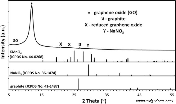
GO 샘플의 상 분석(그림 1)은 미량의 흑연, 질산나트륨 및 환원된 형태의 산화그래핀에서 나오는 불순물의 존재를 나타냅니다.
<사진>
GO 분말의 X선 회절 패턴. GO 샘플의 상 분석은 미량의 흑연, 질산나트륨 및 환원된 형태의 산화그래핀에서 나오는 불순물의 존재를 보여주었습니다.
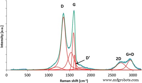
라만 분광법은 산화 그래핀의 구조적 특징에 대한 정보를 제공할 수 있습니다. D 밴드는 구조적 무질서에 기인하는 반면 G 밴드는 탄소 sp
2
의 결합 스트레칭에 기인합니다. 원자 [42]. 추가 밴드(D', 2D 및 D + G 포함)는 탄소 재료의 흑연 구조에 존재하는 결함으로 인해 발생합니다. 나D /나G 비율(D 및 G 밴드의 강도에서 계산됨)은 탄소 재료의 흑연 구조의 무질서를 특성화하는 데 사용할 수 있습니다. 그림 2에서 볼 수 있듯이 GO는 흑연 분말의 산화 과정에서 형성되는 구조에 많은 관능기가 존재하여 매우 무질서한 구조를 가지고 있다. D 밴드의 위치는 1351cm
−1
입니다. 및 G 밴드 1590cm
−1
; 나D /나G 비율은 1.15입니다.
<그림>
D, G, D', 2D 및 D + G 밴드의 제안된 디콘볼루션이 있는 산화 그래핀의 라만 스펙트럼. GO는 흑연 분말의 산화 과정에서 형성되는 구조에 많은 관능기로 인해 매우 무질서한 구조를 가지고 있습니다. D 밴드의 위치는 1351cm
−1
입니다. 및 G 밴드 1590cm
−1
; 나D /나G 비율은 1.15입니다.
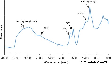
ATR 모드에서 수집된 산화 그래핀의 FT-IR 스펙트럼은 GO가 구조에 많은 작용기를 가지고 있음을 보여주었습니다. 가장 눈에 띄는 피크는 ~ 3500cm
−1
에서 관찰할 수 있습니다. , 주로 물과 수산기에 할당됩니다(그림 3). 약 1080cm
−1
의 매우 집중적인 피크 하이드록실 그룹에 기인할 수도 있습니다. 피크 약 1600cm
−1
일반적으로 흑연 탄소에 존재하는 C=C 결합에 할당됩니다. 그러나 우리의 이전 XPS 연구는 그래핀 옥사이드에 몇 개의 C=C 결합이 있음을 보여줍니다[43]. 따라서 우리는 이 피크를 산화 그래핀에 여전히 존재하는 대부분의 물에 기인합니다. FT-IR 스펙트럼에서 관찰된 다른 피크는 GO가 C=O 결합을 포함하는 그룹(주로 카르복실 그룹)이 풍부함을 보여줍니다. 피크는 약 1720cm
-1
입니다. , 눈에 보이는 피크가 약 1200cm인 에폭시(C–O–C)
−1
, 및 C–H 결합(2800cm 주변에서 피크
−1
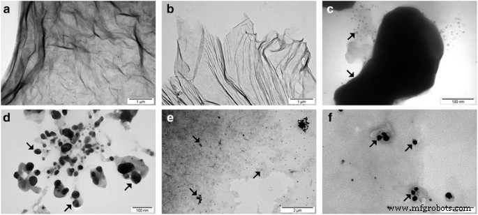
). FT-IR 분석은 히드록실, 카르복실, 에폭시 및 카르보닐 그룹도 확인된 산화 그래핀에 대해 수행된 XPS 측정과 잘 일치합니다[44]. 10분 초음파 균질화 후 GO와 GO를 비교하고(그림 4 및 5), 유사하게 10분 초음파 균질화 후 Ag-NP와 Ag-NP를 비교했습니다(그림 4 및 6). 화합물 형태의 변화를 피하기 위해 모든 화합물을 액체 질소로 빠르게 냉각시키고 동결 건조기에서 건조시켰다. 그림 5a, b는 GO 플레이크를 나타내고 그림 5c, d는 부분 접힘 및 조각화를 겪는 GO 플레이크에 대한 초음파의 영향을 보여줍니다. 그림 6은 재료 형태의 변화가 보이는 Ag-NPs에 대해서도 유사한 효과를 보여줍니다. 그림 6a, b는 Ag-NPs의 현탁액을 안정화하는 데 사용된 건조된 폴리(비닐 알코올)을 보여줍니다. 초음파 균질화의 파괴적인 효과는 폴리비닐알코올 구조가 작은 구형 구멍을 가진 긴 불균일한 부분으로 분해됨에 따라 두드러졌습니다(그림 6c, d).
<그림>
GO에 존재하는 작용기 할당이 제안된 산화 그래핀의 FT-IR(ATR, 감쇠 전반사) 스펙트럼. 가장 주목할만한 피크는 ~ 3500cm
−1
에서 관찰되었습니다. , (물 및 수산기에 기인), ~ 1080cm
−1
(수산기), ~ 1600cm
−1
(흑연 탄소에 존재하는 C=C 결합에 할당됨). FT-IR 스펙트럼에서 관찰된 다른 피크는 GO가 C=O(주로 카르복실기)를 포함하는 그룹이 풍부함을 보여주며 피크는 약 1720cm
-1
입니다. , 눈에 보이는 피크가 약 1200cm인 에폭시(C–O–C)
−1
, 및 C–H 결합(2800cm 주변에서 피크
−1
)
<그림>
응집된 GO 플레이크의 TEM 이미지(a ), 초음파 처리 후 GO 플레이크(b ), 응집된 Ag-NPs(c ), 초음파 처리 후 Ag-NPs(d ) 및 GO-Ag(e , f ). 초음파 처리 후 평균 GO 입자 크기의 감소는 GO 플레이크의 조각 모음 또는 접힘에 의해 발생했습니다. 초음파 처리 후 평균 Ag 크기의 감소는 Ag 덩어리의 조각 모음으로 인해 발생했습니다. 참고:화살표는 Ag-NPs/응집체를 가리킵니다.
<그림>
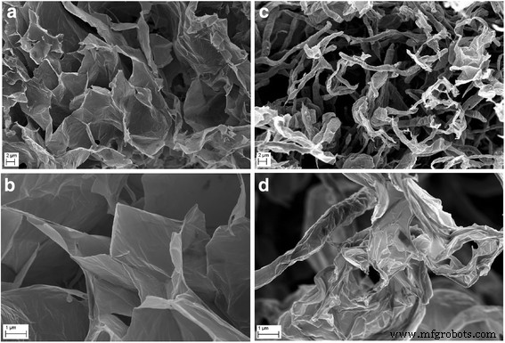
동결건조된 GO 플레이크의 형태 비교(a , b ) 및 초음파 처리 후 GO 플레이크(c , d ) 주사 전자 현미경을 사용하여. 초음파 처리 후 평균 GO 입자 크기의 감소는 GO 플레이크의 조각 모음 또는 접힘으로 인해 발생했습니다.
<그림>
동결건조된 Ag-NP 혼합물의 형태 비교(a , b ) 및 초음파 처리 후 Ag-NP 혼합물(c , d ) 주사 전자 현미경 사용
평균 크기 및 제타 전위
수성 현탁액의 평균 입자/응집체 크기의 결과는 표 2에 나와 있습니다. 평균 크기의 분석은 초음파 균질화를 거치지 않은 농축 현탁액(받은 그대로)과 희석 현탁액에 대해 수행되었습니다. 테스트 전에 희석된 현탁액은 초음파 균질화에 적용되었으며, 균질화 매개변수는 나노물질 층(Ag-NPs, GO)으로 호일을 초음파 코팅하는 동안 사용된 것과 동일합니다. Ag-NP 현탁액의 경우 초음파의 작용으로 평균 입자 크기가 80nm에서 218nm로 증가했습니다. 현탁액에서 초음파 균질화 후 평균 입자 크기가 증가하는 주요 원인은 (Ag-NP 응집 과정을 제외하고) 현탁액 안정화에 사용된 폴리(비닐 알코올)로 Ag-NP가 유입되었기 때문입니다. The large standard deviation of the Ag-NP sample homogenized by ultrasound resulted from the presence of both loose Ag-NPs and Ag-NPs driven into the poly(vinyl alcohol) in the suspension. In the case of GO suspension, the average particle size of the sample subject to ultrasonic homogenization was 263 nm and was ca 7.7 times smaller than the average particle size of the sample that was not subject to homogenization. The obtained results are convergent with the SEM tests (Fig. 5), which show the destructive effect of ultrasounds on GO flakes. The decrease of the average GO particle size was caused by defragmentation or folding of the GO flakes. However, it should be emphasized that the results of the average particle size of GO suspension samples involve an error related to the nanomaterial shape. The results obtained by the DLS method are a hydrodynamic average that is calculated based on the shape of a sphere that has the same diffusion coefficient as the measured particles; however, the shape of GO was flakes, which was confirmed by SEM images.
Test results of the zeta potential analysis of samples are provided in Table 3. The zeta potential of Ag-NPs in a water suspension was merely − 5.9 mV, which resulted in a lack of electrostatic stability of the sample. The sample of Ag-NP suspension was stabilized sterically by preserving Ag-NP distances through poly(vinyl alcohol) addition, which prevented agglomeration/aggregation of Ag-NPs. The zeta potential of the GO suspension sample, in turn, was − 41 mV, which gave a moderate electrostatic stability to the sample. A moderate electrostatic stability of a sample is characterized by slow sedimentation with virtually negligible change of particle size in the period of declared fitness of the suspension. The zeta potential result of the mixture of Ag-NPs and GO was − 7.1 mV, which potentially means that during the action of the ultrasounds, the GO flakes were coated by poly(vinyl alcohol) and Ag-NPs. The obtained zeta potential result of the mixture of Ag-NPs and GO sample in the water suspension implied that electrostatic stability was not present.
Foil Characteristics
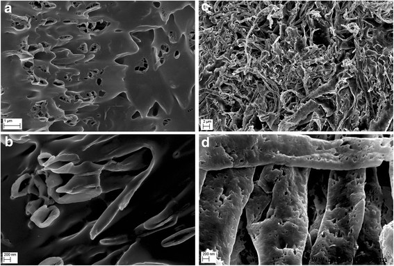
In order to determine the morphology of the created layers, four types of foil samples were compared (Fig. 7):pure polyurethane foil (A, B), GO-coated polyurethane foil (C, D), Ag-NP-coated polyurethane foil (E, F), and GO-Ag mixture-coated polyurethane foil (G, H).
Scanning electron microscopy images of a , b non-coated polyurethane foil with a smooth surface with single impurities; ㄷ , d foil coated with GO, the broken-down GO flakes deposited on the polyurethane foil surface; 이 , f foil coated with Ag-NPs on which grid structures composed of polyvinyl alcohol and Ag-NPs are observed; and g , h foil coated with GO-Ag, which was mixed under the influence of ultrasounds and deposited on the foil surface
Figure 7a, b shows an uncoated polyurethane foil with a smooth surface with single impurities. In Fig. 4c, d, the broken-down GO flakes deposited on the polyurethane foil surface are noticeable. Figure 7e, f shows the foil surface coated with Ag-NPs on which grid structures composed of polyvinyl alcohol and Ag-NPs are observable. Figure 7g, h presents a mixture of GO-Ag composition, which was mixed under the influence of ultrasounds and deposited on the foil surface.
AFM Analysis and Surface Free Energy
AFM and LFM were used to complement the information about the surface morphology investigated by SEM. The investigation confirmed evolution of surface morphology by sonication of the polyvinyl alcohol with Ag-NP, GO, and GO-Ag NP solutions on the foils. Pure polyurethane foil was used as a reference foil in relation to foils coated by the ultrasonic method. The images in Fig. 8 are the AFM phase contrast images made in AC; additionally, cross sections of the GO flakes are attached under the corresponding images. Figure 8a is an image of pure polyurethane film; Fig. 7b depicts the Ag-NP-coated film, where characteristic and homogeneous grid structures are observable, being similar to those in Fig. 8e, f. Figure 8c, d shows GO-Ag-NP-coated film. Figure 8e shows the surface of the foil almost entirely covered with GO flakes; the phase contrast image helps to observe these two phases, the darker area is GO and the lighter area is polymer foil. It was noticed that the morphology of the foils has changed after Ag-NP coating comparing to the not-coated foils. The GO-Ag-NP coating differs from the previous one because it contains also small amounts of GO flakes seen as small black spots on the image, as it was mentioned earlier. Figure 8f depicts magnification of one GO flake made in LFM. The reduced friction confirms that it is, in fact, a GO flake.
AFM phase contrast images and cross sections topographic images of graphene flakes:a non-coated foil polyurethane foil; ㄴ Ag-NPs coated foil where characteristic and homogeneous grid structures are observed; ㄷ , d GO-Ag-coated foil; 이 GO-coated foil, the surface of the foil almost entirely covered with GO flakes; the phase contrast image helps to observe these two phases, the darker area is GO and the lighter area is polymer foil; 에 LFM image of graphene flake. Red marks, area of cross section
The polar component for the GO-coated foil increased in relation to pure foil, from 2.3 ± 0.6 to 68.9 ± 2.8 mJ/m
2
, while the dispersion component decreased from 34.4 ± 1.3 to 8.2 ± 1.2 mJ/m
2
. SFE increased from 36.7 ± 1.4 to 77.0 ± 3.4 mJ/m
2
. A similar effect was not observed on foil surfaces coated with Ag-NPs and GO-Ag mixture. SFE of foil samples coated with Ag-NPs and GO-Ag mixture does not differ statistically (Table 4).
Antibacterial Properties
The antibacterial activity of the different foils coated with GO, Ag-NPs, and GO-Ag were tested with E . coli , S . 구균 , S . epidermidis , 및 C . albicans. Results showed that after co-incubation with bacteria at 37 °C for 24 h, foils inhibited the growth of all tested microorganisms but to various degrees. The maximum antibacterial effect against all tested microorganisms was with foil coated with the GO-Ag nanocomposite. The bacterial growth of the cells treated with foils coated with GO and Ag-NPs was slightly lower than that of cells in the control group whereas the growth of bacterial cells treated with foils coated with GO-Ag was greatly inhibited, 88.6% of E . coli , 79.6% of S . 구균 , 76.5% of S . epidermidis , and 77.5% of C . albicans (Figs. 9, 10, and 11).
Antimicrobial properties of GO-Ag coated foils. The growth of E . coli (b , ㄷ ), S . 구균 (ㄷ , d ), S . epidermidis (이 , f ), and C . albicans (g –i ) colonies is reduced after co-incubation with GO-Ag-coated foils at 37 °C for 24 h. 아 Representative agar plate with GO-Ag-coated foils. Notes:Black arrows point to GO-Ag coated foils; arrowheads point to colonies of microorganisms
Morphology of microorganisms after co-incubation with GO-Ag-coated foils. Scanning electron microscopy images of bacteria and yeast in the control foils (a , ㄷ , e , 지 ) and foils coated with GO-Ag (b , d , f , h ) after incubation at 37 °C for 24 h. 이 . coli (아 , b ), S . 구균 (ㄷ , d ), S . epidermidis (이 , f ), and C . albicans (g , h ) show decreased growth and deformed morphology after co-incubation with GO-Ag-coated foils
Foils coated with nanomaterials decrease E . coli , S . 구균 , S . epidermidis , 및 C . albicans viability. Viability of bacteria and yeast after incubation on foils coated with Ag-NPs, GO, and GO-Ag at 37 °C for 24 h was assessed with Presto Blue assay. C control group (foil without nanoparticles), Ag foil coated with silver nanoparticles, GO foil coated with graphene oxide, GO-Ag foil coated with composite of graphene oxide and silver nanoparticles. Note:The columns with different letters (a–d) indicate significant differences between the concentrations (P = 0.001); error bars are standard deviations
Membrane Integrity
In cases where the cell membrane is damaged, intracellular LDH molecules could be released into the culture medium. The LDH level outside the cells demonstrates the cell membrane integrity. Foils coated with GO, Ag-NPs, and GO-Ag disrupted cell membrane functionality and integrity with significant differences between control groups and the Ag-NPs and GO-Ag groups (Fig. 12). The highest disruption of cell membranes was observed in the GO-Ag groups, 66.3% of E . coli , 59.4% of S . 구균 , 54.8% of S . epidermidis , and 48.5% of C . albicans .
Foils coated with nanomaterials decreased E . coli , S . 구균 , S . epidermidis , 및 C . albicans membrane integrity. Membrane integrity of bacteria and yeast after incubation on foils coated with Ag-NPs, GO, and GO-Ag at 37 °C for 24 h was assessed with LDH assay. C control group (foil without nanoparticles), Ag foil coated with silver nanoparticles, GO foil coated with graphene oxide, GO-Ag foil coated with composite of graphene oxide and silver nanoparticles. Note:The columns with different letters (a–c) indicate significant differences between the concentrations (P = 0.000); error bars are standard deviations
ROS Production
Low levels (or optimum levels) of ROS play an important role in signaling pathways. However, when ROS production increases and overwhelms the cellular antioxidant capacity, it can induce macromolecular damage (by reacting with DNA, proteins, and lipids) and disrupt thiol redox circuits. Foils coated with Ag-NPs and GO-Ag (P < 0.05) increased the ROS production of all tested microorganisms compared to the control group. Foils coated with GO only increased the ROS production of C . albicans . The highest ROS production was observed in the GO-Ag group (Fig. 13).
Effect of foils coated with nanomaterials on the production of reactive oxygen species. 이 . coli , S . 구균 , S . epidermidis , 및 C . albicans were cultured on foils coated with Ag-NPs, GO, and GO-Ag at 37 °C for 24 h. Production of reactive oxygen species was assessed with DCFDA, Cellular Reactive Oxygen Species Detection Assay Kit. C control group (foil without nanoparticles), Ag foil coated with silver nanoparticles, GO foil coated with graphene oxide, GO-Ag foil coated with composite of graphene oxide and silver nanoparticles. Note:The columns with different letters (a–d) indicate significant differences between the concentrations (P = 0.000); error bars are standard deviations
토론
The discovery of antibiotics, natural products produced by microorganisms that are able to prevent the growth of bacteria and thus cure infectious diseases, revolutionized medical therapy; however, the overuse and misuse of antibiotics have been key factors contributing to antibiotic resistance. Now, the era of antibiotics is coming to an end, and new antibacterial agents are needed. In recent years, studies have reported nanoparticles as a promising alternative to antibacterial reagents because of their antibacterial activity in several biomedical applications [19, 45]. Nanoparticles can be an effective way to control many pathogenic and antibiotic-resistant microorganisms. Among many metal nanoparticles, Ag-NPs have been intensely studied because of the distinct properties of their antibacterial activity [7]. Ag-NPs have been proved effective against over 650 microorganisms including bacteria (both gram-positive and negative), fungi, and viruses; however, the precise mechanism of antimicrobial action is not understood completely [46]. Ag-NP exposure to microorganisms could cause adhesion of nanoparticles to the peptidoglycan and the cell membrane [47], penetration inside the cell [48], induction of ROS [49], and damaging of intracellular structures [50]. However, bare Ag-NPs aggregate when they come into contact with bacteria; thus, they lose their active surface area and show weaker antibacterial activity [51]. To overcome this problem, nanocomposites composed of graphenic materials and Ag-NPs or other metal nanoparticles could be fabricated. GO with oxygen-containing functional groups is water soluble and therefore more biocompatible than pristine graphene. As a result, Ag-based GO nanocomposites may be used as antibacterial agents. However, the information about antimicrobial properties of graphene-based composites is limited, and mechanisms of toxicity or lack of toxicity are not fully explained.
The aim of this work was to study the action of graphene oxide-based nanocomposites decorated with Ag nanoparticles on S . 구균 , S . epidermidis , E . coli , 및 C . albicans 성장; membrane integrity; and ROS production. After co-incubation with the bacterial and yeast cells for 24 h, foils coated with GO-Ag nanocomposite inhibited the growth of all tested microorganisms with varying degrees, 88.6% of E . coli , 79.6% of S . 구균 , 76.5% of S . epidermidis , and 77.5% of C . albicans . This action is most probably due to an increase in cell membrane and wall penetration by the nanoparticles. Some researchers suggest that the antimicrobial activity of graphene-based nanocomposites may be due to the disruption of cell membrane integrity and oxidative stress [52].
Foils coated with GO, Ag-NPs, and GO-Ag disrupted cell membrane functionality and integrity with significant differences between the control group and the Ag-NPs and GO-Ag groups. The highest disruption of cell membranes was observed in the GO-Ag groups, 66.3% of E . coli , 59.4% of S . 구균 , 54.8% of S . epidermidis , and 48.5% of C . albicans . However, foils coated with bare Ag-NPs also disrupted cell membranes. It has been proposed that Ag-NPs are able to interact with bacterial membranes by increasing permeability and changing the structure of membranes, which finally leads to cell death [50]. Ag-NPs can cause direct damage to the bacterial cell membrane. Bacteria may be killed by the combined bactericidal effects of the released Ag
+
ions and Ag nanoparticles. Additionally, the antimicrobial potential of Ag-NPs is also influenced by the thickness of the cell wall of the microorganisms [53]. The wall of gram-positive cells contains a thick layer (20–80 nm) of peptidoglycan, which is attached to teichoic acids. In gram-negative bacteria, the cell wall comprises a thin (7–8 nm) peptidoglycan layer and contains an outer membrane. The thicker peptidoglycan layer in gram-positive bacteria, such as S . 구균 그리고 S . epidermidis , may explain why these bacteria are more resistant to the antibacterial effects of GO-Ag.
Many studies have sought to establish a mechanism of action of antibacterial activity exhibited by silver in both its colloidal and ionic form. A disruption of membrane functionality from an interaction between released Ag
+
ions and the cell membrane and extensive cell membrane damage caused by the formation of ROS ultimately causes damage to the cell due to oxidative stress. Additionally, Ag
+
ions could cause dysfunction of the respiratory electron transport chain by uncoupling it from oxidative phosphorylation by inhibiting respiratory chain enzymes [54]. Foils coated with Ag-NPs and GO-Ag increased the ROS production of all tested microorganisms compared to the control group. The biological targets are DNA, RNA, proteins, and lipids. Lipids are one major target during oxidative stress. Free radicals can directly attack polyunsaturated fatty acids in bacterial and yeast membranes and activate peroxidation of lipids. A fundamental effect of lipid peroxidation is a decrease in membrane fluidity, which can significantly disrupt membrane-bound proteins. DNA is also a main target. Mechanisms of DNA damage involve abstractions and addition reactions by free radicals leading to carbon-centered sugar radicals and OH- or H-adduct radicals of heterocyclic bases. The sugar moieties producing single- and double-strand breaks in the backbone, adducts of base and sugar groups, and cross-links to other molecules can block replication. Foils coated with GO increased the ROS production at very low levels. However, Hu et al. [55] demonstrated that GO had a detrimental effect on E . coli due to decreased production of ATP and increased ROS production. Zhao et al. [56] reported the antibacterial activity of GO and reduced GO. Also, Gurunathan et al. [57] presented that GO and reduced GO showed significant antibacterial activity in a concentration- and time-dependent manner. Their results demonstrated that oxidative stress is a key mechanism for the antibacterial activity of GO and reduced GO through ROS generation. Nanda et al. [53] reported the effect of cystamine-conjugated GO against E . coli , S . typhimurium , E . faecalis , and B . subtilis with ROS production and high antibacterial activity.
Kurantowicz et al. [20] confirmed that bacteria could adhere to the GO surface, which results in the highest antibacterial activity. GO is characterized by a high degree of oxygenated functional groups:carbonyl, carboxylate, and hydroxyl. We hypothesize that these groups can be attractive groups for bacterial and yeast attachment. These groups are present on a large range of nutrients (amino acids, fatty acids) which are commonly recognized by microorganisms. In the present study, foils coated with GO induced membrane disruption and ROS production at a lower level than the Ag-NP and Ag-GO groups; however, cell viability was decreased, which is likely connected to the smaller active surface of GO after ultrasonic modifications.
결론
Ag-NPs, GO, and Ag-GO nanocomposites demonstrated the antibacterial activity that is stronger against gram-negative bacteria (E . coli ) versus gram-positive bacteria (S . 구균 그리고 S . epidermidis ) and yeast (C . albicans ). The results showed that the decoration of GO with Ag-NPs promotes a synergistic effect and reduces dramatically the concentrations required to inhibit all tested bacterial and yeast strains. The antimicrobial potential of Ag-GO is also influenced by the thickness of the cell wall of the microorganisms. The thicker peptidoglycan layer in gram-positive bacteria, such as S . 구균 그리고 S . epidermidis , may explain why these bacteria are more resistant to the antibacterial effects of GO-Ag. A disruption of membrane functionality from an interaction between released Ag nanoparticles/Ag
+
ions and the cell membrane and extensive cell membrane damage caused by the formation of ROS ultimately caused damage to the cell due to oxidative stress. In order to explain the mechanism of ROS production, additional studies are needed. Our research indicates the potential applicability of GO-Ag as an antimicrobial agent.
약어
AFM:
원자력 현미경
Ag-NPs:
은 나노입자
DLS:
동적 광산란
이동:
산화 그래핀
GO-Ag:
Graphene oxide decorated with silver nanoparticles