향상된 약물 전달을 위한 후두암 세포 및 면역 세포 내 고분자 나노입자의 세포 내재화에 대한 정량적 평가
초록
폴리(락트산-코-글리콜산)(PLGA) 기반 나노의약품의 임상 번역은 부분적으로는 식세포에 의한 비특이적 식세포 작용으로 인한 낮은 전달 효율로 인해 제한적입니다. 암세포와 면역 세포 사이의 나노입자 상호작용을 이해하는 것은 여전히 어려운 일입니다. 이 연구에서는 단일 배양 또는 공동 배양 시스템에서 단핵구/대식세포가 있거나 없는 후두 암종 세포에 대한 형광 PLGA 입자(100 nm, 500 nm 및 1 µm)의 세포 내재화에 대한 정량적 조사가 먼저 수행되었습니다. 5~20µg/mL 농도의 PLGA 입자는 500nm 및 20µg/mL의 1µm PLGA 입자를 제외하고는 세포 생존력을 약간 감소시키는 것을 제외하고 우수한 생체 적합성을 나타냅니다. 현미경 관찰을 통해 세 가지 크기의 입자가 모두 암세포와 대식세포에 의해 효과적으로 섭취된다는 사실이 밝혀졌습니다. 그러나 정량적 형광 검사는 암 세포의 흡수 지수(대식세포당으로 정규화된 암세포당 평균 세포내 입자 형광)가 단일 배양(1.35–1.05, 100nm, 500nm, 1µm 입자의 경우 각각 1.50–0.59 및 1.4–0.47). 유세포 분석을 사용한 정량 분석은 공동 배양에서 암세포의 흡수 지수 감소를 추가로 확인했지만 대식세포당 입자 수는 더 높았습니다. 또한 대식세포의 융합을 통한 다핵 거대 세포의 형성이 PLGA 처리 후 증가하는 것으로 밝혀졌으며, 이는 종양 약물 전달을 위한 잠재적인 접근으로 추가로 이용될 수 있습니다. 전반적으로, 이러한 발견은 나노입자-면역암 세포의 상호작용에 대한 새로운 통찰력을 제공하며, 이는 후두암 치료를 위한 PLGA 기반 나노운반체의 적용을 촉진할 수 있습니다.
소개
암은 2018년에 약 천만 건의 새로운 사망 사례가 보고된 전 세계 사망의 주요 원인 중 하나입니다[1]. 후두암종(암 세포는 후두에서 발생)은 두경부 편평 세포 암종(HNSCC)의 두 번째로 흔한 악성 종양으로 2018년에 약 180,000건의 신규 사례와 95,000명의 사망을 차지했습니다[2]. 현재 표적 약물은 수술, 방사선 요법, 화학 요법과 같은 전통적인 치료법이 도입한 문제에 대한 선택적 치료법으로 개발되고 있습니다[3]. 예를 들어 새로운 약물 나노운반체(i.e.)를 개발하여 약물 표적화 및 효능을 개선하고 원치 않는 부작용을 줄이기 위한 과학적 노력이 가속화되었습니다. , 미세바늘), 맞춤형 항암제 및 치료용 항체 표적 전달 시스템[4].
나노캐리어는 시스플라틴, 파클리탁셀, 도세탁셀과 같은 항암제를 로딩하여 수용성, 생체이용률 및 안정성을 개선하여 약물 전달 및 효능을 개선하는 데 널리 활용되었습니다[3, 4]. 일반적으로 나노입자(NP) 기반 약물 전달은 광범위한 다른 물질(예: , 단백질, 항체, 백신, 핵산)을 동물 모델과 환자 모두에서 신체의 특정 부위에 전달합니다[4, 5]. 그러나 소위 EPR(Enhanced Permeability and Retention) 효과로 인해 다양한 유형의 암에 따라 표적화 효율성이 크게 달라지게 되었습니다[6]. 최근의 증거에 따르면 나노 약물 운반체(여기서는 금 나노입자)가 종양에 침착되는 것은 물질/NP가 세포를 가로질러 한쪽에서 다른 쪽으로 이동하는 생물학적 세포간 수송의 한 유형인 세포전이 과정[7]에 우선적으로 의존합니다. endocytosis, vesicular transfer 및 exocytosis의 과정을 포함하는 다른 것. 그러나 표적 약물 전달의 메커니즘에 관한 상반된 결과는 시험관 내 세포 배양에서 생체 외 조직 배양에 이르는 다양한 실험 전략을 사용하여 나노 약물 또는 NP와의 세포 상호 작용의 기초를 이해하는 데 매우 중요하다는 점을 강조해야 합니다. 및 생체 내 동물 연구.
리포솜, 알부민 나노입자(NP), 실리카 나노입자, 폴리(락트산-코-글리콜산)(PLGA)과 같은 수많은 나노운반체는 후두암을 비롯한 다양한 유형의 암을 치료하기 위해 임상적으로 사용되었습니다[4]. 미셀 및 PLGA와 같은 고분자 기반 나노입자는 간단한 혼합 또는 공유 결합을 통한 나노 약물의 다양한 제형, 우수한 자기 조립 능력, 높은 약물 로딩 능력 및 생체 적합성으로 인해 생물 의학 응용 분야에서 큰 잠재력을 가지고 있습니다 [8, 9 ]. 예를 들어, 폴리에틸렌 글리콜(PEG) 코팅된 PLGA 나노캐리어에는 독소루비신과 인도시아닌 그린이 포함되어 있어 유방암에 대한 화학 광열 요법을 시너지 효과를 낼 수 있습니다[10]. 또한 시험관 내 및 생체 내 연구 모두에서 시스플라틴이 장착된 미셀이 HNSCC 동소 종양(i.e.)에 대해 우수한 항암 활성을 나타내는 것으로 나타났습니다. , SAS-L1 및 HSC-2) [11]. PLGA 및 미셀과 같은 여러 고분자 기반 나노 약물 운반체가 임상 사용을 위해 승인되었거나 임상 시험에서 평가되고 있지만[4, 6], 더 많은 고분자 나노 약물이 서로 다른 두경부암 세포에 대해 전임상 조사 중입니다. 동물의 계통 및 이종이식 종양 모델 [11,12,13,14].
과학적 증거는 나노 약물 운반체에 대한 숙주 면역학적 반응의 중요성을 강조하고 있습니다. 나노입자가 일단 체내에 들어가면 면역계에 의해 불가피하게 인식되기 때문입니다. 대식세포는 알레르겐, 미생물 및 이물질(예: , nanocarriers) 식균 작용 및 그에 따른 면역 반응 프라이밍을 통해. 새로 설계된 나노운반체의 대부분은 간의 쿠퍼 세포와 비장의 적혈구 대식세포와 같은 단핵 식세포 시스템(MPS)에서 NP가 효율적으로 축적되기 때문에 생체 내 특정 질병 부위 또는 종양으로의 표적 전달에 실패했습니다. [15]. 따라서 단핵구 및 대식세포와 같은 면역 세포에 의한 NP의 세포 흡수 메커니즘을 이해하는 것은 관련 조직 및 생물학적 유체에서 나노운반체의 수명을 결정하기 때문에 중요합니다. 따라서 새로운 시험관 내 세포 공동 배양 시스템은 생체 내 조건을 더 잘 반영할 수 있는 보다 의미 있는 결과를 달성하려는 요구가 증가함에 따라 나노의학 및 독성학 분야에서 점차 주목을 받고 있습니다[16]. 실제로, 공동 배양 시스템은 건강하고 질병에 걸린 조직 상태를 모방하는 현실적인 상황을 나타내는 것으로 입증되었으며[17] NP 세포 흡수 및 약물 흡수 연구[18,19,20,21]에 안정적으로 활용되었습니다. 공동 배양 암 세포 및 면역 세포 모델의 활용은 일반적으로 이러한 나노 물질의 세포 내로의 흡수 경로 및 메커니즘을 조사하는 데 적합한 플랫폼을 제공하며, 이는 동시에 NP 식균 작용을 감소시키면서 암세포를 더 잘 표적으로 하는 나노운반체의 설계를 촉진할 수 있습니다. 따라서 면역 세포가 있는 상태에서 암세포에서 NP의 흡수 효율과 운명을 결정하는 것이 매우 중요합니다. 대부분의 나노운반체는 EPR 효과를 이용하고 혈액 순환을 연장하기 위해 50-200nm의 직경을 갖도록 설계되었으며, 더 큰 NP(> 500nm)는 MPS에 의해 효율적으로 제거되는 것으로 보고되었습니다[6]. 따라서 다양한 크기(100, 500, 1000nm)의 FDA 승인 나노 운반체인 PLGA를 선택하여 UM-SCC-17A(고전적인 후두 편평 암종 세포주)[22] 및 THP- 1(인간 급성 단핵구 세포주) 세포. 여러 시험관 내 연구에서 단일 배양에서 HNSCC 암세포를 죽이는 약물을 전달하기 위해 PLGA 나노운반체를 적용했습니다[12, 13, 23]. 이것은 면역 세포와 후두암 세포가 동시에 NP를 흡수하는 메커니즘을 조사한 첫 번째 연구입니다. HNSCC 치료를 위한 새로운 안전 설계 나노의약 개발을 위한 기본 기반을 제공할 수 있는 다양한 크기의 PLGA를 사용하여 공동 배양 모델.
자료 및 방법
자료
100nm, 500nm, 1000nm를 포함하여 크기가 다른 3개의 상용화된 PLGA 입자(Sigma-Aldrich)가 이 연구에 사용되었습니다. 모든 입자에는 460nm 및 500nm의 광학 여기 및 방출(ex/em) 파장이 있는 녹색 형광단이 로드되었습니다. 모든 입자는 분말 형태로 제공되었으며 추가 사용을 위해 최종 농도가 10mg/mL인 증류수에 현탁되었습니다. 3개 입자의 유체역학적 직경 및 제타 전위는 Malvern Zeta Sizer Nano 기기(Malvern Instruments Ltd., Malvern, UK)로 수행된 동적 광산란(DLS)에 의해 수행되었습니다. 각 입자의 스톡 현탁액은 DLS 측정을 위해 80μl 증류수에 1:100 희석되었습니다. 제조사의 지시에 따라 각 입자에 대해 3번의 반복 측정이 이루어졌습니다.
UM-SCC-17A 및 THP-1의 세포 배양 및 입자 노출
미국 Sigma-Aldrich 및 Shanghai Hengya Biotechnology Company(중국 상하이)에서 각각 구입한 인간 후두 암종 세포주 UM-SCC-17A 및 인간 단핵구/대식세포 세포주 THP-1을 사용하여 시험관 내 모델을 구축했습니다. 이 연구. UM-SCC-17A 세포와 THP-1 세포는 37℃에서 10% FBS(Gibco, Germany) 및 1% Penicillin-Streptomycin 용액(Gibco, Germany)이 보충된 DMEM[22] 또는 RPMI-1640 세포 배양 배지에서 배양되었습니다. °C, 5% CO2. THP-1 세포는 대식세포로 분화하기 위해 세포 시딩 전에 72시간 동안 100nM Phorbol 12-myristate 13-acetate(PMA)(Sigma, USA) 용액에 노출되었습니다. 두 세포 모두 3일마다 0.5% 트립신-EDTA를 사용하여 계대되었으며 세포의 건강을 확인하기 위해 매일 세포 형태를 확인했습니다.
세포를 0.1 × 10
6
의 밀도로 24웰 플레이트에 접종했습니다. WST-1 및 LDH 분석용 단일 배양 세포의 경우 세포/웰, 또는 형광 현미경 검사용 24웰 플레이트의 멸균 유리 커버슬립. 공동 배양 모델의 경우, UM-SCC-17A 세포를 먼저 24웰 플레이트에 50,000/웰의 밀도로 밤새 접종한 다음 50,000/웰 THP-1 세포를 첨가했습니다. 입자를 단일 배양된 UM-SCC-17A 및 THP-1 세포의 경우 각각의 세포 배양 배지 또는 공동 배양된 세포의 경우 1:1 혼합 세포 배양 배지 중 하나에 500μl를 현탁하고 최종 24시간 동안 세포 샘플에 노출시켰다. WST-1 및 LDH 분석의 경우 5, 10 및 20µg/mL 농도 또는 형광 현미경 검사법의 경우 10µg/mL 농도
FACS 측정을 위해, 세포를 250,000개 세포/웰 또는 공동-배양된 모델에 대해 각각 125,000개 세포/웰의 밀도로 이전에 기재된 것과 동일한 방식으로 12-웰 플레이트에 접종하였다. 세포를 1mL 세포 배양 배지에서 밤새 성장시키고 24시간 동안 10μg/mL의 최종 농도에서 PLGA 입자에 노출시켰습니다.
세포 생존 분석
세포 생존율은 제조사의 지침에 따라 세포 증식 시약 WST-1 키트(Roche, Germany)에 의해 결정되었습니다. 간단히 말해서, WST-1 용액은 단일 배양 UM-SCC-17A 또는 THP-1에 대한 각각의 세포 배양 배지 또는 공동 배양된 세포에 대한 1:1 혼합 세포 배양 배지에 1:10 희석되었습니다. 노출 후 상층액을 빼내고 세포를 WST-1 분석의 500μl 작업 용액과 함께 37°C에서 30분 동안 인큐베이션했습니다. 샘플을 수집하고 10분 동안 14,000rpm에서 원심분리하여 입자를 제거했습니다. Infinite
®
를 사용하여 450nm 파장에서 용액의 흡광도(OD 값)를 측정했습니다. F200(미국 테칸). 흡광도 값은 WST-1 working solution만을 포함하는 blank sample의 값을 빼서 보정하였고, 처리되지 않은 control sample과 상대 세포 생존율을 비교하였다.
세포막 누출 분석
LDH(젖산 탈수소효소) 방출은 PLGA 입자에 의한 세포 독성을 결정하기 위해 상용 세포독성 검출 키트(LDH)(Roche, Germany)를 사용하여 측정되었습니다. 노출 24시간 후 세포의 상층액을 수집하고 10분 동안 14,000rpm에서 원심분리하고 200μl 완전 세포 배양 배지에 1:10 희석했습니다. 양성 대조군은 0.2% Triton X-100과 함께 37°C에서 15분 동안 세포를 배양하고 200μl 완전 세포 배양 배지에 1:50으로 희석하여 LDH의 총 방출로 정의했습니다. 샘플을 실온에서 30분 동안 100개의 LDH 분석 작업 용액과 함께 인큐베이션하고 50μl 1% HCl을 사용하여 반응을 중단했습니다. 492 nm 파장에서의 흡광도는 Infinite®F200(Tecan, USA)을 사용하여 측정되었으며, 상대 LDH 농도는 다음 식에 따라 계산되었습니다.
세포는 형광 현미경하에서 입자의 위치를 파악하기 위해 Hoechst 염색에 의해 시각화되었습니다. 입자를 24시간 노출시킨 후 세포를 PBS로 3회 세척하고 실온에서 10분 동안 4% 포름알데히드와 함께 인큐베이션했습니다. 포름알데히드로 고정한 후 세포를 1:1000으로 희석된 Hoechst가 포함된 200µl의 염색 용액과 함께 배양했습니다. 및 30분 동안 PBS 중 1% BSA. 그런 다음, 커버슬립을 거꾸로 유리 슬라이드로 옮기고 시각화를 위해 DAKO 형광 퇴색 방지제 한 방울로 유지했습니다. 세포 형태를 위한 bright field, 세포 핵을 위한 DAPI, 입자를 위한 GFP를 포함한 4개의 광학 채널을 형광 현미경으로 설정했습니다. 각 형광 사진에 대한 입자 채널의 노출 시간을 기록하고 서로 다른 입자에 걸친 형광 강도의 균질화에 사용했으며 ImageJ(https://imagej.nih.gov/ij)에서 무작위로 선택한 영역을 사용하여 형광 강도로 세포 내 입자를 계산했습니다. /). 단일 배양 또는 공동 배양 UM-SCC-17A 세포 간에 서로 다른 입자에 대한 흡수 지수를 비교했습니다. 간단히 말해서, 내재화된 입자의 평균 형광 강도(MFI)는 예를 들어 각 세포 유형에 대해 50개 세포에서 계산되었으며, 이는 총 형광 강도(I총계 ) 특정 영역 및 자가형광(I자동 ) 무입자 영역 방정식 [24, 25]에서 동일한 크기의 영역. 암 세포의 흡수 지수는 단일 배양 또는 공동 배양 모델에서 THP-1 세포의 것으로 정규화된 UM-SCC-17A의 MFI에 의해 결정되었습니다. 계산은 아래 방정식에 따라 수행되었습니다.
3개의 입자의 흡수 능력을 측정하기 위해 형광 활성 세포 분류(FACS)를 수행했습니다. 세포를 PBS로 3회 세척하여 노출 24시간 후 세포 배지와 해리성 입자를 제거하고 200μl 0.5% 트립신-EDTA(Gibco, Germany)와 함께 37°C에서 4분 동안 배양하여 플레이트에서 세포를 제거했습니다. . 그런 다음, 2ml의 완전 세포 배양 배지를 첨가하여 반응을 중지하고, 세포 현탁액을 FACS 측정을 위해 유리관으로 옮기고, 300 × G, 4°C에서 5분 동안 원심분리하였다. 상층액을 부드럽게 배출하고 세포를 PBS 200μl에 현탁하고 얼음에 보존했습니다. 살아있는 세포는 먼저 정방향(FSC-A) 측(SSC-A) 산란을 사용하여 파편과 죽은 세포로부터 게이트되었습니다. 그런 다음, 공동 배양된 세포를 APC 채널 대 FSC-A를 사용하여 분석하여 세포의 크기에 따라 대식세포로부터 UM-SCC-17A 세포를 분리하였다. 각 샘플에 대해 총 30,000개의 세포를 분석하고 각 세포에 대한 평균 형광 강도를 계산하고 서로 다른 입자를 교차하여 정규화했습니다. 한편, 입자를 세포와 24시간 인큐베이션한 후, 세포 배양 상청액, 세척 완충액(세포 표면에 부착된 잔류 입자 제거를 위한 3회 세척) 및 세포(트립신 처리 분해)를 수집하여 마이크로플레이트를 사용하여 형광 강도를 측정하였다. 리더(Infinite®F200, Tecan). 이 접근 방식에 의해 세포에 의해 섭취된 입자의 백분율은 각 그룹에 대해 결정될 수 있습니다. 예를 들어 100nm의 5µg의 용량에서 12개의 웰/플레이트(총 250,000개 세포)의 단일 세포에 약 30,000개의 입자가 노출됩니다. PLGA 입자는 단일 세포에 의해 내부화된 평균 13,000개의 입자를 생성하며 이는 세포에 전달되는 적용된 용량의 약 43%입니다. 이 전달된 백분율은 FACS의 각 유형의 세포에 대한 공동 배양 시스템의 입자 수를 계산하는 데 추가로 사용할 수 있습니다.
세포 융합의 정량화
다핵거대세포(MGC)에 대한 대식세포 융합은 형태학적으로 정상 세포질 내에 2개 이상의 핵을 포함하는 거대 세포로 정의되며, 이는 문헌에 기록된 바와 같이 염색 후 명시야 이미지 및 형광 이미지에서 명확하고 동시적으로 식별될 수 있습니다[26] . 세포 융합의 백분율은 ImageJ(https://imagej.nih.gov/ij/)에서 자동화된 접근 방식으로 결정된 총 세포 수로 정규화된 MGC 핵(수동 계산)의 수로 계산되었습니다.
통계 분석
GraphPad V8.0 소프트웨어(GraphPad Software Inc., San Diego, CA, USA)는 통계 분석 및 결과 시각화에 사용되었습니다. 일원 ANONA에 따라 다중 그룹 결과 또는 두 그룹 결과를 각각 비교하기 위해 Holm-Sidak 방법 또는 t-검정을 수행했습니다. 모든 실험은 독립적인 3중으로 수행되었으며 데이터는 평균 ± 표준편차(STD)로 표시되었습니다. p가 포함된 결과 값 <0.05(*) 및 p <0.01(**)은 유의미한 것으로 간주되었습니다.
결과 및 토론
폴리(락트산-코-글리콜산) 입자의 특성
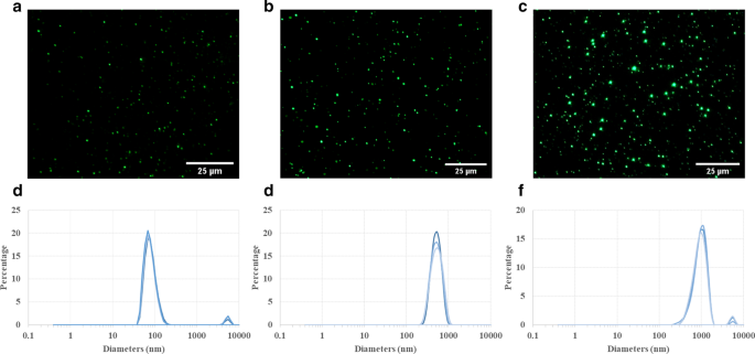
PLGA 입자의 형광 현미경 사진(그림 1a,b,c)은 강력한 형광 강도를 나타내었고 준비 후 14일 동안 형광 신호의 현저한 감소가 없었으며(추가 파일 1:S1), 이는 상대적으로 균일한 크기 분포와 높은 형광성을 나타냅니다. 안정. 공급업체가 명시한 바와 같이 100, 500, 1000nm PLGA 입자는 각각 약 80.6 ± 19.3nm, 542.6 ± 128.3nm, 951.9 ± 237.5nm의 크기를 나타냈습니다. 제타 전위 측정에 따르면 입자의 평균 표면 전하는 0.057, 0.056, 0.0에 대해 0.057, 0.056, 0.0에 대해 0.057, 0.056 및 5의 다분산 지수에서 -20.6 ± 5.3, -17 ± 4.6 및 -16.5 ± 3.5인 것으로 나타났습니다. nm 입자는 각각 높은 단분산 표준을 나타냅니다. 모든 입자를 볼텍싱한 다음 수조에서 5분 동안 초음파 처리하여 입자 응집을 크게 제거했습니다. 그러나 100 및 1000 nm 입자에 해당하는 작은 피크가 직경 약 4-6 µm로 관찰되었는데, 이는 매우 소량(약 3-4%)의 불가피한 입자 응집으로 인해 발생하기 때문입니다.
<그림>
PLGA 입자의 특성화. 형광 현미경 사진은 다양한 크기를 보여줍니다(a 100nm, b 500nm 및 c 1µm)의 PLGA 입자. 이러한 입자는 물 현탁액에서 상대적으로 균일한 입자 크기 분포를 나타내는 Olympus 광학 현미경으로 검출할 수 있습니다. 동적 광산란(DLS) 측정은 100nm(d ), 500(e ), 1μm(f ) PLGA 입자. 100nm 및 1µm PLGA 현탁액에서 입자 응집의 작은 부분(더 작은 피크:3-4%)에도 불구하고 좁은 주 피크로 인해 전체 입자 크기 분포가 상당히 균일합니다.
세포 생존율 및 세포독성 평가
뛰어난 생체 적합성과 생분해성으로 인해 PLGA 입자는 FDA와 유럽 의학청에서 생물 의학 응용 분야에 대한 승인을 받았습니다[27, 28]. 따라서 PLGA 입자는 현재 임상에서 사용되어 왔으며 특정 질병 부위 또는 종양에 약물을 전달하는 나노운반체로서 전임상 연구에 널리 적용됩니다. 그러나, PLGA 입자의 표적화 효율 및 치료 효능은 면역계에 의해 적어도 부분적으로 방해를 받습니다. 예를 들어, 간에서 Kupffer 세포에 의한 높은 입자 식균 작용은 나노 약물 운반체가 종양 부위에 들어가는 것을 크게 제한했습니다[15]. 따라서, 약물 전달의 생체 내 상황을 더 잘 모방하기 위해 암세포, 면역 세포 및 입자 간의 상호 작용을 연구하기 위해 고급 시험관내 모델을 확립하는 것이 매우 중요합니다. UM-SCC-17A는 원발성 후두암 표본에서 분리한 독특한 후두 편평상피암 세포주입니다[29]. 그러나 공동 배양 시스템(예: , 대식세포와 암세포의 동시 배양)은 여전히 불충분하며, 생체 내 반응의 예측 능력을 향상시키기 위해 해결해야 하는 문제입니다. 또한, 입자 크기와 표면 코팅이 동물 모델 및 세포 배양에서 고형 종양, 질병 부위 및 암세포로의 전달 능력에 중요한 역할을 한다는 것이 입증되었습니다[30,31,32,33]. 여기에서 우리는 단일 배양 및 공동 배양 시스템에서 세포 흡수 및 세포 내 분포에 대한 입자 크기의 영향을 조사하기 위해 UM-SCC-17A 암종 세포에 3가지 크기의 PLGA를 적용했습니다.
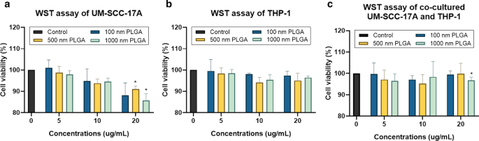
이 연구는 단일 배양 THP-1에 대한 WST-1(4-[3-(4-요오도페닐)-2-(4-니트로페닐)-2H-5-테트라졸리오]-1,3-벤젠 디설포네이트) 방법을 사용하여 세포 생존율을 결정했습니다. 및 UM-SCC-17A 세포, 뿐만 아니라 두 유형의 세포의 공동 배양. 테스트된 모든 농도에서 100 및 500nm PLGA로 처리한 그룹에서 명백한 세포 사멸이 발생하지 않았음에도 불구하고 최고 농도(20µg/mL)에서 500nm 및 1µm PLGA 입자는 단일 배양 UM-SCC-17A에서 세포 생존력을 유의하게 감소시켰습니다. 세포(그림 2a). 예상대로 THP-1 세포 생존율은 5–20µg/mL 농도에서 세 가지 유형의 PLGA와 함께 24시간 배양 후 크게 영향을 받지 않았습니다(생존율이 100%로 간주되는 처리되지 않은 세포와 비교하여 ≥ 95% 생존율). , 그림 2b). 단일 배양 UM-SCC-17A 세포의 결과와 동일하게, 공동 배양 시스템의 세포 생존율은 여기에 사용된 3가지 용량의 100 및 500nm PLGA 입자의 존재에 의해 변경되지 않은 반면, 20개를 처리한 그룹에서는 유의하게 감소했습니다. µg/mL 1 µm PLGA(그림 2c).
<그림>
WST 분석을 활용하여 단일 배양 및 공동 배양 세포에서 세포 생존 능력의 결정. UM-SCC-17A(a ) 및 THP-1 세포(b ) 및 공동 배양된 UM-SCC-17A 및 THP-1 세포(c ) 3가지 크기의 PLGA 입자를 다양한 농도(5~20µg/mL)로 처리했습니다.
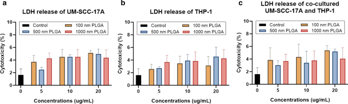
LDH assay를 이용한 입자 세포독성 평가는 세포 외 LDH의 양을 측정하여 세포막의 누출 여부를 판단하는 것이다[9]. 이 세포질 효소의 세포 배양 상청액으로의 방출은 세포막 손상의 특징이며, 이는 돌이킬 수 없는 세포 사멸을 초래합니다. 다양한 용량의 1µm PLGA로 처리된 단일 배양 UM-SCC-17A 세포에서 더 높은 용량으로 LDH 수준이 약간 향상되었음에도 불구하고 20µg/mL의 최고 농도에서도 세포에 대한 뚜렷한 세포 독성은 관찰되지 않았습니다(그림 3a). . 여기에 다양한 용량으로 사용된 3가지 크기의 PLGA 모두 상청액으로 상당한 LDH 방출을 유도할 수 없었으며, 이는 단일 배양 THP-1 세포에 대한 독성 효과가 미미함을 나타내며, 이는 위에서 설명한 세포 생존율 결과와도 매우 일치합니다(그림 2a). . 3b). 공배양 실험에서 농도가 다른 모든 입자는 방출된 LDH 수준 측면에서 두 세포에 대해 우수한 생체적합성을 보였다(그림 3c). 일반적으로 여기에서 사용된 100nm에서 1μm의 넓은 입자 크기 범위는 리포솜, 미셀, 덴드리머, 폴리머 및 미니 셀과 같은 나노운반체(50~200nm)의 일반적인 크기를 포함합니다. 이 범위는 또한 서브미크론 크기의 입자를 포함합니다(500nm 입자는 여전히 NP로 간주될 수 있습니다. 예: 크기가 약 500nm인 입자는 10–100nm 및 마이크론 크기 1µm의 NP와 동일한 제거 경로를 폐에서 가지고 있습니다. 이러한 PLGA 입자 중 어느 것도 단일 배양 및 공동 배양 시스템에서 THP-1 및/또는 UM-SCC-17A 세포에 대해 명백한 세포독성을 나타내지 않았습니다. 단, UM-SCC-에 대한 최고 농도의 500 nm 및 1 µm PLGA 입자는 예외였습니다. 17A 및 공동 배양 세포(그러나 85% 이상의 세포가 생존함)는 약물 전달 시스템에 적용하기에 유리함을 나타냅니다.
<그림>
LDH 분석을 사용하여 단일 배양 및 공동 배양 세포에 대한 세포독성 측정. UM-SCC-17A(a ) 및 THP-1 세포(b ) 및 공동 배양 세포(c )는 PLGA 입자로 처리되었습니다. 단일 배양 및 공동 배양 시스템 모두에서 입자(여기에 사용된 다양한 크기 및 농도 사용) 및 세포의 24시간 배양 후 유의미한 세포 사멸이 관찰되지 않았습니다.
단일 배양 및 공동 배양 시스템에서 PLGA의 세포 흡수 능력
그림 4는 단일 배양 및 공동 배양에서 세포 형태(그림 4a,d,g), 세포 핵 및 100nm NP(그림 4b,e,h) 및 병합된 확대 이미지(그림 4c,f,i)를 보여줍니다. 문화 시스템. 대조군의 미처리 세포에서는 입자가 관찰되지 않았다(추가 파일 1:S2). 거대하고 큰 크기의 100nm PLGA NP 응집체가 대식세포 단일 배양 세포의 세포질에서 관찰되었습니다(그림 4c). 한편, 단일 배양 UM-SCC-17A는 세포막 내부에서 관찰되는 밝은 녹색 형광 신호에 의해 입증된 100nm PLGA의 우수한 흡수 능력을 나타냈습니다(그림 4f). THP-1 또는 UM-SCC-17A 세포에서 PLGA 입자의 세포내 축적과 공동 배양에서 세포외 입자를 더 잘 설명하기 위해 명시야 이미지와 형광 이미지의 오버레이가 추가 파일 1:S3)에서와 같이 적용되었습니다. 공동 배양 시스템에서 두 세포 유형은 명시야 이미지에서 형태 및 세포 크기 측면에서 구별 가능하며, 대식 세포는 둥근 모양과 작은 크기를 나타내는 반면 암세포는 큰 크기와 길쭉한 모양을 나타냅니다. 그러나 암세포는 공동 배양 시스템에서 대식세포에 의한 100nm PLGA의 높은 흡수로 인해 불충분한 세포 섭취를 보였습니다. 이는 대식세포가 외부 미생물, 알레르겐 및 입자를 제거하는 식균작용 기능을 수행하는 효율적이고 특정한 유형의 면역 세포로 간주되기 때문으로 예상된다. 유사하게, 그림 5와 그림 6은 각각 단일 세포 또는 혼합 세포 배양에서 500nm 및 1μm PLGA 입자의 세포 흡수 능력에 대한 정성 분석을 보여줍니다. 여기에 사용된 10mg/mL 농도에서 서로 다른 크기의 PLGA 입자로 처리한 후 두 세포 형태가 모두 변경되지 않았다는 점은 주목할 만합니다(그림 4a,d,g, 그림 5a,d,g 및 그림 6a, d,g). 500nm 및 1µm PLGA 입자는 단독 THP-1 세포(그림 5b,c, 그림 6e,f)보다 단독 UM-SCC-17A 세포(그림 5e,f 및 그림 6e,f)에서 더 강한 형광 강도를 나타냈습니다. . 6b,c). 공동 배양 인큐베이션(그림 5h,i 및 그림 6h,i)에서 입자 형광 강도가 대식세포보다 낮은 것으로 밝혀진 암세포에서 명백한 신호 감소가 있습니다. 대식세포는 모든 유형의 입자를 빠르고 효과적으로 식세포하여 공동 배양 시스템에서 암세포가 충분한 입자를 섭취할 수 없습니다. 다양한 크기의 PLGA 입자에 대한 암세포의 흡수 능력에 대한 식균 작용의 영향을 정량화하기 위해 반정량적 광학 분석을 수행했습니다. 간단히 말해서, 입자 형광 강도는 예를 들어 50개의 개별 입자 함유 세포를 기반으로 계산되어 두 세포 유형에 대한 평균값을 달성하고 암세포의 흡수 지수는 암당 세포내 입자 형광 강도를 평균화하여 결정되었습니다. 대식세포의 세포로 정규화됨(그림 7). 단일 배양에서 100nm PLGA에 대한 암세포 흡수 지수는 약 1.34 ± 0.19로 밝혀졌으며, 이는 단일 세포 배양에서 암세포가 대식세포에 의해 섭취된 입자보다 더 많은 입자를 섭취했음을 시사합니다(그림 7). 이것은 PLGA 입자의 점도가 높고 후두암 세포의 크기가 더 크기 때문에 놀라운 일이 아닙니다. 유사하게, 500 nm 및 1 µm PLGA는 단일 배양에서 각각 약 1.5 ± 0.25 및 1.4 ± 0.31의 흡수 지수를 갖는 암세포에 의해 크게 내재화되었습니다. 큰 입자에 대한 흡수 지수는 공동 배양 시스템에서 대식세포의 존재하에 실질적으로 감소했습니다(0.59 ± 0.12 및 0.47 ± 0.1). 한편, 혼합 세포 배양에서 100nm PLGA 입자를 24시간 배양한 후 두 세포 유형에 대해 동일한 세포 내재화 기능이 나타납니다. Overall, the results accumulated here suggested that the ingestion of particles by cancer cells and macrophages might depend on different routes of uptake pathway in single-cell culture and co-culture environment. Moreover, the presence of macrophages reduced the cellular internalization of PLGA particles by cancer cells particularly for large ones, a biological event that has been observed in in vivo nanomedicine drug delivery studies [3, 15].
Microscopic examination of cellular internalization of 100 nm PLGA nanoparticles in monoculture and co-culture cells. Single type of cells or mixed cells was treated with 100 nm PLGA nanoparticles for 24 h at 37 °C at a concentration of 10 µg/mL. Cells were observed under bright-field (a , d , 지 ) and fluorescent channels (b, e, and h) with particles observed at the green channel and cell nuclei in the blue channel after stained with Hoechst. Massive cellular uptake of 100 nm PLGA nanoparticles can be visualized in magnified images (c , f , i ) for monoculture THP-1 macrophages (yellow arrows) and laryngeal cancer cells UM-SCC-17A (red arrows). In the co-culture system, 100 nm PLGA nanoparticles were still highly ingested by macrophages but less efficient for cancer cells compared to single cultured cells
Microscopic examination of cellular internalization of 500 nm PLGA nanoparticles in monoculture and co-cultured cells. Single type of cells or mixed cells was treated with 500 nm PLGA nanoparticles for 24 h at 37 °C at a concentration of 10 µg/mL. Cells were observed under bright-field (a , d , 지 ) and fluorescent channels (b , e , h ) with particles observed at the green channel and cell nuclei in the blue channel after stained with Hoechst. Massive cellular ingestion of 500 nm PLGA nanoparticles can be visualized in magnified images (c , f , i ) for monoculture THP-1 macrophages (yellow arrows) and laryngeal cancer cells UM-SCC-17A cells (red arrows). In the co-culture system, 500 nm PLGA nanoparticles were still highly uptake by macrophages, while cancer cells had inadequate particle internalization
Microscopic examination of cellular internalization of 1 µm PLGA particles in monoculture and co-culture cells. Single type of cells or mixed cells was treated with 1 µm PLGA particles for 24 h at a concentration of 10 µg/mL. Cells were observed under bright-field (a , d , 지 ) and fluorescent channels (b , e , h ) with particles observed at the green channel and cell nuclei in the blue channel after stained with Hoechst. A large amount of particle uptake and tremendous accumulation of 1 µm PLGA particles can be visualized in magnified images (c , f , i ) for monoculture THP-1 (yellow arrows) and UM-SCC-17A laryngeal cancer cells (red arrows). In the co-culture system, 1 µm PLGA particles were still efficiently uptake by macrophages, while cancer cells had insufficient particle ingestion
Quantitative analysis of PLGA particle internalization in UM-SCC-17A cells in monoculture and co-culture systems. The percentage of particle fluorescent intensity in UM-SCC-17A cells normalized to that in THP-1 cells for solely and mixed cultured cells
Quantification of cellular uptake by flow cytometry
Flow cytometry is widely used to determine of particle-cell interplay in a quantitative manner, for example, the size-dependent uptake of polystyrene particles with sizes ranging from 20 to 1000 nm by dendritic cells in vivo [35]. FACS was thus utilized for analysis of the percentage of NP-positive cells and particle number in those NP-positive cells in monoculture and co-culture systems. Then, average NP counts in a single cell were calculated by determining the total fluorescence intensity in particle-laden cells normalized to the fluorescence of total applied dose. For example, about 70% of 500 nm PLGA was deposited in the cancer cells, whereas the residual was kept in the supernatant of cell cultures (Additional file 1:S4). This fluorescence intensity measured by a Tecan reader was then compared to the average/total FACS signals (FITC signal values of P5 and P6 in Fig. 8,) to estimate the particle number in the co-culture system. As seeded in the co-cultured cells, about half of the cells were recognized as macrophages and the rest were considered as cancer cells. It was observed that approximately 13,000 of single 100 nm PLGA particles accumulated in cancer cells and 9700 particles in macrophages (Fig. 8i) in the monoculture, consistent with the above microscopic examinations. A much lower particle number (164 ± 30 and 45 ± 15) was ingested by monocultured cancer cells for 500 nm and 1 µm PLGA, with a slightly lower particle count in the respective macrophages (Fig. 8i). The number of particles ingested by single cells is in great agreement with the literature records [24]; for instance, an average 2500 of gold NPs coated with cetyltrimethylammonium bromide were deposited in epithelial cells, while PEG-modified gold NPs only had a few tens per cell [36]. In co-culture systems, unexpectedly, there is a slight increase of 100 nm particle number in cancer cells, despite a higher enhancement of NP internalization by macrophage. Comparing to the uptake index in the single-cell to mixed cell culture, it also reduces about 25% for 100 nm PLGA particles. The uptake indices were found to significantly decline with around 2.5 and 3 folds reduction in co-cultured system for 500 nm and 1 µm PLGA, respectively (Fig. 8i). Generally, it was found that the existence of macrophages largely affects the uptake ability of cancer cells especially for large particles, which is also in excellent agreement with the above observations.
Particle uptake quantification in monoculture UM-SCC-17A cells and co-culture with THP-1 cells by flow cytometry. Grating strategy to identify respective cell populations in mixed cell culture (a –h ). Gating is showing one representative experiment of cells exposure to 500 nm PLGA particles. With initial live gating in the y-axis with a side scatter (SSC-A) and x-axis with a forward scattering (FSC-A), P2 a were further gated with FSC-A versus APC-A to differentiate the THP-1 cells in P4 c from UM-SCC-17A cell population in P3 (monoculture cells (b ) and mixed cells (c )). The cell population of P3 further displayed as counts versus FITC plots (P5) in non-exposed cells (d ), monoculture cells (e ), and co-cultured cells (h ). Also, P6 is the counts versus FITC-A plot originated from P4 population in non-exposed cells (g ) and co-cultured cells (f ). Both types of cells efficiently ingested the 500 nm PLGA indicated by the solid histogram completed shifted to the right side of the x -axis, indicating particles are taken up by all exposed cells. Quantification of particle-laden numbers (i ) in both types of cells for mono-culture and co-culture systems
Currently, different uptake pathways by cancer cells in ingesting particles with different sizes and surface modifications like clathrin-mediated endocytosis, caveolae-mediated endocytosis, clathrin- and caveolae-independent endocytosis, micropinocytosis, and macropinocytosis have been claimed in the literature [37]. For instance, it has been proved that iron oxide aggregates with a size of < 200 nm are taken up by MCF-7 cells through the clathrin-mediated endocytosis, whereas larger aggregates tend to be ingested via macropinocytosis [38]. Another study has demonstrated that 100 nm plain polystyrene particles tended to be taken up mainly through macropinocytosis, whereas the internalization of carboxylated polystyrene particles prefer to occur via the clathrin-mediated endocytic route [25]. Phagocytosis is a classical uptake pathway for immune cells such as neutrophils, dendritic cells, and most importantly monocytes/macrophages. The uptake pathways tightly depend on various parameters of drug delivery vesicles, e.g. , the particle size, chemical composition, surface modification, proteins in the culture environment, as well as cell type. Usually, multiple uptake pathways can be involved in particle ingestion such as the caveolae-mediated endocytosis, clathrin-mediated endocytosis, and macropinocytosis, which has been found to participate in cellular internalization of 300–400 nm chitosan NPs in human HeLa cells [39]. It has also been observed that 63 nm cholesterol-modified pullulan NPs enter into the human hepatocellular carcinoma (HepG2) cells via macropinocytosis and clathrin-mediated endocytosis [40]. Several previous studies showed that PLGA particle cell uptake involves different endocytic pathways, in which clathrin-independent endocytosis is the main route responsible for the internalization of PLGA in in vitro models [23, 24, 37]. Nevertheless, once they entered into the cells, PLGA particles applied here were highly potentially entrapped by the endo-lysosomal system consisting of early endosomes, recycling endosomes, late endosomes, and lysosomes [41]. It should be noted, however, that there is a lack of studies showing the cellular uptake mechanisms in co- or tri-cultured systems in vitro, a notion that has to be probe in the future.
Induction of cell fusion by PLGA NPs
Extensive scientific evidence has shown that the fusion of monocytes/macrophages into multinucleated giant cells (MGCs) occurred in a broad range of biological processes [42, 43]. Generally, implantation of biomaterials into the body causes a foreign body response characterized by the fusion of macrophages into MGCs and fibrotic encapsulation [44]. A wide type of human and murine primary cells like alveolar macrophages, splenic macrophages, microglia, bone-marrow-derived macrophages, and blood monocytes, as well as cell lines such as RAW264.7 and UG3 were also frequently observed to form MGCs in vitro [26, 45]. It is well-known that the macrophages form a fusogenic phenotype when they are unable to ingest foreign materials via phagocytosis because of the large size of particles or implants. So far, it is unclear whether in vitro macrophage cell fusion relates to the particle size in the range 100–1000 nm, which can be easily phagocytized. Here, the cell fusions were observed in all groups by microscopic examination (Fig. 9). The cellular fusion was confirmed only when multiple nuclei were found to share the same cytoplasm in both fluorescence images and bright-field images. Spontaneous formation of giant cells by THP-1 cells was occurred in the control group without particle treatment (Fig. 9a), with a fusion percentage of about 10% of the total cells (Fig. 9h), as reported in the literature [46]. Size-dependent cellular fusion was found for 100 and 500 nm PLGA particles, whereas 1 µm PLGA-induced insignificant enhancement of fusions in monoculture. This may be due to the small number of 1 µm PLGA particles exposed to cells (approximately 60 particles exposed to each cell at a concentration of 10 µg/mL). When the macrophages were co-cultured with cancer cells, the percentages of MGC formation were increased in all groups in comparison with the corresponding monoculture groups. In particular, there is a significant increment of cellular fusion for 1 µm PLGA (19%) in the mixed cell culture. The most prominent increase in fusion occurred in co-cultured 500 nm PLGA particles, which might be attributed to the large size of particles and sufficient particle numbers ingested per cell.
Visualization and quantification of THP-1 cell fusion in monoculture and co-culture systems. Cell fusion occurred in all groups including the control group without particle treatment for THP-1 cells (a ). Monocultured (b , ㄷ ) 100 nm and 500 nm NP-induced significant cell fusion compared to 1 µm PLGA group (d ), which has a slightly higher level than that of the control group. In co-cultured systems, 100 nm (g ) and 1 µm (e ) PLGA had an identical percentage of cellular fusion, which is apparently smaller than that of 500 nm group (f ). Quantification of cell fusion for all groups after 24 h incubation was displayed in h
The molecular machinery involved in macrophage fusion has been widely probed, achieving substantial progress [43]. For example, the formation of macrophage fusion receptors CD47and CD44 together allowed mediating the process of macrophage fusion, and subsequent the differentiation of giant cells [42] and miR-223 delivery by a NP vesicle permitted attenuating it [47]. Hence, further study should focus on the determination of key molecules in regulating particle-induced macrophage fusion and the dominant receptors expressed in the giant cell membrane. Another important concern is the fate of MGCs. It is believed that MGCs fused with particles or stimuli might subsequently experience the process of apoptosis or necrosis [48]. This may lead to the release of undigested particles or other giant materials to the other cells in vitro or biological tissues in vivo, further inducing the long-term inflammatory response or granulomas. On the other hand, the macrophage fusion itself may be benefit for NP drug delivery, for example, in tumorous tissues, the re-release of drugs from macrophages to the cancer cells might represent an enhanced tumor killing ability. More importantly, future nanomedicine study should address how to exploit the application of macrophage fusion for nanocarrier drug delivery to improved disease diagnosis and therapy. This is because not only macrophages an abundant type of immune cells in the body with the excellent uptake ability of nano-drugs, but also they may be used in targeted gene delivery for repair of injured tissues.
Conclusion
To the best of our knowledge, this study first reported the competitive cellular uptake of PLGA particles with sizes ranging from 100 nm to 1 µm in co-cultured UM-SCC-17A laryngeal cancer cells and THP-1 monocytes/macrophages. The data collected here proved that immune cells may alter/lower the particle internalization by cancer cells in vitro, which is similar to the previous findings in in vivo nanocarrier drug delivery studies. Size-dependent and cell culture-related macrophage cellular fusion caused by PLGA particles has also been demonstrated here. Future studies should probe the uptake mechanism in co-cultured systems and design novel approaches to achieve a higher uptake index for laryngeal cancer cells in the presence of phagocytes. Moreover, elaborate evaluation of intracellular trafficking and fate of nano-drug-carriers in co- or tri-cultured systems in 3D, which better mimic the in vivo conditions, is needed prior to the utilization of animal studies in vivo and even in clinical trials.
데이터 및 자료의 가용성
이 연구 동안 생성되거나 분석된 모든 데이터는 이 출판된 기사[및 추가 정보 파일]에 포함됩니다.