다른 La 함량(1:0, 3:1, 1:1, 1:3 및 0:1의 몰비의 Ce 및 La)을 갖는 1차원(1D) Ce-La 나노로드는 열수 공정에 의해 합성되었습니다. Au/Ce-La 나노로드 촉매는 수정된 증착-침전 방법으로 얻었다. 샘플은 N2로 특성화되었습니다. 흡착-탈착(BET), ICP, X-선 회절(XRD), SEM, TEM, EDX, X-선 광전자 분광법(XPS), UV-vis 확산 반사 분광법(UV-vis DRS) 및 온도 프로그램 환원 (H2 -TPR). La가 LaOx로 존재함을 밝혔다. 1D 나노로드에서. 촉매 작용 결과는 혼합된 이원 Ce-La 나노막대 산화물이 금 촉매에 대한 좋은 지지체가 될 수 있음을 보여주었습니다. La의 함량은 Au/Ce-La 나노막대 촉매의 촉매 성능에 중요한 영향을 미쳤다. 촉매 중 Ce/La 몰비가 3:1일 때 1.0%Au/Ce0.75 -라0.25 300°C에서 전처리된 나노로드는 30°C에서 CO를 완전히 전환할 수 있는 CO 산화 촉매 중 가장 좋은 활성을 보였습니다. 촉매는 또한 40, 70 및 200°C의 반응 온도에서 CO 산화에 대해 고온 내성과 우수한 안정성을 수행했습니다.
<섹션 데이터-제목="배경">
배경
CO는 매우 유해한 가스로서 혈액 헤모글로빈의 철 원자와 강하게 결합하여 산소 방출을 방지할 수 있습니다. 따라서 실내에 존재하면 짧은 시간에 사람과 동물의 죽음을 초래할 수 있습니다. 그것은 점점 더 심각한 오염 문제가 되었습니다. 촉매적 CO 산화는 이러한 심각한 환경 문제를 해결하기 위한 CO 제거를 위한 가장 효과적인 솔루션 중 하나입니다[1,2,3,4,5,6,7,8]. 또한 최근 자동차 배기 정화용 공해 제어 장치, 실내 공기 정화 및 저온 CO 센서 분야에서 학계의 큰 주목을 받고 있다[6,7,8,9,10]. 많은 경우에 귀중한 Au는 CeO2와 같은 높은 산소 저장 용량을 가진 특정 금속 산화물에 분산됩니다. , TiO2 및 Fe2 O3 CO 산화에 대한 매우 효과적인 후보이다[11,12,13]. 지난 수십 년 동안 저온에서 CO 산화를 위한 지지된 금 촉매에 대한 연구는 예상치 못한 결과를 낳았습니다. Au 촉매의 촉매 활성은 금 입자 크기, Au 금속-지지체 상호작용 및 지지체 환원성과 같은 지지체의 특성과 Au 나노입자의 특성에 크게 의존한다는 것이 일반적으로 인정됩니다[14,15,16 ,17,18].
가장 중요한 희토류 산화물 중 하나인 CeO2 독특한 물리적 및 화학적 특성으로 인해 3원 촉매에서 효율적인 촉매 지지체로 널리 사용되었습니다[6, 8, 15, 17]. CEO2 Ce
4+
를 전환하는 능력으로 인해 우수한 산소 저장 및 방출 용량을 가집니다. /Ce
3+
, CEO2 다양한 산화 환원 촉매 반응에 사용되는 다양한 산화 촉매의 활성 산화물 성분이 됩니다[17,18,19,20,21,22,23,24,25,26,27,28,29,30,31,32]. 표면적, 메조포러스 구조, 격자 결함 및 다른 도펀트와의 시너지 효과는 모두 세리아 나노물질의 촉매 특성을 촉진할 수 있습니다[3, 22]. Au-CeO2의 성능을 더욱 향상시키려면 CO 산화 반응을 위한 촉매로서 Au-O-Ce 구조의 상호작용을 제어하고 최적화하기 위해 사용되는 증착-침전, 공침 및 요소-겔화 공침을 포함한 제조 방법과 같은 많은 전략이 시도되었습니다. 세리아의 크기와 모양 [33,34,35]. 지지대의 표면 개질에 의한 시도도 있었다[4, 5, 22, 24, 26, 36, 37, 38]. 지지체로 이원 혼합 산화물의 사용이 금 나노입자의 안정화를 위한 좋은 솔루션을 제공할 수 있음이 밝혀졌습니다. 더욱이, 귀금속 또는 전이금속에 의한 촉진은 세리아 환원성을 향상시키고 표면 산소 결손의 형성을 촉진한다. 한편, 전이금속 양이온으로 도핑하는 것은 촉매 활성과 같은 1차원(1D) 나노구조 나노물질의 물리화학적 특성을 증진시키는 효과적인 방법임이 입증되었다[38,39,40]. Wang et al. [5] Au/CeO2의 표면 수정 고도로 분산된 CoOx 저온 CO 산화에서 우수한 촉매 활성을 보여주었다. Ma et al. [37] CaO, NiO, ZnO, Ga2 O3 , Y2 O3 , ZrO2 금-티타니아 촉매에 대한 희토류 첨가제는 CO 산화에 유익하며 도핑된 촉매는 500°C 노화 후 주변 온도에서 상당한 활성을 나타낼 수 있습니다. Park et al. [38] CeOx 수정된 TiO2 지지체는 수성 가스 이동 반응에 대한 좋은 촉매입니다. CO 촉매 산화를 위한 혼합 금속 산화물에 대한 많은 연구가 있었다. 이러한 도핑된 금속 이온은 산화물 입자의 형태로 지지체 표면에 증착되거나 별도의 산화물 상을 형성할 수 없는 지지체 격자로 증착됩니다. 이 연구의 목적은 비페로브스카이트 또는 고용체형 혼합 산화물인 1D 바이너리 Ce-La 나노로드를 준비하는 것입니다. 즉, 1차원 나노로드 구조에서는 두 가지 금속 산화물이 공존하여 두 조성의 장점을 결합하여 시너지 효과를 극대화한다. 잠재적인 기술 응용으로 인해 지난 몇 년 동안 나노로드, 나노와이어 및 나노튜브를 포함한 많은 1D 나노물질이 광범위하게 조사되었습니다[2, 4, 41, 42]. 이러한 1차원 나노구조 물질, 특히 1차원 나노막대 물질은 촉매, 광학, 전기화학 분야에서 태양전지에 사용되는 잘 제어된 실리콘 나노와이어와 같은 중요한 지지체 또는 활성 성분으로 연구되어 왔다[42]. 촉매 활성과 같은 1D 구조 재료의 특성은 종종 결정 구조 및 모양과 밀접한 관련이 있음이 밝혀졌습니다. 결과적으로 전자 및 촉매 특성을 맞춤화하기 위한 1D 나노막대 재료의 개발은 흥미롭고 가치 있는 것으로 판명되었습니다.
여기에서 우리는 일련의 혼합 Ce-La 나노로드 복합 재료를 준비하기 위한 간단한 solvothermal 전략을 보고합니다. 합성 과정에서 LaOx 그리고 CEO2 한 막대에서 함께 자랄 수 있습니다. 최종 제품의 형태는 영향을 받지 않았습니다. XRD 및 TEM 결과는 La 양이온이 LaOx 형태로 존재함을 보여줍니다 . LaOx의 도펀트가 금-세리아 촉매의 활성에 긍정적인 영향을 보였다. 금/세륨0.25 -라0.75 나노로드는 CO 산화에 대한 우수한 촉매 활성을 나타냈습니다.
실험
이 문서에 있는 모든 화학 물질은 분석 등급이며 정제 없이 받은 그대로 사용되었습니다.
지원 준비
Ce-La 나노로드는 기존의 열수 방법으로 합성되었습니다. 일반적인 합성에서 NaOH(9mol/L) 및 Ln(NO3 )3 (Ln=Ce, La, 0.8mol/L)을 혼합하고 실온에서 30분 동안 격렬한 교반을 유지했습니다. 생성된 현탁액을 테플론 라이닝된 스테인리스강 오토클레이브에 부었다. 오토클레이브를 밀봉하고 110°C에서 14시간 동안 보관한 다음 실온으로 공랭했습니다. 생성된 생성물을 여과하고, 탈이온수 및 무수 알코올로 세척하고, 80°C에서 12시간 동안 건조한 다음, 5°C min
-1
의 가열 속도로 공기 중에서 400°C에서 소성했습니다. 금 나노 입자를 지원하기 전에. La 함량이 다른 최종 생성물(1:0, 3:1, 1:1, 1:3 및 0:1의 몰비의 Ce 및 La)은 Ce 나노로드, Ce0.75로 표시되었습니다. 하위> -라0.25 나노로드, Ce0.50 -라0.50 나노로드, Ce0.25 -라0.75 nanorods 및 La nanorods.
촉매 준비
Au/Ce-La 나노로드 촉매를 제조하기 위해 증착-침전 공정을 수행하였다. 간단히 말해서, 필요한 양의 Ce-La 나노로드를 100mL 탈이온수에 분산시킨 다음 일정량 0.01mol/L HAuCl4와 혼합합니다. 해결책. 최종 HAuCl4의 pH로 용액은 지지체의 염기도 및 HAuCl4의 산성도와 관련된 약 7이었습니다. , 용액의 pH가 조정되지 않습니다. 현탁액을 12시간 동안 계속 교반하고 100°C에서 4시간 동안 환류했습니다. 침전-침전 절차 후 침전물을 원심분리하고 물로 세척하여 Cl
-
을 제거했습니다. 이온을 처리하고 80°C에서 12시간 동안 공기 중에서 건조합니다. 금의 농도는 질량 함량으로 백분율로 표시되었습니다.
특성화 기법
Au/Ce-La 나노막대 촉매의 금 함량은 유도 결합 플라즈마-원자 방출 분광법(ICP-9000, USA Thermo Jarrell-Ash Corp)에 의해 결정되었습니다. Ce-La 나노로드 샘플의 BET(Brunauer-Emmett-Teller) 표면적은 Micromeritics Tristar II 3020 장치를 사용하여 - 196°C에서 질소 흡착에 의해 측정되었습니다. XRD 연구는 Rigaku D/Max-2500 X선 회절계(Kα λ =0.154 nm) 2θ 3–80°의 범위. 촉매의 UV-visible DRS는 UV-vis NIR 분광 광도계(JASCO Corp V-570)에서 수집되었습니다. TEM 관찰 및 에너지 분산 X선 분석(EDX)은 200kV에서 작동하는 JEM-2100 투과 전자 현미경을 사용하여 얻었습니다. 15kV에서 작동하는 JSM-7500F 주사 전자 현미경으로 SEM 데이터 및 요소 매핑 이미지를 얻었습니다. 단색 Al Kα를 사용하여 Kratos Axis Ultra DLD X선 광전자 분광계에서 촉매의 화학적 조성과 산화 상태를 확인하기 위해 XPS를 기록했습니다. 150W에서 작동되는 소스. 결합 에너지는 C 1s를 사용하여 보정되었습니다. 피크는 284.6eV에 위치합니다. 온도 프로그래밍 감소(H2 –TPR)은 H2를 측정하기 위해 PX200 장치에서 수행되었습니다. 소비. H2 이전 -TPR 분석, 샘플은 300°C에서 1시간 동안 He 흐름에서 전처리되었습니다. 50°C로 냉각한 후 촉매는 10vol% H2로 환원되었습니다. /분당 10°C의 속도로 최대 900°C까지 가열하여 Ar 가스 흐름
촉매 활성 테스트
촉매 활성 평가는 내경이 8mm인 고정층 유동 밀리 반응기에서 수행되었습니다. 반응 전에 촉매 200mg을 화학적으로 불활성인 석영 모래 17.6g으로 희석했습니다. 그 후, 공기와 균형을 이룬 10% CO 혼합물을 36.3mL min
-1
의 총 유속으로 반응기에 도입했습니다. . 30분 동안 반응 온도에서 유지한 후, 기체 생성물을 COx로 온라인 분석했습니다. 분석기(GC-508A 가스 크로마토그래피). CO 변환은 다음 방정식에 따라 계산되었습니다.
여기서 [CO] 및 [CO2 ] 콘센트 CO 및 CO를 나타냅니다2 농도는 각각. 샘플 촉매 활성의 온도 의존성은 10°C min
−1
의 램프 속도로 30–200°C 범위에서 기록되었습니다. .
결과 및 토론
Au/Ce-La Nanorod 촉매의 특성
ICP
Au/Ce-La 나노로드 촉매에 존재하는 금의 양은 ICP-AES에 의해 결정되었습니다. 표 1에 나타난 결과는 모든 촉매에서 실제 금의 양이 명목상의 금보다 적음을 보여주었다. 준비 절차에 따르면 금은 퇴적-침전 과정에서 손실되어야 합니다.
베팅
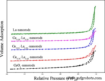
N2 두 CeO2에서 BET 표면적과 평균 직경을 측정하기 위해 흡수 측정이 사용되었습니다. 나노로드 및 Ce-La 나노복합체. 그림 1에서 보는 바와 같이 Ce-La 나노막대에 대한 흡착 등온선은 유형 IV이며 H3 히스테리시스 루프의 특성을 나타냅니다. 모든 샘플은 N2의 매우 강력한 증가를 보여줍니다. - 0.85보다 큰 상대 압력에서 흡착된 부피는 상당한 양의 메조포러스 존재의 특징이며, [2, 22] 슬릿 같은 기공을 형성하는 응집체(느슨한 집합체)로 구성된 Ce-La 나노복합체를 나타냅니다. La의 도펀트와 함께 히스테리시스 루프는 약 0.95의 상대 압력으로 이동했으며, 이는 Ce-La 합성물의 감소에 따라 기공의 크기가 작아질 것임을 의미합니다. 표 2와 같이 CeO2의 비표면적 나노로드는 99.7m
2
입니다. /g, 74.1m
2
로 감소 /g는 La가 3:1의 Ce/La 몰비로 도핑된 경우입니다. La 함량이 증가함에 따라 Ce-La 나노복합체의 표면적이 지속적으로 감소했습니다. 이것은 주로 CeO2의 격자에 포함되지 않는 La의 내용으로 인해 발생합니다. 분리된 LaOx로 존재 Ce-La 나노복합체의 형태는 거의 차이가 없습니다. 모든 나노로드는 80–100m
2
의 유사한 표면적을 가지고 있음을 관찰할 수 있습니다. /G. Ce0.75의 기공 부피 -라0.25 나노로드는 0.23cm
3
입니다. /g는 Ce 나노로드와 유사하고 다른 Ce-La 나노로드보다 큽니다. BJH 분석에서 추정된 기공 직경은 Ce-La 나노복합체의 메조포러스 특성을 확인했습니다. 촉매적 CO 산화에 대한 이점일 수 있습니다.
<그림>
La 함량이 다른 Ce-La 나노막대의 질소 흡탈착 등온선
XRD
합성된 샘플은 분말 X선 회절 분석을 거쳐 구조적 특성을 분석했습니다. 산화세륨의 결정도 피크(그림 2a)는 (111), (200), (220), (311) 회절면에 해당하는 2θ =28.6°, 33.1°, 47.6°, 56.3°에서 관찰되었으며, CeO2의 입방형 형석 구조를 확증 크리스탈(JCPDS 번호 34-0394). La의 함량이 0.25at.%일 때 La-Ce 복합재료의 회절 피크가 넓어진다. 2θ =30.0°, 46.0°, 52.0° 및 53.6°에 중심을 둔 피크는 격리된 La2의 회절 평면에 해당합니다. O3 . La(OH)3에 할당된 피크 없음 감지될 수 있습니다. 그러나 함량이 낮고 회절 위치가 근사하여 LaOx의 존재를 식별하기가 쉽지 않습니다. . La 함량이 증가함에 따라 La2에 대해 몇 가지 두드러진 피크가 관찰됩니다. O3 또는 La(OH)3 나노복합체에서. La2의 주요 회절 피크 O3 2θ =30.0°(101), 39.6°(220), 46.2°(110) 및 66.8°(112)에 존재하며, 이는 6각형 위상에 할당될 수 있습니다(JCPDS 카드 05-0602). La(OH)3의 주요 회절 피크 2θ =15.7°(100), 27.3°(110), 27.9°(101) 및 39.4°(201)에 존재하며, 이는 6각형 위상에 할당될 수 있습니다(JCPDS 카드 36-1481). 결과는 La가 고립된 La2로 존재할 수 있음을 보여줍니다. O3 또는 La(OH)3 합성에서. 금이 증착된 후에는 순수한 면심 결정 구조의 금으로 표시될 수 있는 회절 피크가 없었습니다(그림 2b). 이는 금 나노 입자의 함량이 낮거나 입자 크기가 작기 때문일 수 있습니다.
<그림>
La 함량이 다른 1% Au/Ce-La 나노막대의 XRD 패턴(0–100 at.%)(a ) 및 금/세륨0.75 -라0.25 300°C에서 2시간 동안 소성된 다양한 Au 로딩을 갖는 나노로드(b )
SEM 및 TEM
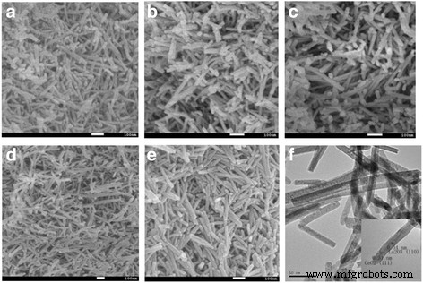
그림 3a-e는 CeO2의 SEM 사진을 보여줍니다. 및 다른 농도의 La
3+
에서 얻은 Ce-La 나노복합체 이온. 모든 Ce-La 나노복합체는 막대형 구조를 나타냄을 알 수 있다. 분명히 많은 막대가 Ce-La 번들로 쌓여 크기가 다른 슬릿 같은 기공이 형성됩니다. 결과는 N2와 일치했습니다. 흡착-탈착 등온선. 그림 2a와 같이 제품은 주로 직경 5~10nm, 길이 100~300nm의 나노로드로 구성되어 있습니다. 도 3e에서는 직경 약 12.5nm의 나노로드가 대량으로 명확하게 관찰되었으며 평균 직경이 약 8.0nm인 짧은 나노로드도 소량 존재하였다. 그림 3b-d에서 La
3+
의 도핑 농도가 증가함에 따라 , 샘플은 항상 나노로드 형태를 나타냅니다. 그러나 도핑 농도가 25mol%인 반면, 얻은 샘플은 모든 샘플 중에서 직경 5~20nm, 길이 100~300nm로 가장 균일한 나노로드를 나타냈습니다. 그림 3f는 얻은 개별 Ce-La 나노로드의 TEM 이미지를 표시합니다. 질소 흡탈착 등온선에서 알 수 있듯이 지지체에 많은 기공이 있음을 알 수 있습니다. Ce-La 나노로드의 HRTEM 이미지는 구조적으로 균일하고 본질적으로 단결정인 것으로 나타났습니다. 그림 3f에 삽입된 격자 무늬는 CeO2의 (111), (110) 평면과 일치하는 두 개의 면간 간격 값, 즉 0.31 및 0.34 nm를 보여줍니다. 그리고 라2 O3 , 각각 [3, 15, 43]. La
3+
이온이 La2로 효과적으로 생성되었습니다. O3 , 이는 XRD 스펙트럼과 일치합니다.
<그림>
La 함량이 다른 Ce-La 나노로드의 SEM 이미지:0(a ), 25%(b) ), 50 at.%(c ), 75%(d) ), 100 at.%(e ); Ce0.50의 TEM 이미지 -라0.50 나노로드(f ); 삽입된 이미지는 해당 HRTEM 이미지를 보여줍니다.
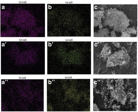
요소 매핑 및 EDS 분석을 사용하여 Ce-La 샘플의 화학적 조성을 결정했습니다(그림 4 및 표 3). 결과는 합성에서 예상된 값과 잘 일치하는 균일한 La/Ce 몰 비율을 보여주었습니다. Au/Ce0.75의 TEM 이미지 -라0.25 300°C(그림 5a) 및 400°C(그림 5c)에서 소성된 샘플은 금 첨가 후 Ce-La 나노결정의 모양이 본질적으로 변하지 않았음을 분명히 보여주었습니다. Ce-La 나노로드에서 TEM에 의해 금 입자가 관찰되지 않았습니다. 고도로 분산된 금 클러스터의 존재(d <1nm)는 요소 매핑 및 EDX 분석에 의해 입증되었습니다(그림 5b, e 및 f 삽입). 일치하게, 이 샘플에 대해 수행된 XRD 분석(그림 2)은 금 입자가 너무 작아 감지할 수 없기 때문에 금과 관련된 피크를 나타내지 않았습니다. 이것은 Ce-La 나노로드 표면이 금 원자를 나노미터 이하 클러스터(TEM 보이지 않음)로 분산시키고 안정화시킬 수 있음을 나타냅니다. 이것은 문헌[2, 44,45,46]과 일치합니다. 그러나 일부 큰 금 입자 덩어리(평균 d ~ 7 nm) Au/Ce0.75에서 관찰되었습니다. -라0.25 금속 Au의 (1 1 1) 평면에 할당된 0.236nm 간격의 프린지로 인해 400°C에서 소성된 나노로드(금 입자 덩어리가 표시된 그림 5c). 높은 소성 온도와 함께 금 입자가 분명히 성장하여 촉매 활성의 손실로 이어지는 것을 알 수 있습니다.
<그림>
Ce 및 La의 요소 매핑 이미지, La 함량이 25at.%인 Ce-La 나노막대용 혼합 샘플의 SEM 이미지(a –ㄷ ), 50 at.%(a '–ㄷ '), 75 at.%(a ′′–ㄷ ″)
<그림>
0.5% Au/Ce0.75의 TEM 및 STEM 이미지 -라0.25 300°C에서 소성된 나노로드(a –b ) 및 400°C(c ), EDX 분석(e –f )의 (a 이미지) –b ) Au 신호의 존재를 나타냄
XPS
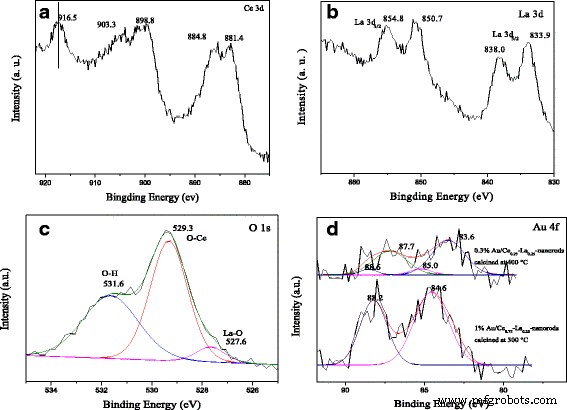
그림 6의 XPS 스펙트럼은 1%Au/Ce0.75의 화학적 합성 및 상태를 조사하기 위해 수행되었습니다. -라0.25 nanorod 샘플은 300°C에서 2시간 동안 소성되었습니다. Ce 3d의 XPS 스펙트럼 3d의 뚜렷한 피크를 보여줍니다. 3/2 회전 궤도 상태 및 3d5/2 그림 6a의 스핀 궤도 상태. 알려진 바와 같이 피크는 일반적으로 Ce
4+
의 존재를 감지하는 분광 마커로 사용되는 약 899, 903 및 916eV의 결합 에너지에 위치합니다. 상태. 우리의 경우 Ce 3d 코어 수준은 4가 Ce
4+
상태의 특징적인 피크인 3개의 스핀 궤도 이중선을 보여줍니다. . 약 882.8, 888.1, 898.4eV에 위치한 피크는 Ce 3d에 할당됩니다. 5/2 , 그리고 약 901.3, 907.0, 916.7 eV에 있는 것들은 Ce 3d에 할당됩니다. 3/2 , Ce(IV) 화합물의 스핀-궤도 분할 이중선에 해당합니다. 관찰된 결과는 일반적으로 보고된 문헌[19, 28, 29, 32]과 일치합니다. 샘플이 Ce
4+
상태에 있음이 분명합니다. Ce
3+
불순물 없음 상태. 그림 6b는 La 3d의 XPS 스펙트럼을 보여줍니다. 1%Au/Ce0.75 지역 -라0.25 나노로드 샘플. 스핀-궤도 분할 3d5/2 그리고 3d3/2 수준은 이중 피크 구조를 보였다. 3d 사이의 스핀-궤도 분할 3/2 그리고 3d5/2 수준은 약 17.0eV이고 위성과 주 피크 사이의 간격은 4.1eV로 보고된 La
3+
값과 일치합니다. 화합물 [11, 47]. 예상대로 La는 + 3 산화 상태로 빠져나가 촉매 활성에 중요한 영향을 미칠 수 있습니다. O 1s XPS 스펙트럼(그림 6c)은 비대칭이며 각각 529.3, 531.6 및 527.6eV로 분해됩니다. 529.3eV의 피크는 격자 산소에 할당되고 약 531.9eV의 피크는 지지체 표면의 수산기에 할당됩니다[27, 28, 32]. 527.6eV의 작은 숄더 피크는 La-O에 기인하며 LaOx의 존재를 나타낼 수도 있습니다. 촉매에서 [11, 48]. 분명히 높은 피크 강도에 따라 지지체 표면에 많은 수의 하이드록실 그룹이 있습니다. Au 4f의 XPS 스펙트럼 300 및 400°C에서 소성된 촉매 영역은 그림 6d에 나와 있습니다. 그림 6d에서 300°C에서 소성된 촉매는 Au 4f7/2 84.6eV에서 에너지 신호를 결합합니다. 신호는 양이온성 Au
+
에 대해 특징적이었습니다. 종 [14, 15, 31]. 이에 비해 촉매가 400°C에서 소성된 후 Au 4f7/2 피크는 83.6eV의 결합 에너지 및 Au 4f에 위치했습니다. 5/2 87.7 eV의 결합 에너지에 위치했습니다. 금속성 Au
0
의 존재 분명히 관찰됩니다. 산화된 금 종에 해당하는 85.0 및 88.2 eV 주변에 위치한 작은 피크도 감지되었습니다. 300°C에서 소성된 촉매는 실질적으로 주로 양이온성 Au
+
를 나타냄이 분명합니다. 종(> 90%의 Au
+
종). 대조적으로, 400°C에서 소성된 샘플은 Au
0
의 90%를 갖습니다. 및 Au
δ+
의 10% . 금속 Au에서 지지체로의 전자 밀도 전달은 Au의 부분 산화 및 금과 지지체 사이의 강한 상호 작용을 초래했습니다. Au
δ+
의 존재 지지면의 부분적 감소를 담당합니다. 따라서 Au
δ+
Au
0
보다 더 활동적인 것으로 간주됩니다. CO 산화 [11, 21]. 우리의 경우 300°C에서 소성된 촉매는 더 많은 Au
δ+
를 가졌습니다. 400°C에서 소성된 촉매보다 300°C에서 소성된 촉매가 400°C에서 소성된 촉매보다 활성이 더 높았으며 이는 활성 결과와 일치한다고 추론하는 것은 어렵지 않습니다.
<그림>
1%Au/Ce0.75의 XPS 스펙트럼 -라0.25 300°C에서 2시간 동안 소성된 나노로드:Ce 3d 피크(a ), 라 3d 피크(b ) 및 O 1s 피크(c ). Au 4f 피크(d ) 금/세륨0.75 -라0.25 300 및 400°C에서 2시간 동안 소성된 나노로드
자외선 가시
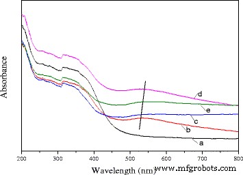
Ce0.75의 UV-vis 확산 반사 스펙트럼 -라0.25 나노로드 및 0.5% Au/Ce0.75 -라0.25 다른 온도에서 소성된 나노로드는 그림 7에 나와 있습니다. 이 그림에서 볼 수 있듯이 지지체의 스펙트럼과 비교하여 다른 온도에서 소성된 촉매의 스펙트럼은 금속 금 나노 입자의 표면 플라즈몬 공명(SPR) [21, 24, 49]. SPR은 광학 여기에 대한 응답으로 전자의 집합적 진동에 기인할 수 있으며, 이는 Uv-vis 영역에서 빛을 흡수하게 됩니다. 표면 플라즈몬 공명의 위치는 분산된 금 입자의 크기, 입자의 모양 및 주변 물질의 유전 특성에 영향을 받습니다. 본 연구에서 소성 전처리는 흡수 밴드의 큰 적색 편이를 일으켰고, 흡수 밴드(500-600 nm)의 위치는 소성 온도가 증가함에 따라 적색 이동을 보였다. 교대 등급은 80°C→200°C→300°C입니다. 소성 온도가 400°C로 추가 증가함에 따라 흡수 밴드가 단파장으로 이동했습니다. 피크 위치 이동에 대한 설명에 대한 여러 보고가 있었습니다[24, 50,51,52,53]. 금 입자의 직경이 <2 nm인 반면, 확장된 이동 피크 위치는 주로 금속 유전 기능의 크기 의존적 감쇠로 인해 발생했습니다. 또한 주변 금속 산화물과의 화학적 상호작용으로 인해 금 입자의 전자 밀도가 감소했는데, 이는 적색 편이를 추가로 유도하는 메커니즘을 설명할 수 있습니다[52]. 금 입자의 크기가 증가하면 흡수 피크(평균 직경이 25nm보다 작음)에서 파란색 이동이 발생하고 큰 입자(평균 직경이 25nm보다 큰 경우)의 경우 반대 효과가 관찰되었습니다[53]. TEM 데이터에 따르면 촉매의 금 입자 크기는 300°C에서 소성된 촉매의 경우 <1 nm였습니다. 그러나 소성 온도가 400°C로 추가 증가함에 따라 금 입자가 성장했으며 평균 크기는 약 7nm였습니다. 앞서 언급한 바와 같이 플라즈몬 띠의 위치는 금 입자의 모양과 크기에 따라 크게 좌우된다. 현재의 경우 이러한 큰 이동은 금 입자 크기의 차이로 설명될 수 있습니다. 데이터는 촉매 활성 테스트의 결과와 일치했습니다. 또한 금 나노 입자가 지지체 표면에 잘 분산되어 있음을 나타냅니다.
<그림>
순수 Ce0.75의 UV-vis DRS -라0.25 nanorod 지원 (a) 및 0.3% Au/Ce0.75 -라0.25 80°C(b), 200°C(c), 300°C(d) 및 400°C(e)에서 소성된 나노로드
H2 -TPR
그림 8a는 순수 및 혼합 산화물 샘플에 대한 TPR 프로파일을 보여줍니다. 순수한 CEO의 경우2 nanorods의 경우, 약 저온(410 °C) 및 고온(620 °C)을 중심으로 한 환원 피크는 CeO2의 표면 및 벌크 산소 종의 환원에 기인할 수 있습니다. , 각각 [1, 32]. 순수한 La 나노로드의 경우 벌크 La2의 환원에 할당된 ~ 700°C에서 명백한 환원 피크가 감지될 수 있습니다. O3 . Ce-La 나노로드의 ~ 500°C에서 감소 피크가 나타난다는 사실을 발견하는 것은 흥미로웠습니다. 25%, 50% 및 75% at.% La 도핑이 있는 세 가지 샘플의 감소 피크는 La 도핑 시 약 20°C만큼 더 높은 온도로의 이동을 보여줍니다. La 함량이 25at.%일 때 520°C의 강한 환원 피크 온도가 관찰되었습니다. 새로운 환원 온도로 순수 CeO2에 비해 현저히 상대적입니다. 나노로드. Reference와 비교하여, La-O와 Ce-O 사이의 상승적 상호작용으로 인해 Ce-La 나노로드의 환원 온도는 순수한 CeO2보다 높았다. [31, 54]. 이원 산화물은 독립적인 CeO2를 가져야 함을 알 수 있습니다. 및 LaOx . 그림 8b에서 볼 수 있듯이 금 증착 후 Au/CeO2에 대해 매우 낮은 온도(100–200°C)에서 새로운 환원 피크가 나타납니다. 및 Au/La-Ce 나노로드. 여기에서 XPS 결과로 인해 촉매가 300°C에서 소성된 후 Au는 주로 Au
δ+
였습니다. , 따라서 ~ 200°C에서 환원 피크는 높은 원자가의 Au 종의 환원에 기인합니다[21]. ~ 350°C에 중심을 둔 작은 피크는 Ce-La 나노막대의 환원과 관련될 수 있습니다. 또한 1% Au/Ce0.75의 경우 -라0.25 nanorods, 약 230°C에서 또 다른 환원 피크는 CeO2의 금 촉진 환원에 기인할 수 있습니다. . 1% Au/Ce0.75 -라0.25 나노로드는 촉매 중 가장 낮은 환원 온도를 가지므로 CO 산화에 가장 활성인 촉매가 될 수 있습니다. 이는 활동 결과와 일치했다. Since the surface reduction peaks for all oxide supports are significantly decreased after gold deposition, it indicates that most available oxygen is reduced at this lower temperature and suggests that H2 dissociation on gold and spill-over onto the adjacent oxide surface are more likely to be responsible for the strong low-temperature reduction peak [31]. TEM and XPS data indicated that the cationic gold particles with small size highly dispersed on the surface of the supports. The presence of LaOx could also help stabilize cationic Au. This is beneficial for the strength of gold-support interaction [11]. The strong interaction between gold and support promoted the reduction of Au/Ce-La nanorods shifted to low temperature. The results indicated that the reducibility of Ce-La nanorods is strongly affected by the gold deposition.
H2 -TPR profiles of a La-Ce nanorods with different La content (0–100 at.%) and b 1% Au/La-Ce nanorods with different La content calcined at 300 °C
Catalytic Activity
Effect of La Content
As shown in Fig. 9, catalytic activity results for Au/Ce-La nanorod samples ranging from pure CeO2 to 100 at.% La-content nanorod supports. The most striking feature in the figure is the high activity of the Au/Ce0.75 -La0.25 nanorod catalyst with the 100% conversion at temperature as low as 30 °C. In contrast, the other Au/La-Ce catalysts showed lower activity compared to Au/Ce0.75 -La0.25 nanorods catalysts under the same reaction conditions. The results indicated that La doping has very much impact on this high CO conversion activity with a La content of 25 at.%, while a further increase results in a significant drop in activity. This again closely mirrors the trends seen in the reducibility of the samples, where an increase of La content from 25 at.% results in a strong loss of reducibility.
Catalytic activities of 1% Au/ La-Ce nanorods with different La content calcined at 300 °C for 2 h
In consideration of the preparation methods, gold loadings, gold particle size and distribution on different Ce-La nanorods supports, XRD, TEM and XPS data showed that all the catalysts should have the same number and type of active Au sites. So this high activity of the Au/Ce0.75 -La0.25 nanorods catalysts correlates well with the reducibility data discussed above. H2 -TPR results indicated that Au/Ce0.75 -La0.25 nanorods has the lowest reducibility temperature and highest reducibility in the region of 50–400 °C, especially in the region of 50–150 °C, which could exactly approach the region of reaction temperature. In the process of reaction, the Ce0.75 -La0.25 nanorod support served as oxygen carrier. The reducibility of Ce0.75 -La0.25 nanorods could promote the formation of active oxygen. That is to say high reducibility of the catalyst, good activity the catalyst has. Au/Ce0.75 -La0.25 nanorod catalyst subsequently has the best activity.
Effect of Gold Content
The catalytic activities for CO oxidation were measured from low conversion to 100% conversion for the Au/Ce0.75 -La0.25 nanorod catalysts calcined at 300 °C for 2 h with a series of low gold contents. As shown in Fig. 10, all of the catalysts showed high catalytic activities. The CO conversion increased greatly with increasing gold content. The complete CO conversion could be attained at 50 °C over 0.5% Au/Ce0.75 -La0.25 nanorod catalyst. The size and chemical states of gold nanoparticles are generally thought to be vital for the performance of supported gold catalysts. It has been reported that its gold nanoparticles with the diameter of < 5 nm would be suitable for the supported gold catalysts in the catalytic CO oxidation [27, 28]. The XPS data proved that gold in Au/Ce0.75 -La0.25 nanorod catalyst exists in the form of cationic Au
+
. TEM images of the samples were also shown to investigate the diameter of gold nanoparticles in the catalysts. Consequently, the gold particles of Au/Ce0.75 -La0.25 nanorods were detected as sub-nanometer. Taking into account the particle size, mass content, and chemical states of the gold nanoparticles, gold particles with small diameter highly dispersed on the surface of Ce0.75 -La0.25 nanorods and interacted strongly with the support [17, 21, 23]. The strong interaction between gold particles and the support would help improve CO adsorption and accelerate active oxygen spillover to gold particles from the support, so 0.5% Au/Ce0.75 -La0.25 nanorods which had relatively high content of gold should exhibit the best CO oxidation activity. In fact, 0.5% Au/Ce0.75 -La0.25 nanorods indeed present high performance. The results demonstrated the activity of supported gold catalysts is strongly dependent on the gold nanoparticle size, chemical states, and the quantity of the active species, an increase of which implied an increase of the catalytic activity. In the case of Au/Ce0.75 -La0.25 nanorod catalyst, catalysts with low gold content could also exhibits high activity at low temperature, which would promote the progress of supported gold catalyst. The results indicated that supported gold catalysts prepared by deposition-precipitation with pH value of 6–10 for HAuCl4 solution could have high catalytic activity due to small diameter of gold nanoparticles, corresponding with the references [8,9,10].
Catalytic activities of Au/Ce0.75 -La0.25 nanorods with different gold goadings calcined at 300 °C for 2 h
Effect of Calcination Temperature
The effect of calcination temperature on the catalytic activity of 0.5%Au/Ce0.75 -La0.25 nanorods is also shown in Fig. 11. The results indicated an increase in the activity of catalyst with the calcination temperature from 80 to 300 °C. The 0.5% Au/Ce0.75 -La0.25 nanorod catalyst calcined at 200 °C could convert CO to CO2 completely at 80 °C. While for 0.5% Au/Ce0.75 -La0.25 nanorod catalyst calcined at 80 °C, the temperature increased to 100 °C. The results showed that CO conversion increased with increasing calcinations temperature. Then, for the sample calcined at 400 °C, about 90% CO can be converted to CO2 at 100 °C and CO could be converted to CO2 completely at 120 °C. The sample calcined at 300 °C possessed the best catalytic activity. The catalytic performance of supported gold catalysts strongly depends on gold nanoparticle size and metal-support interaction due to “synergic effect” at the gold-support interface [10, 13, 15, 18]. The gold-support interaction largely depended on calcination temperature of catalysts. The electron could transfer from gold to the support [10]. Thus, with increasing calcination temperature, the charges on gold particles became increasingly positive, which is good for the enhancement of catalytic activity for CO oxidation. Here, as shown in the Fig. 5, size of gold particles in the catalysts calcined at 300 °C was small. The XPS data also indicated that gold was main Au
δ+
after calcination at 300 °C. Thus, the stronger metal-support interaction could account for the relative good catalytic performance for catalysts calcined at 300 °C. From 80 to 300 °C, the higher the calcination temperature is, the stronger interaction exists between gold particles and support. As a consequence, from 80 to 300 °C, the activity of catalysts increased. However, after the 0.5% Au/Ce0.75 -La0.25 nanorod catalyst calcined at 400 °C, complete conversion temperature increased. The main reason might be that the high-temperature treatment led to increased mobility and growth of gold nanoparticles, which correspondingly led to the loss of catalytic activity. The XPS also suggested that the catalysts calcined at 400 °C, Au was mainly Au
0
. It could be concluded that the activities of supported gold nanoparticles were influenced by both the oxidation state and the size of gold nanoparticles, and the appropriate calcination temperature was 300 °C.
Catalytic activities of 0.5% Au/Ce0.75 -La0.25 nanorods calcined at different temperatures
Stability Observations
The stability of the 0.3% Au/Ce0.75 -La0.25 nanorod catalyst during CO oxidation at different reaction temperatures was measured, as shown in Fig. 12a. When the reaction was carried out at 70 °C, the initial CO conversion over 0.3% Au/Ce-La catalyst can reach 100% and has almost no change with continuously increasing reaction time. 0.3% Au/Ce-La catalyst with 60% CO conversion rate at 40 °C is also attained even after 10-h running period, and no obvious decline in CO conversion is observed. Although the catalytic activity of 0.3% Au/Ce0.75 -La0.25 nanorod catalyst at 40 °C was lower than that of 0.3% Au/Ce0.75 -La0.25 nanorod catalyst at 70 °C, the conversion of CO over the catalysts at both temperatures still seemed to be stable over 10 h on stream. It is thought that the catalyst was of good durability. It was clear that the activity over 0.3% Au/Ce0.75 -La0.25 nanorod catalyst did not strongly depend on the reaction temperature. As the reaction temperature decrease the activation rate barely becomes little slower and then finally reaching a steady state in which the CO conversion was still around 90%. For comparison, the stability of 0.5% Au/Ce0.75 -La0.25 nanorod catalyst at the reaction temperature of 40 °C with initial conversion of 100% was also provided in Fig. 12b. It was obvious that in 10 h, no decrease of CO conversion for 0.5% Au/Ce0.75 -La0.25 nanorods was detected. The results depicted that with the change of gold content, Au/Ce0.75 -La0.25 nanorods could still perform good stability.
The stability of 0.3% Au/Ce0.75 -La0.25 nanorods, reaction temperature:40 and 70 °C (a ) and 0.5% Au/Ce0.75 -La0.25 nanorods, reaction temperature:40 °C (b ) for the CO oxidation
As engine efficiency increases and automotive exhaust temperatures decrease, traditional supported gold catalysts would be insufficient to meet emission regulations. And there are also a number of industrial catalytic processes which (e. g., the catalytic oxidation of CO in automotive exhaust gas) are sometimes carried out at high temperatures. Thus, the development of new catalysts that are active at lower temperature, yet still stable at periodic high temperatures, will be vital. In the two regards, catalysts with good activity at low temperature that are stable at high reaction temperatures are desirable. It is necessary to investigate their catalytic performance for CO oxidation at a certain high temperature which is a very stringent test for the stability of gold nanocatalysts against sintering. In the present work, the stability of 0.3% Au/Ce0.75 -La0.25 nanorod catalyst was also measured at 200 °C (100%) for high-temperature treatment. As shown in Fig. 13, no decline of catalytic activity was observed within 50 h indicates that the catalyst keeps good stability within 50 h. Remarkably, very few serious gold sintering occurred during the reaction. It indicated that 0.3% Au/Ce-La catalyst can exhibit good catalytic stability at both low and high reaction temperatures.
The stability of 0.3% Au/Ce0.75 -La0.25 nanorods at the reaction temperature of 200 °C for CO oxidation
Reaction Mechanism Speculate
Combined with H2 -TPR and XPS experiments, it suggested that CO oxidation over LaOx -doped CeO2 -supported Au catalysts might follow the Langmuir–Hinshelwood + Redox mechanism [1, 20, 26, 30, 32]. The XPS results suggest that there are Ce
3+
and Ce
4+
on the surface of the catalyst. H2 -TPR data also proved that reducibility of this binary Ce-La nanorod oxides could be promoted by Au deposition. The reducibility of Au/Ce-La nanorods was much higher than pure Au/CeO2 or Au/LaOx catalysts. This would help the produce of oxygen vacancies. The oxygen vacancy is a very lively activity center. The active center can promote the activation of O2 . Thus, the CO oxidation reaction could become more easily. There are also amount of adsorbed oxygen species on the surface of catalyst. Usually adsorbed oxygen species play an important role in the oxidation of CO. The O2 of the reaction will form the chemisorbed oxygen, and the oxygen vacancy would be replenished by O2 of reaction gas to form new active lattice oxygen. XPS data also proved that gold in the catalysts was mainly Au
δ+
species, which would accelerate the adsorption of CO. The possible reaction mechanisms of Au/Ce-La nanorod catalyst could be described as follows. Firstly, CO and O2 were chemisorbed on the surface of the catalysts. Then, the chemisorbed oxygen directly reacts with CO, or the active lattice oxygen of the catalyst reacts with CO, and the catalyst produced the oxygen vacancy with oxygen from gas-phase O2 . At last, CO was oxidized into CO2 (shown in Fig. 14).
Proposed CO reaction pathways over the catalysts, Au/Ce-La nanorods
Conclusions
In summary, a series of mixed Ce-La nanorods with various amounts of La was prepared via a simple hydrothermal reaction at high concentration of NaOH and without surfactant. Gold was loaded by deposition-precipitation. After La doping, the composite could retain the initial rod morphology. As a result, Ce-La nanorods with 25 at.% La maintained the optimal nanorods with the length of 0.6 um and the diameter of 3–5 nm. Gold particles were dispersed well on the support. The reducibility of Ce-La nanorods could be affected significantly by LaOx doping. The deposition of gold had important influence on the reducibility of catalyst. Thus, the CO oxidation activity of Au/Ce-La nanorods was essentially changed in comparison with pure Au/CeO2 and Au/La nanorods. One percent Au/Ce0.75 -La0.25 nanorods could convert CO to CO2 completely at 30 °C. Further increase in La content results in decreased activity due to the decrease in reducible oxygen sites. The Au/Ce0.75 -La0.25 nanorod catalyst with low gold concentration also showed high activity. With the increase of gold content, there is an increase in the activity. The stability test of 0.3% Au/Ce0.75 -La0.25 nanorods indicated that the catalyst not only kept 100% conversion after continuous operation for 10 h under 70 °C but also showed no deactivation after 10 h on stream at 40 °C. As expected, the activity of 0.3% Au/Ce0.75 -La0.25 nanorods also retained a 100% CO conversion during 50 h at 200 °C. The results revealed that LaOx as the dopant could grow together with CeO2 in one rod. The 1D binary mixed Ce-La nanorods could be a good support for precious metal group catalysts with low content of gold.