첨단 기능성 소재로서 은 나노입자는 주로 다양한 특성을 기반으로 하는 광전, 바이오 센싱, 촉매, 항균 및 기타 분야와 같은 다양한 분야에서 잠재적으로 유용할 수 있습니다. 그러나 은 나노 입자의 특성은 일반적으로 크기, 모양 및 주변 매질에 의해 결정되며 다양한 합성 방법으로 조절할 수 있습니다. 이 리뷰에서는 다양한 모양과 특정 크기의 은 나노 입자를 합성하는 제조 방법을 자세히 설명합니다. 또한, 은 나노 입자의 해당 특성과 응용도 이 백서에서 논의됩니다.
<섹션 데이터-제목="배경">
배경
독특한 광학 및 전기적 특성을 가진 금속 나노 입자는 지난 수십 년 동안 광범위하게 조사되었습니다. Ag 나노입자(AgNPs)는 독특한 특성과 응용으로 인해 가장 집중적으로 연구된 금속 나노입자입니다[1,2,3,4,5]. AgNPs의 특성은 모양, 크기 및 주변 매질을 포함한 입자의 형태에 크게 의존합니다. 은 나노입자의 합성 방법과 형태학적 조절에 많은 노력을 기울였습니다.
최근 연구자들은 광전[6], 촉매[7], 항균[8,9], 바이오센서[10], 표면강화라만산란(SERS)과 같은 은 나노입자의 우수한 기능에 대한 심층 연구를 수행하였다. ) [11]. 지금까지 AgNPs는 화학적 환원[12,13,14,15,16], 광 환원[17, 18] 및 레이저 합성[19] 등을 통해 성공적으로 준비되었지만 이러한 방법은 일반적으로 시간과 에너지를 소모합니다. 동시에 엄격한 준비 조건과 AgNP의 크기가 불균일하다는 단점도 있습니다. 따라서 AgNPs의 크기, 모양 및 크기 분포를 미세하게 조절할 수 있는 간단하고 경제적인 방법의 개발이 시급하다. 보호제를 사용하는 것은 안정성과 분산성이 좋은 AgNP를 만드는 효율적인 방법입니다. 한편, 입자 사이의 응집은 보호제에 의해 방지될 수 있다. 따라서 보호제는 AgNPs 합성에 사용되는 것이 중요합니다[20].
본 연구에서는 나노큐브, 나노와이어, 나노스피어 등 다양한 형태의 은 나노입자 제조에 대해 자세히 살펴보았다. 1-10 nm AgNPs, 10-100 nm AgNPs의 모양과 크기가 다른 은 나노 입자를 준비하는 대표적인 작업은 이전에 검토되었습니다. 우수한 환경 보호 특성과 간단한 조작으로 복잡한 화학 합성 절차를 대체할 수 있는 은 나노 입자를 얻기 위한 새로운 생합성 방법이 강조점으로 선정되었습니다. 한편, 항균, 형광, 촉매, 표면 플라즈몬 공명 등의 AgNPs의 특성과 응용에 대해 구체적으로 살펴보면 다음과 같다. 이번 리뷰에서는 나노센서에 사용할 수 있는 은나노입자의 중요한 응용을 강조했습니다.
이 연구는 AgNP의 조사에 중요한 포괄적인 접근 방식을 제공합니다. 그러나 혁신적인 준비 방법과 응용 프로그램 혁신이 여전히 탐구되어야 한다는 점은 주목할 가치가 있습니다.
합성 방법
은 나노입자는 종자 성장법[21] 및 단계적 환원법[22]과 같은 다양한 방법으로 합성되었다. 각 방법에는 장점과 한계가 있습니다. 따라서 효과적인 준비 방법을 개발하는 것은 여전히 어려운 일입니다. 독특한 특성과 광범위한 응용으로 인해 은 나노 입자의 합성 방법은 최적화할 가치가 있습니다. 본 연구에서는 새로운 생합성 방법을 포함하여 6가지 유형의 제조 방법을 요약하였다. 이 분야에 종사하는 근로자들에게 작은 도움이 되기를 기대합니다.
다양한 유형의 AgNP 준비
최근 연구자들은 형태 의존적 특성으로 인해 AgNPs의 형태 조절에 초점을 맞추었다[23, 24]. 한편, 현재의 응용을 확대하기 위해 산호 모양[25], 새장[26], 삼각형 나노결정[27]과 같은 다양한 모양의 은 나노입자 제조는 광범위한 과학적 연구를 촉발시켰다. 은 나노 입자의 형성 메커니즘과 다양한 제조 방법이 오랫동안 연구되었습니다.
Ag 나노큐브의 합성
Xia et al. [28,29,30] 폴리비닐피롤리돈(PVP)이 있는 상태에서 질산은을 에틸렌 글리콜로 환원하여 은 나노큐브의 단분산 샘플을 대량으로 준비했습니다. 합성과정에서 PVP는 분산된 은나노입자를 안정화시키고 덩어리를 방지할 수 있는 보호제로 사용되었다. 동시에, PVP 첨가량은 AgNP의 형태에도 영향을 미칠 수 있습니다. 따라서 합성 중에는 PVP를 사용하는 것이 필수적입니다. 가열은 에틸렌 글리콜의 환원성을 증가시키는 데 유익한 더 많은 반응 에너지를 제공할 수 있다는 것은 잘 알려져 있습니다. 하이드록실 이온이 있는 경우 Ag
+
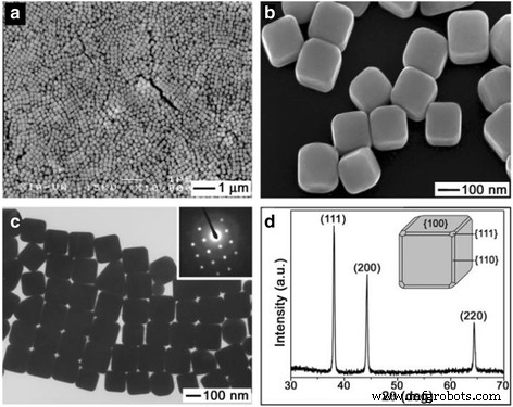
환원되어 은나노큐브를 형성한다. 이 연구의 장점은 균질한 단결정 나노 큐브를 제조하는 데 활용할 수 있다는 것입니다. 나노미터 규모에서 금속은 대부분이 면심입방체(fcc)인 금속 표면이 나노미터 규모에서 가장 낮은 에너지 면으로 경계를 이루기 때문에 핵 생성 및 쌍정 및 다중 쌍정 입자(MTP)로 성장하는 경향이 있습니다[31] . 또한, 이 구조는 포토닉스, 촉매, SERS 기반 센싱 분야에 적용하기에 유리하다. 그림(Fig. 1)은 은나노큐브의 SEM, TEM, XRD 이미지를 보여준다. 이 은 나노큐브의 평균 가장자리 길이는 175nm이고 표준 편차는 13nm입니다. 표면은 매끄러웠고 이 입자의 모든 모서리와 가장자리가 약간 잘렸습니다. 이 구조는 잘린 모서리에 약물을 주입하여 약물 전달 시스템에 사용할 수 있습니다.
<그림>
아 낮음 및 b 약간 잘린 은 나노 큐브의 고배율 SEM 이미지. ㄷ 동일한 배치의 은 나노큐브의 TEM 이미지. d 순수한 fcc 은의 형성을 확인하는 동일한 배치의 샘플에 대한 XRD 패턴 [28]
새로운 은 나노 입자는 Yam et al.에 의해 발표되었습니다. [32] 수용액에서 Cetyltrimethyl Ammonium Bromide (CTAB)를 계면 활성제로 사용했습니다. 브롬 이온은 은 암모니아 착물([Ag(NH3 ) 2 ]
+
) AgBr 침전을 생성하고 은 이온은 후속 반응에서 천천히 방출됩니다. 동시에, 잔류 은 이온은 포도당에 의해 환원되었고 코팅된 계면활성제로 ~ 55 nm 크기의 나노은 큐브가 형성되었습니다. 계면활성제 CTAB는 물리적 흡착에 의해 AgNPs의 표면에 흡착될 수 있습니다. 이러한 이유로 AgNPs의 응집 및 규모 성장은 억제에 의해 효과적으로 제어될 수 있습니다. CTAB가 존재하기 때문에 균일한 분산과 적당한 크기의 AgNP를 얻을 수 있다.
Xia와 Yam 합성의 방법으로 보고된 나노은 큐브를 준비하는 데 오랜 시간이 걸립니다. 그러나 은 나노 입자는 마이크로파 방식으로 빠르게 생성할 수 있습니다. Saraf et al. [33]은 고분자 전해질과 60-120초 동안 마이크로파 가열이 있는 상태에서 다량의 금 종자를 사용하여 은 나노큐브를 제조했습니다. 실험은 고분자 전해질이 특정 결정학적 방향으로 입자의 성장을 유도하여 면처리된 입자, 즉 나노큐브를 생성함을 나타냅니다. 현재, 폴리올법에 의한 은 나노입자의 제조는 보다 성숙되어 있다.
Ag 나노와이어 및 나노로드의 합성
Murphy et al. [34]는 AgNO3를 줄이기 위해 아스코르브산을 사용하여 나노막대와 나노와이어를 성공적으로 준비할 수 있다고 보고했습니다. Ag 종자, 미셀 주형 CTAB 및 NaOH의 존재하에. Ag 종자의 평균 직경은 4 nm입니다. 이 작업에서 종자의 농도와 Ag
+
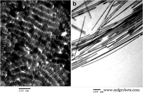
의 염기 상대 농도 더 큰 종횡비의 나노 물질을 만드는 데 중요한 역할을 합니다. CTAB는 또한 높은 수율의 막대를 준비하는 데 필요합니다. TEM 사진(Fig. 2)은 나노로드와 나노와이어의 형태를 보여준다.
<그림>
아 0.06mL 종자가 있는 제제에서 모양이 분리된 은 나노로드. ㄴ 모양이 분리된 은나노와이어 [34]
은 나노로드는 Lee et al. [35]. 종자매개 성장법에서는 먼저 작은 금속 입자를 준비하고 나중에 나노막대 제조를 위한 종자로 사용합니다. 은 종자는 안정제로서 시트르산나트륨 이수화물의 존재하에 수소화붕소나트륨으로 은 이온을 환원시켜 제조하였다. 이 은 종자를 은염, 아스코르브산(약환원제) 및 CTAB를 더 많이 포함하는 용액에 첨가했습니다. 이 연구에서 반응 온도와 pH는 생성된 막대의 종횡비와 균일성을 제어했습니다. 반응 온도의 증가는 은 나노로드의 종횡비를 감소시키고 단분산 입자의 크기를 증가시켰다. 또한 pH의 증가도 유사한 결과를 보였다. 반응온도와 pH가 증가할수록 은의 환원율은 더욱 증가하였다. 실험에서는 30 °C, pH 10.56 조건에서 높은 종횡비와 단분산성을 갖는 은 나노로드를 합성하였다. 은 나노로드는 AgNO3 수용액에서 전기화학적 방법으로 합성되었습니다. Zhu et al.의 폴리에틸렌 글리콜(PEG) 존재 하에 [36]. AgNO3의 농도가 PEG는 나노로드의 형성에 영향을 미쳤습니다.
Murphy et al. 은 나노와이어를 제조하는 더 나은 방법을 제공했지만 Sun의 [37, 38] 합성 방법이 더 정교합니다. 그들은 AgNO3를 환원하여 은 나노와이어를 합성했습니다. 종자 및 PVP의 존재하에 에틸렌 글리콜로. 반응 메커니즘은 다음과 같습니다.
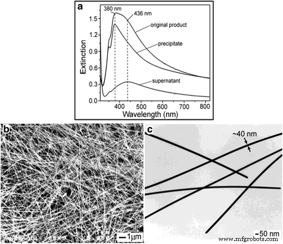
다음으로 AgNO3 및 PVP는 반응 시스템에 추가되어 은의 핵 생성 및 성장을 허용하고 균일한 모양과 크기의 나노와이어를 형성합니다. 직경이 30~40 nm이고 길이가 최대 ~ 50 μm인 은 나노와이어가 이러한 방식으로 생성되었습니다. 다양한 반응 조건(온도, 반응 시간, 씨뿌리기 조건)이 형태와 크기에 미치는 영향이 이 리뷰에서 논의되었습니다. 그림 3은 정제된 나노와이어의 모양과 크기를 보여줍니다.
<그림>
아 원심분리 및 분리 3주기 전후의 최종 제품의 UV-가시광 소광 스펙트럼. ㄴ 검색엔진 마케팅 및 c 은 나노와이어의 정제된 샘플의 TEM 이미지 [37]
UV-vis 스펙트럼(그림 3a)에서 은 나노와이어는 정제 후 원래 샘플과 비교하여 UV-vis 흡수에서 명백한 청색 이동을 보여줍니다. UV-vis 흡수 피크는 380 nm에서 나타납니다. 그림 3c는 이러한 나노와이어의 너비가 40nm임을 보여줍니다. 동일한 폭의 나노와이어를 얻을 수 있다는 것이 이 연구의 가장 큰 장점이다. 이러한 나노와이어는 전도성 필름[39] 및 효율적인 유기 태양 전지[40] 등을 제조하는 데 사용될 수 있습니다.
PVP가 은 나노와이어와 어떻게 반응하는지 추가 연구를 통해 Xie et al. [41,42,43]은 PVP 단층이 Ag-O 결합을 통해 Ag 나노와이어와 반응한다고 결론지었습니다. 이를 바탕으로 Xie et al. [44, 45]는 다중 쌍정에 대한 Xia가 Ag 나노와이어 형성의 핵심 요소 중 하나임을 입증한 실험에서 다중 쌍정의 존재를 관찰했습니다. 질산은의 초기량을 조절하거나 질산은의 초기 환원율을 낮추는 것은 용액에서 은 나노와이어의 형성에 도움이 된다[46, 47]. 그들이 사용한 구체적인 방법은 반응 용액에 염소 이온을 첨가하거나 은 이온의 방출 속도를 감소시켜 금속염과 질산은의 반응을 제어하는 것입니다.
Tanget al. [48] 이온 농도가 더 높은 시스템에 스테인리스 스틸 메쉬를 추가하여 크기가 조절된 은 나노와이어를 합성했습니다. 주로 스테인레스 스틸 메쉬가 질산과 반응하여 다중 결정립의 부식을 방지하는 데 도움이 될 수 있습니다. 그들은 염화물 이온이 있는 상태에서 열수법, 마이크로웨이브 방법 및 기타 실험 방법을 사용하여 균일한 은 나노와이어를 제조했습니다[49, 50]. 황화은 나노 입자는 황 이온과 은 이온의 반응을 통해 쉽게 합성되는 새로운 유형의 반도체입니다. 황화은 나노 입자는 전자를 제공하고 은 이온을 표면에 흡착시켜 코어 및 환원제로 작용할 수 있습니다. 동시에 은 원자도 Ag2 표면에 증착될 수 있습니다. Ag2를 형성하는 S S@Ag 종자 및 자체 촉매 환원의 역할을 하여 은 나노와이어 형성에 도움이 됩니다[51].
Ag 나노스피어의 합성
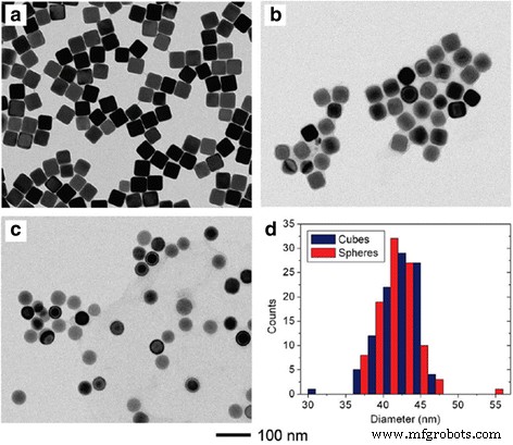
일반적으로 화학적 환원법에 의해 합성되는 준구형 은 나노입자는 은 나노입자 합성 과정에서 은 원자가 구형 구조를 형성하기 쉽기 때문에 많은 연구가 보고되고 있다. 화학적 환원 과정에서 일반적으로 사용되는 환원제로는 수소화붕소나트륨[52], 시트르산나트륨[53], 히드라진 수화물[54], 아스코르브산[55], 수소[56]가 있습니다. 모든 연구 팀 중 Xia의 팀은 연구에서 가장 상세하고 포괄적입니다. 고품질의 단결정 Ag 나노구체를 얻기 위해[57], 그들은 화학적 환원과는 다른 습식 에칭에 기초한 새로운 방법을 사용한다. 균일한 Ag 나노큐브의 현탁액을 소량의 질산제2철 또는 페리시아나이드 기반 에칭 용액과 빠르게 혼합하여 날카로운 모서리와 가장자리를 잘라 둥근 나노큐브를 형성하거나 원래와 동일한 직경을 갖는 날카로운 특징 없이 나노구를 얻을 수 있습니다. 큐브. 이전 합성 방법은 ~ 35 nm보다 큰 균일한 구체를 준비할 수 없기 때문입니다. 특히, 이 방법은 광범위한 크기의 균일한 Ag 나노구를 생성하고 SERS에 대한 기본 연구를 위한 새로운 가능성을 여는 데 사용될 수 있습니다. 이 작업에서 Ag 구체는 25nm의 가장 작은 크기와 142nm의 가장 큰 크기로 준비될 수 있습니다. 그림 4는 은 나노큐브가 준 나노구로 에칭되었음을 보여줍니다.
<그림>
TEM 이미지(a–c ) 다른 부피의 0.5mM 질산제2철로 에칭할 때 PVP로 덮인 42nm Ag 큐브. 아 0 μL. ㄴ 10μL. ㄷ 100μL. da 부분의 100개 입자에서 계산된 크기 분포 및 c [57]
이 방법으로 제조된 AgNP는 규칙적인 모양과 균일한 크기를 가지고 있습니다. 규칙적인 모양과 균일한 크기를 가진 이러한 은 나노구는 생물학적 표적 약물 전달에 사용할 수 있는 균일한 금 나노케이지를 제조하는 데 사용할 수 있습니다[58].
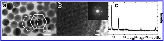
Liang et al. [59]는 단분산 은 나노입자를 제조하는 새로운 기술을 보고했다. PEG는 용매 및 환원제로 사용되며 PVP는 단분산 은 나노 입자 합성을 위한 캡핑제로 사용됩니다. 평균 직경이 54 nm인 균일한 나노구를 얻기 위해 Liang은 PVP/AgNO3 260 °C에서 8의 비율로 몰. 그림 5는 은 나노구의 TEM, HRTEM 및 XRD 이미지를 보여줍니다.
<사진>
아 TEM 및 b AgNO3에 대한 PVP의 몰비로 260°C에서 24시간 동안 제조된 은 나노구의 HRTEM 이미지 8의 SAED 패턴(삽입), 직경이 약 50 nm인 개별 은 나노구. ㄷ 동일한 샘플 배치에서 가져온 분말 XRD 패턴 [59]
TEM 이미지에서 Ag 나노스피어의 크기가 균일함을 알 수 있다. 또한, 합성 방법이 간단하여 대량 생산에 적용할 수 있습니다. 물론 구형 나노은 연구에 대한 다른 많은 논문들도 배울 가치가 있습니다. 그러나 이 작업에서 우리는 그것들을 반복하지 않을 것입니다. 다음 섹션에서는 다양한 크기의 은 나노 입자를 제조하는 세 가지 유형의 제조 방법에 대해 설명합니다. 규모와 성과의 효과를 연구하는 직장인들에게 조금이나마 도움이 되었으면 합니다.
다양한 크기의 AgNP 준비
크기가 다른 은 나노 입자가 재료의 성능에 큰 영향을 미친다는 것은 보편적으로 알려져 있습니다. 그럼에도 불구하고 우리는 크기가 다른 은 나노 입자의 제조 방법을 체계적으로 설명하는 논문이 거의 없음을 발견했습니다. 그래서 확실한 사이즈를 얻고자 하는 분들에게 도움이 되었으면 하는 마음으로 다음 섹션에서 합성 방법을 소개했습니다.
1-10 nm AgNP의 제작
작은 크기의 은나노입자는 일반적으로 수소화붕소나트륨을 환원제로 사용하는 급속환원 공정을 통해 제조되었으며, 생성된 입자의 크기와 모양이 균일하지 않았다. Shekhar et al. [60]은 환원제로 사용되는 다양한 비율의 수소화붕소나트륨과 시트르산나트륨을 혼합하여 5-10 nm 은 나노입자를 준비했습니다(빠른 핵형성을 우선적으로 줄이기 위해 수소화붕소나트륨을 사용하고 꾸준한 성장을 유지하기 위해 다시 시트르산나트륨 환원 사용). 이 방법으로 AgNP의 균일한 크기와 모양을 얻을 수 있었습니다. 다음 표 1은 다양한 크기의 은나노입자 합성을 위해 설계된 조건을 나타낸 것이다.
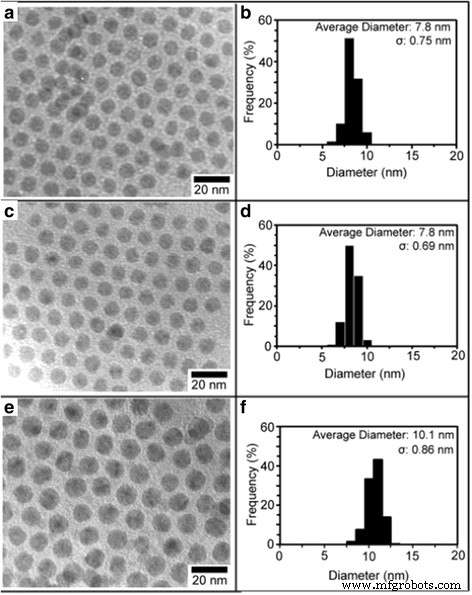
Linet al. [61]은 2003년에 모양과 크기가 균일한 7-10 nm 은 입자를 제조했다. 간단한 합성 방법은 은 트리플루오로아세테이트의 열 환원에 의한 크기 선택 공정을 사용하는 것보다 좁게 분산된 은 나노 입자를 직접 제조하는 것을 선호한다고 기술되었다. 올레산의 존재하에 이소아밀 에테르. 이 직접 합성은 합성적으로 제어하기 쉽고 직경이 7-10 nm 범위이고 크기 분포가 좁은 AgNP를 얻을 수 있습니다. 용매에 은염 전구체와 환원제를 포함하는 전통적인 접근 방식을 사용하는 대신, 유기 용매에 있는 단일 소스 전구체가 실험에 사용되었습니다. 이러한 이유로 그들은 쉽게 구할 수 있고 다양한 온도에서 은 금속으로 열적으로 환원될 수 있기 때문에 단일 소스 전구체로 은 트리플루오로아세테이트를 선택했습니다. 마지막으로, 그들은 올레산 대 은 트리플루오로아세테이트의 몰비를 조정하여 AgNPs의 직경을 변형시켰다. 다음 그림 6은 (A, B) 30, (C, D) 기간 동안 10:1의 올레산/은 트리플루오로아세테이트 몰비에서 얻은 명시야 TEM 이미지와 AgNP의 해당 입자 크기 분포 분석을 보여줍니다. ) 90, (E, F) 150분
<그림>
명시야 TEM 이미지 및 (a 기간 동안 올레산/은 트리플루오로아세테이트 몰 비율 10:1에서 얻은 AgNPs의 해당 입자 크기 분포 분석 , b ) 30, (c , d ) 90 및 (e , f ) 150분 [61]
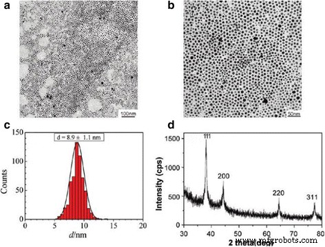
고농도에서 직경이 10nm 미만인 단분산 은 나노 입자를 합성하는 간단한 방법은 Yang et al. [62]. 그들은 아닐린을 환원제로 사용하고 도데실 벤젠 설폰산(DBSA)을 안정제로 사용하는 방법을 개척했습니다. DBSA 아닐린 AgNO3에 과량의 NaOH 첨가 시 시스템에서 은 나노입자의 형성은 90°C에서 단 2분 만에 거의 완료되었습니다(수율 94%). 또한, 생성된 은 나노입자의 평균 크기는 8.9 ± 1.1 nm이며, 콜로이드는 상온에서 1년 이상 보관할 수 있습니다. Fig. 7은 AgNPs의 TEM, DLS, XRD 이미지이다.
<그림>
아 , b 90℃에서 1시간 동안 NaOH를 첨가한 후 반응 시스템에서 수집된 은 나노입자의 2배율에서의 TEM 이미지. ㄷ 은 나노 입자 크기 분포의 해당 히스토그램. d 은 나노입자의 XRD 패턴 [62]
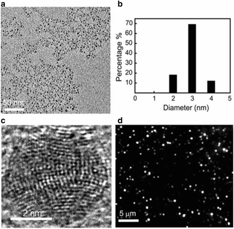
상술한 작은 크기의 은나노입자 합성방법은 모두 액상계이다. 그러나 Zheng et al. [63] 고체상 시스템에서 직경 2-4 nm의 은 나노 입자를 합성했습니다. 그들은 열 환원 방법을 이용하여 발광 및 라만 활성 은 나노 입자를 합성했습니다. 그림 8은 고체상 열분해에 의해 생성된 3nm 은 나노입자의 크기 분포, 구조 및 발광 방출을 보여줍니다.
<그림>
고체상 열분해를 사용하여 생성된 3nm 은 나노입자의 크기 분포, 구조 및 발광 방출. 아 이 나노 입자의 저해상도 TEM 이미지. ㄴ TEM에서 결정된 나노 입자의 크기 분포. ㄷ 이러한 작은 은 나노입자의 고해상도 TEM 이미지는 고도로 다중 도메인 구조를 보여줍니다. d ~ 10 W/cm
2
의 488nm 레이저 여기에서 촬영한 이 작은 은 나노입자의 발광 이미지 [63]
10–100 nm AgNP 제작
6 MeV 전자 조사에 의해 직경 10-60 nm의 AgNPs가 Bogle et al에 의해 합성되었습니다. 질산은과 PVP의 혼합물에서. 이 방법은 제조 효율이 높고 생산성이 높으며 부산물이 적다는 장점이 있습니다. Abid et al. [65] 위의 작업과 유사한 레이저 조사를 이용하여 은 나노입자를 제조하였다. 차이점은 질산은과 혼합하기 위해 캡핑제로 소듐 도데실 설페이트(SDS)를 사용하고 13-16 nm 크기의 은 나노입자를 제조할 수 있다는 점입니다. 입자 크기는 레이저 강도와 SDS 계면활성제의 초기 농도에 의해 제어됩니다. Ascorbic acid 환원을 사용하여 30-72 nm 크기의 구형 은 입자가 Qin et al.에 의해 합성되었습니다. [66]. 한편, 은 나노입자의 크기는 반응계의 pH가 6.0에서 10.5로 증가함에 따라 감소하였다. Ajithaet al. [67]은 14-31 nm AgNP를 얻기 위해 PH를 조정하여 화학적 환원을 활용했습니다. 그들은 에탄올을 용매로, 수소화붕소나트륨을 환원제로, 폴리비닐알코올(PVA)을 캡핑제로 사용했습니다. 그림 9는 이러한 은 나노입자의 형성 메커니즘을 보여줍니다.
<그림>
화학적 환원법을 사용한 크기 조절 AgNP 합성의 도식적 표현[67]
유사하게, 직경이 15-21 nm인 Ag 입자는 Silvert, P. Y. et al. 그들은 특정 온도 조건에서 질산은을 줄이기 위해 에틸렌 글리콜-PVP 용액을 사용했습니다. 이 방법으로 균일한 준나노구를 합성하였다. 크기가 다른 은 나노입자의 용해도를 감지하기 위해 Rui Ma 등은 10-80 nm Ag 입자를 준비했습니다. [69]. 그들은 성숙한 제조 방법인 폴리올 공정에 의해 분산 콜로이드은을 제조하였다[70]. 그들의 제조 방법은 보호제의 유형을 변경하여 Silvert, P.Y를 기반으로 합니다. 최근 녹색 합성 연구는 매우 열광적이며 연구자들은 일반적으로 아미노산 또는 박피 환원 Ag
+
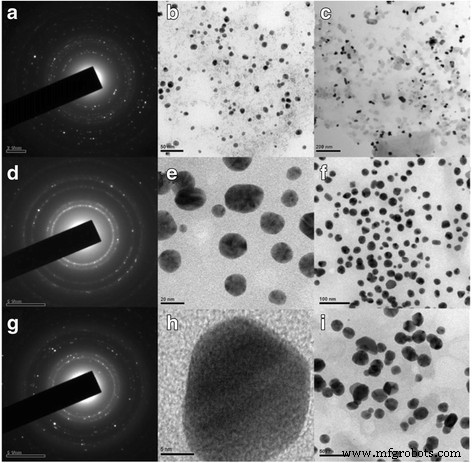
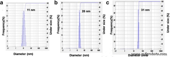
를 활용합니다. 은 나노 입자를 합성합니다. 환경 친화적인 합성 방법은 물리적, 화학적 제조 방법에서 유독 물질이 생성되는 문제를 극복할 수 있기 때문입니다. 그 중 Maddinedi et al. [71]은 티로신을 환원제 및 캡핑제로 활용하여 PH를 12에서 10으로 조정하여 13-33 nm 은 입자를 제조했습니다. Mandal et al. [72] 같은 결과를 얻었다. 그들은 Cinnamomum tsoi의 잎 추출물을 환원제 및 캡핑제로 사용하여 Cinnamomum tsoi의 잎 추출물의 내부 부피를 조정하여 11-31 nm 은 입자를 제조했습니다. 그림 10은 AgNP의 TEM 및 SAED 패턴을 보여줍니다.
<그림>
콜로이드의 TEM 이미지 및 SAED 패턴 잎 추출물 4ml(Ct4)의 부피(a –ㄷ ), Ct3(d –f ) 및 Ct1(g –나 ) 나노 입자 [72]
그림 11은 잎 추출물의 부피가 1, 3, 4 ml로 변화된 AgNPs의 동적 광산란(DLS)을 보여줍니다.
<그림>
AgNPs Ct1에 대해 얻은 평균 입자 크기(a ), Ct3(b ) 및 Ct4(c ) [72]
물론, 1-100 nm 은 입자를 제조하는 다른 많은 방법이 있습니다. 위의 논문은 전형적인 것입니다. 우리가 이 작업을 하는 이유는 정해진 크기를 합성하려는 누군가에게 도움이 되기를 바라는 마음 때문입니다. 결론적으로, 은 나노입자의 제조는 친숙한 합성 및 제어 가능한 크기로 안내되어야 합니다.
생합성 방법에 의한 AgNP의 제조
생물학적 시스템을 이용한 금속 나노입자의 생합성은 나노생명공학의 중요한 영역으로 발전하고 있다. 생합성 방법은 제조 공정에 환경 친화적인 기술이 채택되고 제품이 생체 응용에 적합하기 때문에 AsNP를 준비하기 위한 더 나은 후보입니다. 여기에서 생합성 방법은 개발 및 연구에 대한 전망이 있습니다. 따라서 일부 합성 사례에 대한 자세한 논의가 있습니다. 1999년, Klaus et al. [73] 처음 사용된 Pseudomonas stutzeri 200나노미터 크기의 은나노결정 합성 그 후 Aspergillus flavus 및 trichoderma와 같은 다른 균주를 사용하여 은 나노 입자를 제조하는 방법이 크게 개발되었습니다. 및 Kazemi et al. Geotricum sp.를 활용하여 Ag 나노 입자를 성공적으로 합성했습니다. 지오트리쿰 sp. Sabro Dextrose Agar(SDA) 배지에서 25 ± 1 °C에서 96시간 동안 성장했습니다. 균사체는 질산은 용액을 나노은으로 변환하는 데 사용됩니다. 이 균류(Geotricum sp.)를 사용하여 은 나노입자를 세포외에서 합성하였다. 이 효율적이고 친환경적이며 간단한 합성 방법은 30-50 nm의 Ag 나노 입자를 합성하는 데 사용할 수 있습니다. 상온 조건을 사용하고 유해한 환원제가 없기 때문에 이 방법은 환경 친화적이며 저렴한 비용으로 간주할 수 있습니다. 최근에는 laryssa et al. 선충류 Duddingtonia flagrans의 무세포여과액을 이용하여 은나노입자를 제조하였다. 이 연구에서 그들은 선충 균인 D. flagrans를 사용하여 AgNPs를 합성하는 간단한 생물학적 과정을 보고했습니다. 저렴하고 친환경적이며 고수율인 생합성에 비해 입자를 살아있는 세포에서 분리하기 위한 별도의 처리가 필요 없는 세포외 합성은 보다 간단한 과정이다. 생합성되고 기능화된 AgNP는 우수한 안정성과 높은 수율을 가지며 항균, 항진균, 항바이러스 및 항암의 탁월한 특성으로 인해 치료 응용 분야에서 유망한 미래를 갖게 되어 곰팡이 D. flagrans 사용에 대한 새로운 실험 설계를 강화할 수 있습니다.
생물학적 미생물의 종류는 나노은 연구의 최신 연구 방향이 될 것임을 알 수 있습니다.
AgNP의 속성 및 응용
항균에 대한 AgNP의 특성 및 응용
최근에는 Ag 나노 물질의 항균 특성이 점차 사람들의 관심을 불러일으키며 많은 항균 응용이 보고되었다[76, 77]. Helmlinger et al.은 모양이 다른 항균 AgNP를 연구했습니다. [78]. By studying the cytotoxicity and antibacterial effect of four types silver nanometals, it can be seen that silver nanoparticles with different shapes own equal cytotoxicity, but it has different antibacterial effect. Meanwhile, particles with a higher specific surface area are more toxic for bacteria than particles with smaller specific surface areas. The dissolution kinetics is correlated to the estimated specific surface area of the particles where particles with a higher specific surface area dissolve faster than particles with a smaller one. The difference in the dissolution rate may be exploited to synthesize silver nanoparticles with a relative higher antibacterial effect and a lower cytotoxic effect towards tissue. However, Helmlinger et al. did not give a further detail study on the antibacterial effect of different sizes of AgNPs.
The antibacterial properties of silver particles with different sizes were studied by Agnihotri et al. [60]. It can be seen that 5 nm nanoparticles have the best antibacterial properties. It was found that the smaller particles exhibited the better antibacterial properties. The Fig. 12 shows the antibacterial properties of the different-sized silver nanoparticles.
Disk diffusion tests for different-sized silver nanoparticles against the E. coli MTCC 443 strain. The zone of inhibition is highlighted with a dashed circle indicating a noticeable antibacterial effect [60]
Silver extends its antibacterial properties by combining with other materials. Research about combining with other materials included SiO2 @Ag [79], PLLA microcapsules combined with silver nanoparticles [80], electrodeposited chrome/silver nanoparticles (Cr/AgNPs) [81], graphene quantum dot/silver nanoparticles [82], Ag-decorated polymeric micelles with curcumin [83] and so on.
All the above studies are about the antibacterial properties of AgNPs. Next, we introduced the silver nanoparticles for antimicrobial application. It was found that the silver nanoparticles can be directly utilized as antibacterial agents which have been also testified by Kujda et al. [84]. It is shown that silver particles attach to the bacteria surface inducing disintegration, which enables their penetration inside the bacteria. In the future, the antibacterial properties of silver nanoparticles should be applied in industry by combining with other materials. For example, Meng et al. [85] made silver nanoparticles adhered to multilayered film-coated silk fibers with the aim to get antibacterial application. The as-prepared silk could effectively kill the existing bacteria and inhibit the bacterial growth, demonstrating the antimicrobial activity. Moreover, the release of Ag
+
for the modified silk can last for 120 h, rendering the modified silk sustainable antimicrobial activity. This work may provide a novel method to prepare AgNPs-functionalized antimicrobial silk for potential applications in textile industry. Figure 13 shows the surface morphologies of pristine silk fiber and coated morphologies of silk. By the EDS analysis, we can make sure that nanosilver was coated with silk.
Surface morphologies of pristine silk fiber (a ), (PAA/PDDA)8 film-coated silk fiber (b ), and AgNPs-(PAA/PDDA)8 film-coated silk fiber (c ). Inset:SEM image with higher magnification. (d ) EDS spectrum of AgNPs-(PAA/PDDA)8 film-coated silk. The arrow indicates the point randomly selected for the EDS analysis [85]
Other people like Zulfiqar Ali Raza et al. [86] investigated single-bath fabrication and impregnation of silver nanoparticles on enzymatic pretreated cotton fabric by using starch both as reducing as well as stabilizing agent under the autoclave conditions of 103.42 kPa, 121 °C for 15 min. The silver nanoparticles impregnated cotton fabrics showed good durable antibacterial activity against Escherichia coli and Staphylococcus aureus strains. Figure 14 shows the formation mechanism of impregnation of silver nanoparticles on cotton fabric.
Schematic diagram of impregnation of silver nanoparticles on cotton fabric [86]
Recently, silver nanoparticles were coated with zirconia by Yamada et al. [87] for antibacterial prosthesis. In view of the pronounced antimicrobial properties and small toxicity of AgNPs, the biocompatible AgNPs-coated yttria-stabilized zirconia can be potentially utilized to control dental caries and periodontal disease. Maybe the inspiration about wound repair will be obtained by this study. The excellent antibacterial properties of silver nanoparticles can be revealed by the above studies. Moreover, this work will help someone who wants to do further research on antibacterial.
Properties and Applications of AgNPs on Fluorescence
Because nanomaterials with fluorescent property have a great application prospect. Many efforts have been devoted to study the fluorescent property [88, 89]. Research on fluorescent nanoparticles mainly concentrates on semiconductor particles, which are usually referred to as quantum dots. Among these, CdSe particles and ZnS particles have stronger fluorescent intensity. In spite of their broaden applications, quantum dots frequently still have some problems which are related to the intrinsic blinking of their luminescence and to toxicity issues that limit their applications in the health sciences [90]. Silver is expected to have lower toxicity and can be readily prepared reproducibly and with excellent solution stability. At the same time, Ag is readily detectable in the visible spectral region [91]. Because silver has the abovementioned advantages, the preparation of highly fluorescent silver nanoparticles is needed. Highly fluorescent silver nanoparticles were prepared by Maretti et al. [92] with a facile photochemical method, which can yield these materials with excellent long-term stability in just a few minutes. The method is used photogenerated ketyl radicals which can reduce Ag
+
from silver trifluoroacetate in the presence of amines. The conclusion they obtained is that the luminescence arises from particle-supported small metal clusters (predominantly Ag2 ). Typically, silver nanoparticles show a distinct plasma band which has been between 390 and 420 nm in their past work. Due to the presence of small silver clusters, the study of the absorption band obtained was closer to 450 nm. Figure 15 shows the UV-vis absorption spectra of silver nanoparticles. Figure 16 shows the absorption (red), emission (green), and excitation (blue) spectra of Ag particles after 4 min of irradiation in tetrahydrofuran (THF) under the conditions of Fig. 15 and resuspension in toluene. From Fig. 16, we can draw the conclusion that the silver nanoparticles can emit green light. This property can be used for fluorescence diagnosis in biomedical field [93].
UV-vis absorption spectra following irradiation (350 nm, four lamps) of a toluene solution containing 2 mM silver trifluoroacetate, 2 mM I-2959, 2 mM cyclohexylamine. Reaction performed and monitored directly in a 0.7 × 0.3 cm quartz cuvette [92]
Absorption (red), emission (green), and excitation (blue) spectra of Ag particles after 4 min of irradiation in THF under the conditions of Fig. 15 and resuspension in toluene [92]
In order to distinguish these ultra-small particles, these nanoparticles which are smaller than 2 nm are usually called nanoclusters. In this size regime, metal nanoclusters become molecular species and size-dependent strong fluorescent emission can often be observed upon photoexcitation in the UV-visible range [94]. In particular, Ag nanoclusters, which show higher fluorescent intensity than Au nanoclusters in solutions, received considerable attention in the past few years owing to their great promise in a wide range of applications [95]. Fluorescent Ag nanoclusters were found to have wide applications in bio-imaging [96], chemical sensing [97, 98], fluorescence labeling [99], and single-molecule microscopy [100].
Properties and Applications of AgNPs on Catalysis
Since the addition of silver nanoparticles into reaction, the catalytic performance of the reaction has been significantly improved. Thus, nanocatalysis of silver nanoparticles has been a rapid growing research area which involves the use of nanoparticles as catalysts. As we all know, metals such as Ag, Au, Pt, and other metal ions can catalyze the decomposition of H2 O2 to oxygen [101]. Guo et al. found that when the AgNP colloid was added into the solution of luminol-H2 O2 , the chemiluminescence (CL) emission from the luminol–H2 O2 system could be greatly enhanced. AgNPs exhibited a better catalytic performance of CL than gold and platinum nanoparticles. The AgNPs-enhanced CL was ascribed to that AgNPs could catalyze the decomposition of H2 O2 to produce some reactive intermediates such as hydroxyl radical and superoxide anion. Figure 17 shows the effect of Ag colloid, Au colloid, Pt colloid, and filtrated solution of precipitated Ag colloid on luminol–H2 O2 CL [102].
Effect of Ag colloid (solid line), 38 nm Au colloid (dashed line), Pt colloid (dash-dot-dot line), and filtrated solution of precipitated Ag colloid (dotted line) on luminol–H2 O2 씨. The blank (filtrated solution of precipitated Ag colloid) signal was amplified by 100 times. Conditions:luminol, 1 × 10⁻⁴ mol/L; H2 O2 , 0.15 mol/L; pH 9.32 carbonate buffer for Ag, pH 12.0 NaOH for Au, pH 10.3 carbonate buffer for Pt [102]
Silver is the most popular catalyst when it has interaction with oxygen, water, carbon dioxide, ethylene, and methanol [103]. From the study that the catalytic properties of silver nanoparticles have accordingly changed can be realized. Jiang et al. [104] enhanced the catalytic properties of Ag by combining silver nanoparticles with silica spheres, and they also applied it to the detection of dye reduction. The technique to support silver particles on silica spheres effectively avoids flocculation of nano-sized colloidal metal particles during a catalytic process in the solution, which allows one to carry out the successful catalytic reduction of dyes. Figure 18 shows how the absorbance spectrum of the dyes decreases when the dyes are reduced.
아 Silver nanoparticles immobilized on silica spheres are illustrated. ㄴ The absorbance spectrum of the dyes decreases as the dyes are reduced by sodium borohydride. This process is catalyzed by silver nanoparticles. The arrow marks the increase of reaction time [104]
In addition, the catalytic properties of silver also have important applications in other areas, for example, wet-spun fibers [105].
Properties and Applications of AgNPs on Surface Plasmon Resonance
In 1902, Wood found the SPR phenomenon for the first time in an optical experiment and made a brief record about that, but until in 1941, a scientist named Fano explained the phenomenon of SPR. Over the next 30 years, the theory about SPR has not been further explored nor has it been put into practical application. In 1971, Kretschmann put forward prism coupling structure that settled the foundation for the structure of SPR sensor, and SPR theory started to be widely achieved for experiments. On this basis, the surface plasma resonance effect of silver nanoparticles was explored deeply. The most successful part of the applications of plasmonic structures was in the detection of molecules. This technique has been commercialized for propagating surface plasmons (PSPs) on continuous metal films. The films are chemically functionalized to selective bind target molecules like DNA strands or proteins. Upon binding the target molecule, the dielectric environment is altered around the surface of the metal film. Consequently, binding can be monitored by measuring the change in coupling geometry (i.e., the angle) between the metal film and the excitation source needed to generate PSPs [106, 107]. This technique plays a key role, and a number of commercially available instruments are widely used today in the biological sciences [108].
Recently, the combination of silver nanoparticles with other materials to improve their surface plasmon resonance performance is another way of development. The nanosilver particles were bonded with starch by Vasileva et al. [109], and the materials were applied as a surface plasmon resonance-based sensor of hydrogen peroxide. Figure 19 shows the change of hydrogen peroxide decomposition.
아 Change of the LSPR absorbance strength with time due to the introduction of 10–3 mol/L H2 O2 solution in the as-synthesized Ag-NPs solution at a volume ratio 1:1.5; the inset shows the bubbles from H2 O2 decomposition generated by the catalytic reaction between hydrogen peroxide and starch-stabilized Ag-NPs. ㄴ UV-vis absorption spectra recorded 15 min after the introduction of hydrogen peroxide solution with different concentrations in the solution of Ag-NPs at a volume ratio 1:1.5. ㄷ relevant photographs of Ag-NPs dispersions 60 min after the introduction of hydrogen peroxide with different concentrations [109]
SPR has a wide range of applications in other fields such as life science, medical testing, drug screening, food testing, environmental monitoring, and forensic identification.
The SPR technology becomes an indispensable part in the field of biological chemistry, food, and drug monitoring. The applications of SPR biosensors will be more diversified. And especially its emerging application in small molecule detection and lipid field will make it play an increasingly important role in the film and biology. In recent years, its development is particularly rapid. With the continuous improvement of SPR instruments and the continuous enhancement of biological membrane construction capability, SPR biosensor has a bright future.
Applications of AgNPs on Nanosensors
Due to the great research prospect of silver nanoparticles in nanosensors, many researchers have devoted to study it [110, 111]. So, we pick three representative examples to write in detail. Among them Zhu et al. [110] fabricated rhombic silver nanoparticles for biosensing. The rhombic silver nanoparticles were prepared by follow method. The mixed solution (polystyrene nanospheres and glass nanospheres with fluorocarbon surfactant) was coated onto the glass substrate to form a deposition mask, and then followed by hydrofluoric acid etching to remove the glass nanospheres. After that, the Ag metal thin film was deposited through the nanosphere masks using thermal evaporation or electron beam evaporation. After removal of the polystyrene nanospheres by sonication in absolute ethanol for 3 min, well-ordered rhombic AgNPs array was finally obtained on the substrates. The rhombic AgNPs array was single particle dimension of 140 nm in-plane width and 47 nm out-of-plane height. To prepare the biosensing, the Ag nanorhombuses are firstly functionalized using the self-assembly monolayer technique. Then assisting with 1-ethyl-3-[3-dimethylaminopropyl] carbodiimide hydrochloride, we covalently attached biotin to the carboxylate groups. The advantage of this biosensor is that the rhombic AgNPs array-based sensor with more hot spots has higher sensitivity than that of the traditional Ag triangular nanoparticles-based sensor. A detection of high sensitivity of the bio-molecule in lower concentration has been realized by means of the LSPR-based nanobiosensor. This type of biosensor will have potential applications in many fields such as medical science and biological technology. Meanwhile, M. Ghiaci et al. [111] utilized silver nanoparticles compounds as new electrochemical sensors for glucose detection. These electrochemical sensors were prepared based on synthesizing of two amine compounds bounded to silica support. The size of used AgNPs is 10 nm. The electrochemical sensor prepared by this method has a lower limit of glucose detection than other electrochemical sensors. This type of nanosensors will be more conducive to diabetes detection and treatment. Silver nanoscale sensors can also be used for environmental detection such as Li et al. [112] synthesized aza-crown ether (ACE)-modified silver nanoparticles as colorimetric sensors for Ba
2+
. What is more, colorimetric sensors merely need minimal instrumentation, achieve high sensitivity, and thus can make on-site detection even easier. The colorimetric sensors were synthesized by silver nanoparticles efficiently conjugated with CS2 –ACE. ACE-modified AgNPs have good recognition of Ba
2+
, with the detection limit of 10
− 8
mol/L.
In addition to the abovementioned, silver nanosensors also have other different applications that are worth us to explore.
Other Applications
Ag nanomaterials also have many other applications in various fields, such as nanoscale detection [113] and solar cells.
Silver nanoparticle and its complex can be used for solar cells to enhance photoelectric conversion efficiency and photovoltaic performances [114,115,116].
Shen et al. [114] enhanced photovoltaic performances of polymer solar cells by incorporating Ag–SiO2 core–shell nanoparticles in the active layer. They creatively incorporated Ag–SiO2 core–shell nanoparticles (Ag–SiO2 -NPs) into photo−/electro-active layers consisting of poly(3-hexylthiophene) (P3HT) and phenyl-C61 -butyric acid methyl ester (PCBM) in polymer solar cells (PSCs). By this way, the photovoltaic performance of PSCs have largely been enhanced. The results demonstrate a 13.50% enhancement of short-circuit photocurrent density and a 15.11% enhancement of power conversion efficiency as the weight percent of doped Ag–SiO2 -NPs is 1.5 wt% in the active layer of corresponding PSCs. In the later research, bare silver nanoplate (Ag-nPl) were spin-coated on indium tin oxide and silica capsulated Ag-NPs were incorporated to a PBDTTT-C-T:PC71BM active layer by Shen et al. [115]. As a result, the devices incorporated with Ag-nPl and Ag@SiO2 -NPs showed great enhancements. With the dual effects of Ag-nPl and Ag@SiO2 -NPs in devices, all wavelength sensitization in the visible range was realized; therefore, the power conversion efficiency of PSCs showed a great enhancement of 14.0 to 8.46%, with an increased short-circuit current density of 17.23 mA cm
− 2
. Importantly, the methodology of multiple shape combination of metallic nanoadditives improves the photovoltaic performance of PSCs very effectively compared to the single-shape method.
Thus, Ag is a promising material for the conversion of solar energy into electricity and good detection. In addition to the abovementioned, Ag also has many other applications, but it still needs people to further explore it.
Conclusions
This work reviewed the development progress of Ag nanomaterials on synthesis methods and applications. Different shapes of Ag nanostructures had been synthesized such as cubic, rod-shaped, and sphere-shaped, Ag nanostructure obtained by chemical synthesis and microwave methods were successfully prepared. In addition, different size of AgNPs have been synthesized such as 1–10 nm, 10–100 nm, AgNPs obtained by chemical synthesis, laser ablation, and green synthesis. Meanwhile, it has been successfully applied to many fields, such as antibacterial, fluorescence, catalysis, SPR, and nanosensors, and it is expected to use in other fields. In fact, there are still limitations for their practical applications in photoelectric and medical fields because it often requires complex preparation process, and the yield is very low. In most cases, AgNPs are easy to agglomerate, which will greatly reduce its optical properties. Therefore, it is necessary to utilize surface active agent to achieve a good effect. Although, there are so many challenges, the advances in nanoscience and nanotechnology of silver still promise a better future for many kinds of industries. In conclusion, the future research of silver nanoparticles should be directed towards biosynthetic, size controllable, and uniform shape preparation. And the future application of AgNPs-based will be utilized in new energy battery or wearable intelligent equipment by its excellent localized surface plasmon resonance effect and antibacterial activity. In addition, AgNPs-based materials can be further utilized for applications in nanodevices by self-assembly and molecular molding technology.