발광 재료는 독특한 광학적 특성으로 인해 전 세계적으로 관심을 받고 있습니다. 빛에 투명한 실리카는 발광 재료에 이상적인 매트릭스입니다. 발광 실리카 나노입자(LSN)는 향상된 화학적 및 열적 안정성으로 인해 광범위한 응용 분야를 가지고 있습니다. 다양한 크기의 실리카 구체는 특정 요구 사항을 충족시키기 위해 다양한 방법으로 합성할 수 있습니다. 다양한 발광 염료는 다양한 응용 가능성을 가지고 있습니다. 소광제 등 여러 요인으로 인해 성능이 그다지 만족스럽지 못했습니다. 따라서 이 검토에서는 분류, 합성 및 적용을 포함한 LSN의 개발에 대해 논의합니다. 실리카가 발광 염료의 특성을 향상시키는 방법과 실리카가 시스템에서 어떤 역할을 하는지가 하이라이트입니다. 또한 생물학, 디스플레이 및 센서에서의 응용 프로그램에 대해서도 설명합니다.
소개
발광 재료는 특수한 광학적 특성 때문에 널리 적용됩니다[1]. 그러나 낮은 소수성 및 생체적합성과 같은 많은 제한이나 높은 독성, 열악한 생체적합성, 낮은 흡광도와 같은 단점으로 인해 적용이 제한된다[2,3,4,5]. 따라서 실제 응용 요구 사항을 충족시키기 위해 발광 재료를 수정할 필요가 있습니다.
특성이 개선된 LSN은 생물학[6, 7], 조명[8], 센서[9]에서 점점 더 많은 관심을 끌고 있습니다. 그들의 특징적인 광학적 특성으로 인해 광학 재료에서 고유합니다[10]. 실리카는 빛에 투명하여 실리카를 형광 물질의 매트릭스로 이상적인 후보로 만듭니다. 열역학적 및 화학적 안정성도 매트릭스와 실리카가 이러한 기본 요소와 일치하므로 중요한 요소입니다[11]. 또한, 실리카의 표면은 쉽게 변형될 수 있어 다양한 요구 사항에 적응할 수 있도록 다양한 작용기로 추가 작용화가 가능합니다[12]. 위의 많은 장점을 가진 실리카는 자연적으로 발광 물질의 특성을 향상시키기 위한 이상적인 기질이다[13]. 다기능 나노시스템은 서로 다른 공정을 사용하여 실리카 나노입자의 표면과 내부에 하나 이상의 서로 다른 나노물질을 조립, 캡슐화 또는 통합하여 만들 수 있습니다[11]. 발광 물질을 변형시키는 것으로 우수한 특성을 갖는 LSN이 프론티어 연구에서 점점 더 주목받고 있다[14]. Montalti et al. 유기 염료 도핑된 실리카를 사용한 의료 영상의 많은 우수한 연구를 요약했습니다[6]. 실리카는 형광체를 위한 안정적이고 다기능적인 플랫폼을 제공하지만 장기적인 독성에 대한 연구가 필요합니다. Michael Schäfrling은 발광 센서 기술을 시연했습니다[9]. 선택성과 감도는 센서 재료의 핵심입니다. Zou Hua et al. 유기 실리카 개질 수단에 대해 자세히 설명했습니다. 나노복합체는 구성요소를 분리하는 우수한 특성을 가지고 있습니다[15]. 생물학과 같은 특정 영역에 초점을 맞춘 경이로운 리뷰가 많이 있지만[6, 7, 16], LSN에 대한 체계적인 소개와 다른 분야에서의 우수한 성능이 부족합니다.
이 검토는 합성 방법에 따라 LSN의 분류로 시작합니다. LSN의 범주는 발광 물질의 분류를 기반으로 체계적으로 설정됩니다. 화학적 특성 및 발광 메커니즘 측면에서 유기 분자 염료, 발광 금속 및 양자점(QD) 도핑된 형광체는 3가지 대표적인 형광체이며, 모두 고유한 발광 메커니즘과 LSN을 대표하는 장점이 있습니다[17,18,19 ]. 하이라이트는 실리카가 형광체의 특성을 향상시키는 방법입니다. 발광 재료 응용 프로그램의 부족으로 LSN에 대한 성능을 향상시키기 위한 가능한 전략이 논의됩니다. 여기에는 생물학적 응용뿐만 아니라 디스플레이 및 센서도 포함됩니다.
LSN 분류
다양한 밝기를 방출하는 발광은 물질 분야에서 큰 가치를 지닌다[20]. 신호 대 잡음비, 안정성 및 잠재적 응용 분야에 대한 환경 적응성을 향상시키는 방법에 대해 발광 재료의 수정에 대한 많은 연구가 수행되었습니다. 발광 성능을 향상시키기 위해 란탄족 착물에 안테나 리간드를 도입하는 것은 변형의 전형적인 예입니다. 실리카는 기능이 다르고 화학적 특성이 다른 재료를 혼합하는 데 좋은 매트릭스입니다. 인광체는 실리카 매트릭스에 도핑되어 자연적 결함을 수정하고 특성을 개선하며, 이는 쉽게 개질되고 무독성인 실리카 표면과 발광 염료 보호가 있는 광범위한 응용 분야에 유리합니다. 다기능 및 조정 가능한 적응성을 갖춘 LSN은 점점 더 많은 관심을 끌고 있습니다. 모든 발광 인광체 중에서 유기 발광 분자, 발광 금속 도핑 인광체 및 QD는 강조할 가치가 있는 가장 대표적인 3가지 범주입니다. 따라서 위의 세 가지 염료는 실리카와 함께 일반적인 LSN으로 표시됩니다. 대표적인 예가 표 1에 나와 있습니다.
유기 발광 분자 도핑된 LSN
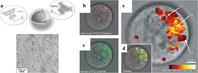
유기 발광 분자는 π-공액 고리 구조와 작은 크기를 갖는 중요한 발광 물질이다[16]. 그러나 비특이적 라벨링 및 표백은 적용을 방해합니다. 유기 염료 도핑된 실리카 나노 입자는 우수한 안정성, 선택성 및 생체 적합성을 가지고 널리 연구되고 있습니다[52, 53]. Van Blaaderen et al. [21]은 발광 실리카 구체를 합성하기 위한 예비 시도를 했다. Fluorescein isothiocyanate(FITC)는 APS((3-aminopropyl)triethoxysilane)의 도움으로 실리카 표면에 코팅되어 염료를 공유 결합으로 실리카와 결합하는 실행 가능한 방법을 제공했습니다. 이 과정에서 영감을 받아 Andrew et al. [22] 2개의 층을 가진 이중 방출 형광 실리카 나노 입자를 합성했습니다. 테트라메틸로다민 이소티오시아네이트(TRITC)와 FITC라는 두 가지 염료가 무수 질소 환경에서 APS를 통해 실리카에 접합되었습니다. 개략도 및 SEM 이미지(그림 1a)는 나노입자의 나노구조를 보여주었다. 이중 방출 나노입자의 코어로 TRITC를 포함하는 실리카를 먼저 합성하고 코어 표면에 FITC를 추가 테트라에톡시실란(TEOS)과 접합시켰다. 합성된 이중 방출 형광 실리카 나노 입자는 그림 1b-d에서 쥐 호염기성 백혈병 비만 세포(RBL-2H3)에서 세포 내 pH 값을 성공적으로 조사했습니다.
<그림>
아 참조 염료(TRITC) 및 센서 염료(FITC)를 포함하는 이중 방출 형광 실리카 나노입자의 형성 다이어그램 및 주사 전자 현미경(SEM) 이미지; RBL 비만 세포의 공초점 형광 현미경 이미지(TRITC 실리카 입자로 빨간색, AlexaFluor 488-콜레라 독소 B로 녹색); pH 센서로서의 RBL 비만 세포의 공초점 형광 현미경 이미지. ㄴ 참조 채널의 경우 c 센서 채널의 경우 d 오버레이된 이미지의 경우 e 실험에 따라 계산된 pH 값에 대한 가색 비율 측정 이미징 [22]
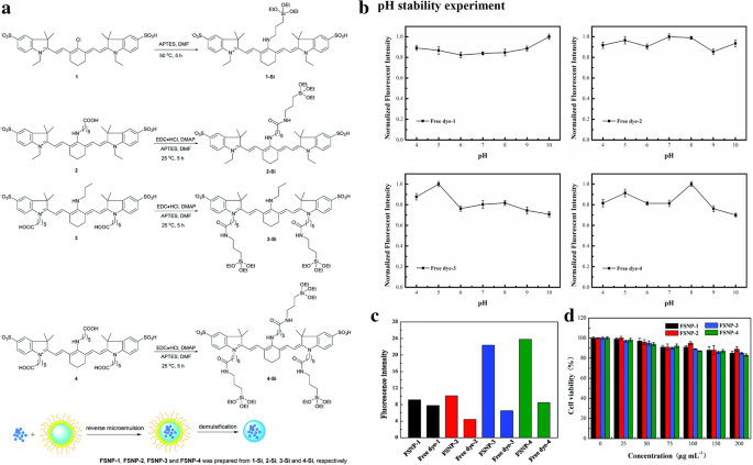
유기 형광단의 광안정성을 향상시키기 위해 실리카 캡슐화는 일반적으로 사용되는 변형 방법입니다. Long Jiao et al. [23]은 근적외선(NIR) 형광 프로브로 4개의 아미노시아닌 염료를 선택하고 일반적인 실란 커플링제인 3-아미노프로필트리에톡시실란(APTES)과 상응하게 커플링했습니다. 마이크로에멀젼 시스템에서 처리된 NIR 염료로 가수분해된 TEOS. 원심분리 및 세척 후에 시아닌이 로딩된 형광 실리카 나노입자(FSNP)를 얻었다. 전체 프로세스는 그림 2a에 나와 있습니다. 그림 2b에서 볼 수 있듯이 실리카에 캡슐화된 4개의 FSNP는 유리 염료보다 형광 강도 pH 안정성이 더 우수했습니다. 4개의 FSNP는 동시에 밝기를 개선했습니다(그림 2c). 그들은 공초점 레이저 주사 현미경(CLSM)으로 살아있는 세포에서 광안정성을 테스트했습니다. FSNP-3 및 FSNP-4(고정 위치가 많음)는 유리 염료보다 광안정성이 개선된 반면 FSNP-1 및 FSNP-2는 개선되지 않았습니다. 더 많은 고정 사이트는 염료 분자의 구조를 강화했습니다. 단단한 구조의 염료는 비방사성 붕괴가 적고 분자 내 회전이 단단하여 염료를 더 밝게 만듭니다. 실리카 층은 분자 구조가 강화된 캡슐화된 물질을 보호하고 광표백 없이 밝기를 향상시킬 수 있습니다. FSNP-3 및 4도 그림 2d의 MTT(methyl tetrazolium) 방법에 따라 생물학적 독성이 낮았습니다. 생체 적합성은 실리카의 또 다른 장점입니다.
<그림>
아 FSNP-1, FSNP-2, FSNP-3 및 FSNP-4의 메커니즘. ㄴ 모든 샘플의 다양한 pH 값에서 정규화된 강도. ㄷ FSNP 및 자유 염료의 방출 강도. d 24시간 동안 FSNP와 함께 배양한 후 원시 264.7 대식세포의 생존 가능성을 보여줍니다[23]
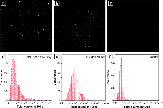
응집은 대부분의 발광 염료에 대한 담금질의 주요 원인 중 하나입니다. 형광체는 실리카와 함께 적정 농도를 꾸준히 유지할 수 있습니다. AIEgens(Aggregation-induced emission luminogens)는 기존 발광단과 달리 이 문제를 겪지 않습니다. 반대로, 응집은 강력한 방출로 이어집니다[54]. 생물학적 분야에서 AIEgen의 성능을 향상시키기 위해 많은 고분자 매트릭스가 AIEgen을 캡슐화하는 데 사용됩니다. 또한 물과 산소와 같이 AIEgens 퀜칭을 유발할 수 있는 몇 가지 다른 문제가 있어 적용에 부정적인 영향을 미칩니다. 실리카는 소광제에서 이들을 방지할 수 있습니다[55]. 위의 분석을 바탕으로, AIEgen인 TPETPAFN(2개의 테트라페닐에틸렌 펜던트와 분자내 전하 전달 코어로 구성된 전형적인 형광체)은 F127(폴리(에틸렌 글리콜)-블록-폴리(프로필렌 글리콜)-블록-폴리)에 의해 생체 기능화되었습니다. 에틸렌 글리콜)) 코어 미셀[24]을 형성합니다. TEOS를 가수분해하여 수정 졸겔법을 통해 코어 미셀에 코팅된 실리카 쉘을 얻었다. 그림 3에서 알 수 있듯이 합성 TPETPAFN-F127-SiO2 나노입자는 실리카 껍질의 보호로 인해 더 나은 광발광 특성을 나타냈습니다.
<그림>
아 , d TPETPAFN-F127-SiO2의 형광 이미지와 광자의 히스토그램 표시 NP, 해당 b , e TPETOAFN NP 및 c용 , f 상업용 QD655용 [24]
발광 금속 도핑 LSN
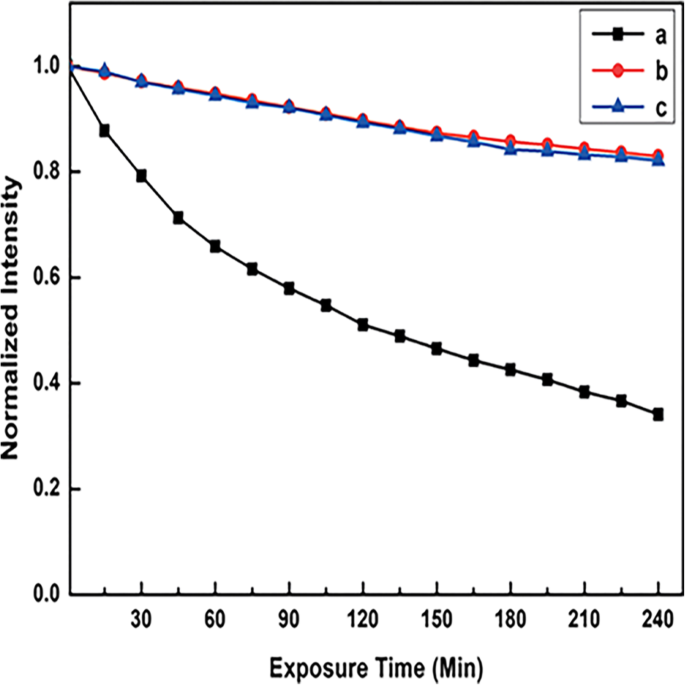
희토류 금속[56]과 전이 금속[57]은 전하 이동 전이를 기반으로 하는 일반적인 발광 금속 재료입니다. 리간드와의 착화 후 발광은 이 물질의 가장 명백한 특징입니다. 금속 발광의 두 가지 주요 메커니즘, LMCT(리간드에서 금속으로의 전하 전이) 및 MLCT(금속에서 리간드로의 전하 전이)가 있습니다. 란탄족 금속과 전이 금속이 각각 그 대표적인 예입니다. 풍부한 전자 에너지 준위로 인해 다양한 발광을 갖는 발광 분야에 적용할 수 있는 큰 잠재력을 가진 다양한 발광 금속이 있습니다[58]. LSPR이 있는 귀금속은 향상된 발광 재료에 널리 사용되었으며 이 섹션에 포함됩니다. 그럼에도 불구하고 낮은 감광 효율과 소광은 발광 금속의 적용을 제한합니다[59]. 광안정성과 생체적합성을 향상시키기 위해 Francis et al. 추가 수정을 위해 리간드에 치환된 실릴기를 추가했습니다[30]. Eu@Si-OH 나노입자는 역마이크로에멀젼법을 통해 실리카로 실릴기 개질된 Eu 착물을 코팅한 후 얻었다. 제품은 APTES로 Eu@Si-NH2로 기능화된 아민을 마침내 얻었습니다. 나노 입자. 실리카 층은 소광제(OH 및 NH2 여러 떼). 그 결과 그림 4에서 둘 다 더 나은 광안정성을 보였다. Eu@Si-NH2 나노입자는 바이오이미징에서 좋은 성능을 보였다.
<그림>
365nm 조사에서 노출 시간에 따라 변하는 형광 강도 곡선, a CH2Cl3의 부모 Eu 착물 솔루션, b Eu@Si-NH2 , 및 c 인산완충식염수(PBS) 완충용액의 Eu@Si-OH 나노입자 [30]
Ezquerroet al. Ir 착물, MLCT 발광 착물을 실리카 프레임워크에 통합하여 졸-겔 공정을 통해 안정성과 광물리적 특성을 향상시켰습니다[60]. 이 형광체는 실리카의 보호로 주변 조건뿐만 아니라 백색 발광 다이오드(WLED)에 추가로 적용되는 가혹한 환경에서도 우수한 안정성을 보여주었습니다.
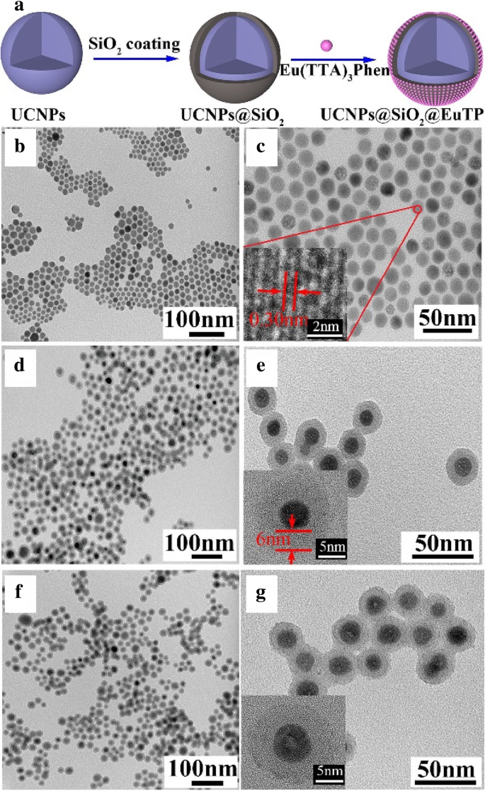
Y. Li et al. 합성된 올레산 안정화된 상향변환 나노입자(UCNP). 그런 다음 그들은 마이크로 에멀젼 방법을 통해 UCNP 위에 실리카 층을 코팅하여 수용성 UCNP를 얻었다. Eu(TTA)를 소개합니다3 phen 복합체를 시스템에 넣고 NaGdF4를 합성했습니다. :Yb,Er@SiO2 @Eu (TTA)3 펜(UCNPs@SiO2 @EuTP) 나노스피어. 실리카 코팅 후 표면 소광이 억제되어 그림 5와 같이 방출 강도가 향상되었습니다. 실리카를 사용하여 두 가지 다른 방출을 갖는 수용성 나노 입자를 얻었습니다.
<그림>
아 UCNPs@SiO2의 형성 @EuTP 및 TEM(투과 전자 현미경) 샘플의 이미지; ㄴ , ㄷ UCNP의 경우 d , e UCNPs@SiO2용 , 및 f , 지 UCNPs@SiO2용 @EuTP [31]
Chen et al. [42] WLED에 탄소점(CD)과 희토류 이온을 성공적으로 사용했습니다. 그들은 각각 470nm에서 최대 청색 방출과 251nm 및 364nm에서 2개의 여기 피크를 갖는 one-pot 유기 열분해 방법으로 CD를 합성했습니다. 백색 발광 합성물을 얻기 위해 청색 발광 코어로 CD를 사용하고 Sr2 시5 N8 :Eu
2+
형광체는 주황색 방출 성분으로 사용되었습니다. CD는 Stöber 시스템에 넣어졌습니다. TEOS가 가수분해됨에 따라 CD는 적색 형광체가 있는 실리카 층으로 코팅됩니다. 탄소점-실리카-형광체 복합체(CDSP)는 원심분리, 건조 및 분쇄 후에 합성되었습니다. CDSP는 UV(자외선)에서 노란색 영역(200–600nm)에 이르는 광범위한 흡수를 가지며 특히 UV 영역에서 강력합니다. 다양한 파장에서 여기를 테스트한 후, 그들은 CDSP가 CIE(Commission Internationale de l'Eclairage) 좌표(0.32, 0.32)를 그림 4의 400nm 여기에서 순수한 백색광(0.33, 0.33)의 좌표로 얻었다는 것을 발견했습니다. 6. 그리고 CD와 형광체의 질량비를 조정하여 CDSP의 방출을 얻으려는 좋은 시도였습니다. 400nm 여기에서 백색 방출에 가장 가까운 질량비(3.9%(0.32, 0.32) 및 5.1%(0.34, 0.32))를 얻었습니다. CDSP는 CD&P(CD와 형광체를 직접 혼합)(0.28, 0.29)보다 발광다이오드(LED) 패키징에서 더 나은 백색 방출(0.30, 0.31)을 보였다. 두 성분이 실리카로 균일하게 분산되어 응집 및 상분리 가능성을 감소시켰습니다. 마지막으로 UV 다이오드 칩(375nm)에 CDSP 분말이 포함된 WLED를 얻었고 백색광(0.30, 0.33)을 얻었습니다. 연색지수(CRI)는 약 94로 YAG:Ce 기반 상업용 WLED(CRI <75)보다 높았다.
<그림>
CDSP 기반 WLED의 성능:방출 스펙트럼 및 사진 [42]
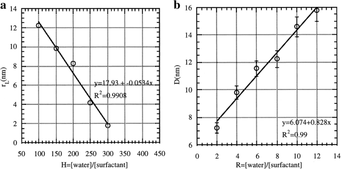
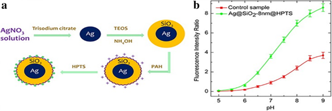
실리카는 일반적으로 형광성을 향상시키기 위해 귀금속과 적절한 거리를 유지하기 위해 발광 물질의 보호층으로 사용됩니다. 이것은 빛에 의한 자유 전자의 정상 진동 때문입니다. 발광성을 높이려면 염료와 귀금속 입자 사이에 적절한 거리를 유지해야 합니다. 고귀한 강화 물질의 경우 금속 나노입자 자체가 발색단 소광(5nm 이내)을 유발할 수 있지만 형광은 최대 100배(10nm 부근) 향상될 수 있습니다. 초기 연구에서 Tuo Li et al. 마이크로에멀젼 매트릭스(Ag/SiO2)에 실리카 쉘이 있는 합성된 Ag 나노입자 나노 입자). 실리카를 생산하는 데 필요한 시약(TEOS 및 사이클로헥산)은 은이 환원된 후 마이크로에멀젼에 주입되었습니다. 그들은 Ag/SiO2에 대한 다양한 조건(R의 경우 물/계면활성제 및 H의 경우 물/TEOS)의 영향을 주의 깊게 연구했습니다. 나노입자 및 그 결과는 Fig. 7과 같다. 마이크로에멀젼 시스템으로 Ag 뿐만 아니라 다른 나노입자도 코어에 균일하고 두꺼운 실리카 층을 코팅하는 좋은 경로이다. What Zhenhua Bai et al. [25] 가 좋은 예이다. 8-Hydroxypyrene-1,3,6-tresulfonic acid(HPTS)는 형광성 pH 민감성 염료의 일종으로 고유한 장점으로 인해 세포 내 pH 센서로 만들기에 적합합니다. 그러나 극한의 pH 조건은 그것을 둔감하게 만들었습니다. 용액이 산성이면 형광 효율이 크게 감소합니다. HPTS 흡착 Ag@SiO2 나노 입자(그림 8a)는 귀금속 강화 형광 효과를 기반으로 준비되었습니다. Ag@SiO2 -8 nm@HPTS는 특히 극한 pH 조건에서 더 나은 형광 강도를 나타냈습니다.
<그림>
아 H(R =4 및 X =1); ㄴR일 때 가변, Ag 클러스터의 크기 변화 [61]
<사진>
아 HPTS 흡착 Ag@SiO2 합성 진행 나노 입자. ㄴ Ag@SiO2의 형광 강도 비율 -8 nm@HPTS(녹색) 및 대조 샘플(빨간색) [25]
QD가 도핑된 LSN
양자 구속 효과로 인해 QD는 반도체 QD, 탄소 QD 또는 기타 유형에 관계없이 우수한 발광 특성을 나타냅니다. 최근 광소자에서의 양자점의 응용에 대한 많은 연구가 이루어지고 있다. 때로는 속성이 복잡한 응용 프로그램을 적용하기에 충분하지 않습니다. 필요한 수정이 필수적이며 실리카가 적합한 매트릭스[1]입니다.
생체표지와 자기공명영상의 결합을 실현하기 위해 자성 Fe2에 CdSe QD를 코팅했습니다. O3 NH2가 있는 실리카 층에 의한 코어 그룹. 관련 사진 및 특성은 그림 9에 나와 있습니다. 결합된 NH2 생체 고정막(BAM)이 있는 그룹, 4 T1 마우스 유방암 세포막은 BAM-SiO2로 특이적인 표지를 나타냄 -CdSe MQD[44]. 생체 적합성 및 자성을 갖춘 다기능 발광 나노 입자는 의학 분야에서 폭넓게 응용될 것입니다.
<그림>
자기 변형을 증명하기 위해 일반 조명 아래에서 사진(a , b ). ㄷ 자성 및 발광 특성을 모두 증명하기 위해 자외선 아래에서 사진. d 녹색 및 주황색 자기 양자점(MQD)의 UV 사진. 이 , f SiO2의 발광 스펙트럼 -PBS 솔루션의 MQD(e 흡수 및 f 방출용) [44]
QD 적용을 확대하기 위해서는 수용성 및 무독성의 수정이 필요합니다. 실리카는 QD의 변형에 큰 잠재력을 보여줍니다. Yunfei Ma et al. [43] 수제 상전이 시약(adenosine 5'-monophosphate, AMP)과 실란 커플링제(3-mercaptopropyltrimethoxysilane, MPS)를 Stöber 시스템에 도입했습니다. 지용성(초기 CdSe/CdS/ZnS 양자점), 알코올 용해성(AMP-QDs) 및 수용성(QD 주변의 TEOS 가수분해)은 용해도 변화의 전체 진행이었습니다. QD@SiO2 초기와 동일한 광발광 효율(50-65%)을 가졌습니다. QD@SiO2의 장점은 더 넓은 pH 범위(pH 4–8 ~ 2–13), 전해질의 향상된 안정성, 향상된 열 안정성, Hela 세포의 향상된 생체 적합성입니다. .
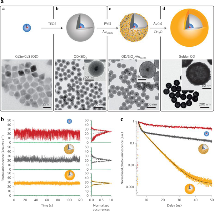
광학 소자에서 양자점의 안정성을 확보하기 위해서는 깜박임의 영향을 줄이는 것이 필요하다. 깜박임은 QD 광학 장치의 안정성에 영향을 미치는 무작위 간헐 발광 현상입니다[62]. 깜박임의 효과를 줄이기 위해 Botao Ji et al. [45] CdSe/CdS 양자점을 코어 물질로 생산하고 역 마이크로에멀젼 방법에 의한 초기 소수성 리간드의 치환을 기반으로 이 양자점을 실리카 쉘로 캡슐화하였다. 그리고 실란 커플링제로 폴리(1-비닐이미다졸-코-비닐트리메톡시실란)(PVIS)를 사용하여 실리카 표면의 Au 층에 의해 QD가 추가로 개질되었습니다. 나노 크기의 금 껍질은 QD에 향상된 광학 상태 밀도를 제공하는 플라즈몬 공진기 역할을 했습니다. 하이브리드 레이어로 인해 로컬 환경의 급격한 변화에도 QD의 속성을 유지할 수 있습니다. 그 결과 QD의 광안정성이 높아졌습니다. QD 형광 수명은 금 코팅 후 123에서 20ns로 감소했습니다. 황금 양자점은 효율적인 다중 여기자 방출을 보였고 그것의 중성 광발광 강도는 양자점보다 높았다. 안정성 테스트 결과는 그림 10에 나와 있습니다. 또한 광발광 강도는 몇 시간(심지어 24시간) 동안 안정적으로 유지될 수 있었습니다. 발광 안정성 테스트에서는 베어 QD의 발광이 단 1시간 후에 극적으로 떨어지는 것으로 나타났습니다. 실리카 층은 QD의 성능을 약간 향상시켰지만 다음 Au 층이 플라즈마 강화 효과를 나타내기 위한 적절한 간격을 제공했습니다.
<사진>
아 양자점/SiO2의 개략도 /Au 하이브리드(황금 QD) 및 각 단계의 TEM 이미지(CdSe/CdS QD, QD/SiO2 QD/SiO2 /Au종자 및 황금 QD). ㄴ 시간에 따른 광발광 강도의 변화. CdSe/CdS의 경우 빨간색, QD/SiO의 경우 회색2 /Au종자 , 황금 QD의 경우 주황색입니다. ㄷ (b에서 3개의 나노입자의 광발광 감쇠 곡선 ) [45]
LSN의 합성 방법
LSN의 제조를 위해서는 형광체의 선택과 합성 경로의 설계가 핵심 내용입니다. 인광체는 LSN의 방출 범위를 결정하고 합성 경로는 구조와 기능을 설정합니다. LSN의 모든 합성 경로는 실리카를 기반으로 합니다. 졸-겔 방법, 역 마이크로에멀젼 방법 및 직접 미셀 보조 방법은 LSN에서 사용된 균일하고 규칙적인 실리카 구를 얻기 위한 세 가지 주요 접근 방식입니다. 그림 11은 언급된 방법의 개략도입니다.
<그림>
다른 방법으로 다른 LSN의 도해. 아 Stöber 방법의 경우. ㄴ 역 마이크로에멀젼 방법의 경우 c 직접 미셀 보조법
Sol-Gel 방법
Stöber 방법이라고도 하는 졸-겔 방법은 단분산 실리카 나노구를 얻는 편리하고 실행 가능한 방법입니다. Stöber[63]는 암모니아 촉매 하에서 알콕시실란 가수분해를 사용하여 50nm-2μm 범위의 크기별 실리카 구의 합성을 신중하게 연구했기 때문에 실리카 나노구를 합성하는 것이 이상적입니다. 졸겔법을 통해 에탄올과 물의 비율, 암모니아의 양, 온도 등의 합성 조건을 조절하여 다양한 크기(10~수백 나노미터)의 균일한 실리카 구를 쉽게 얻을 수 있습니다. Stöber 방법을 사용하여 Van Blaaderen과 A. Vrij Langmuir는 반응 시스템에 (APS)를 추가하여 염료(FITC) 도핑된 실리카를 성공적으로 합성했습니다[21]. APS의 아민 그룹을 사용하면 그림 11a와 같이 실리카 구체가 FITC를 쉽게 포착했습니다. 지금까지 염료 외에도 많은 다른 물질이 Stöber 방법을 통해 실리카에 연결되었습니다. Luis M. Liz-Marzan et al. Stöber 방법을 개선하고 (3-아미노프로필)-트리메톡시실란(APTS)을 계면활성제로 사용하여 금-실리카 코어-쉘 입자를 합성했습니다[64]. APTES는 금 코어와 결합하여 실리카 캡슐화를 위한 화학적 결합 브리징을 제공합니다. 알칼리성 조건은 널리 사용되는 Stöber 시스템으로서 균일한 실리카 구를 생성하며 알콕시실란의 산 촉매 가수분해는 발광 염료를 실리카로 캡슐화하는 실행 가능한 방법이기도 합니다[65].
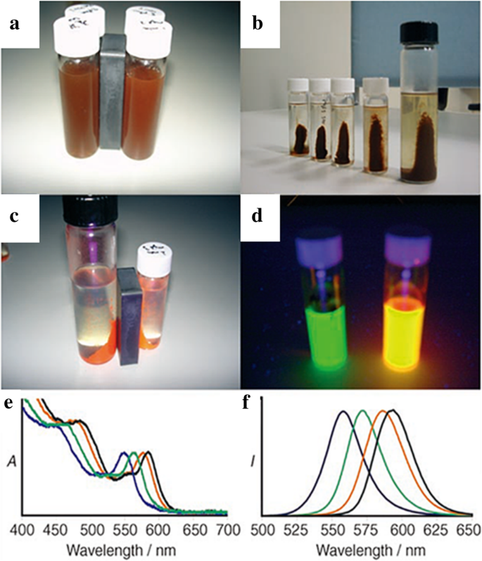
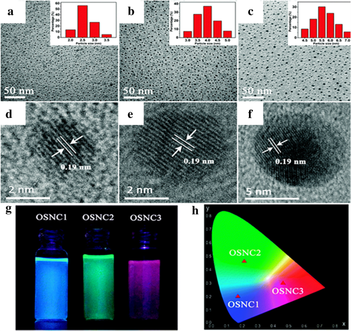
Stöber 방법을 기반으로 새로운 종류의 LSN이 합성되었습니다. Lingang Yang et al. [50] 비닐 그룹의 π-π 적층에 기반한 Stöber 방법으로 결정 실리카를 성공적으로 합성했습니다. A Stöber는 VTES(vinyltriethoxysilane)를 전구체로 사용하고, 염산으로 중화하고, 용매를 제거하기 위한 진공 증류 및 tetrahydrofuran을 사용한 추출은 유기 규소 나노 결정(OSNC)의 전체 절차입니다. 3개의 OSNC가 그림 12d–f와 같이 동일한 결정 구조로 합성되었지만 크기는 다릅니다. VTES의 증가로 인해 유기실리카 나노결정(OSNC)의 크기가 점차 증가하고 있습니다. 그 결과 그림 12g, h(자외선 아래에서 파란색, 녹색, 빨간색)에서 볼 수 있는 것과 같이 서로 다른 발광 특성을 나타냈습니다. OSNC는 우수한 광안정성과 pH 안정성을 가지고 있는 것이 특징입니다. 다이아몬드 입방정 구조에서 비닐기의 에피택셜 성장은 π-π 적층으로 인해 나타납니다. 순서대로 쌓인 비닐 그룹은 최종적으로 양자 구속 후 형광을 갖는 큰 π 공액 시스템을 형성합니다. 이 OSNC는 자체 발광 실리카 재료를 얻기 위한 새로운 접근 방식을 제공한 실리카의 특성으로 인해 광학 분야에서 큰 잠재력을 가지고 있었습니다.
<그림>
OSNC의 특성:a –ㄷ TEM 이미지 및 d –f 고해상도 투과전자현미경(HRTEM) 이미지로. 지 UV-light 조명을 받는 OSNC 샘플의 사진. 아 CIE(Commission Internationale de l'Eclairage) 색도 다이어그램의 분포 [50]
역 마이크로에멀젼 방법
Stöber 방법은 LSN을 합성하는 간단하고 편리한 방법이지만 제어되지 않는 반응 조건 및 초기 입자는 발광 염료에 제한을 둡니다. 이러한 한계를 극복하기 위해 Bagwe와 Khilar[66]는 실리카 나노복합체로 코팅된 은을 합성하는 동안 유중수 마이크로에멀젼 시스템[67]을 도입했습니다(그림 11b). TEOS를 포함하는 은 나노입자의 초기 알칼리 수용액은 계면활성제를 사용하여 물방울에 캡슐화되었습니다. TEOS의 가수분해 진행은 Stöber 방법과 동일하였다. 그러나 전체 진행은 잘 제어된 시스템과 단분산된 실리카 나노 입자로 이어지는 계면 활성제로 둘러싸인 물방울로 제한되었습니다. 실리카의 크기는 다양한 계면활성제, 용매를 선택하고 계면활성제 대 물의 비율을 변경하여 잘 제어되었습니다. 형광단이 수용성인 경우 액적 내 분자 내 표면에 균일한 실리카 층을 형성하기 쉽습니다. Nianfang Wang et al. 역 마이크로에멀젼 방법을 통해 발광 실리카 코팅된 CdS/CdSe/CdS 나노 입자를 합성했습니다. 그림 13은 합성 QD 및 QDs@SiO2의 TEM 이미지를 보여줍니다. . 보호된 QD는 우수한 산 및 열 안정성을 보였다. 응용 프로그램에 대한 특별 요구 사항을 충족하기 위해 추가 수정 가능성을 제공했습니다.
<그림>
CdS 얇은 쉘이 있는 CdSe/CdS 코어/쉘 QD의 TEM 이미지(a ) 및 실리카 CdSe/CdS@SiO2로 코팅한 후 (d ); CdS 쉘이 있는 CdSe/CdS 코어/쉘 QD(b ) 및 실리카 CdSe/CdS@SiO2로 코팅한 후 (이 ); CdS/CdSe/CdS 코어/쉘 QD(c ) 및 실리카 CdS/CdSe/CdS@SiO2로 코팅한 후 (f ) [46]
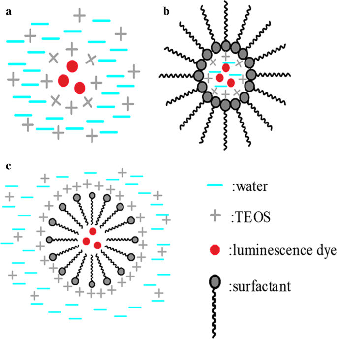
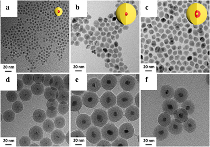
Direct Micelles Assistant Method
Reverse microemulsion method require the water-soluble luminescence dyes. Inversely, liposoluble initial micelles are the major features of direct micelle method, and the hydrolysis progress takes place around of the micelles (Fig. 11c). A precursor is indispensable for the agglomeration of silica. As a common progress, the luminescent dye is modified with the silane coupling agent, such as APS, to form the assistant micelles. The initial modified micelles ensure that the TEOS condensation occurs around them. Using Rhodamine B conjugated to APTES as the original micelle, Kumar et al. [26] successfully synthesized Rhodamine-conjugated organically modified silica nanoparticles in oil in water system and modified them with different function groups (such as sulfhydryl, amino, and carbonyl) which can be used as cell fluorescence probe.
The role of the surfactant is not only reflected in the silica synthesis but also in the synthesis of mesoporous silica. A common method of synthesizing mesoporous silica is calcination. Large specific surface area and modifiable surfaces make the mesoporous silica nanoparticles perfect carriers. In addition to the known application value in the field of medical drug loading, it also has important application prospects in the field of loading phosphors. Li Wang et al. [68] mixed up CDs with hollow mesoporous silica microspheres with good photochemical stability which can be used for oxygen detection in the whole range. Mesoporous structure makes them unique. Bin Xie et al. [69] incorporated the CdSe/ZnS core-shell QDs into mesoporous silica microspheres by a swelling and evaporation method. Coated with a mesoporous silica layer on the surface of Gd2 O3 :Eu phosphors via modified Stöber method is also feasible according to the Ali Aldalbahi et al. [70]. Because of the encapsulation of silica, the modified Gd2 O3 :Eu nanoparticles showed excellent solubility and biocompatibility.
Other Methods
There are also other methods to synthesize LSNs such as chemical vapor deposition (CVD) [71], hydrothermal method [51], and amino acid-catalyzed seed regrowth technique [72, 73].
Lianzhen Cao et al. [71] synthesized SiC/SiO2 by CVD and thermal annealing processes. Si was used to coat on the SiC core by thermal CVD and then SiO2 shell was obtained after oxidizing. The annealed SiC/SiO2 nanoparticles showed narrow luminescence in the blue-green region. The synthetic method provided a new way to synthesize core-shell nanomaterials.
Chandra et al. [51] synthesized smaller fluorescent silica nanoparticles (1 to 2 nm) with silicon tetrabromide (SiBr4 ) and APTS. Heating to 200 °C in an autoclave was the core step of the whole reaction. The final products were obtained after further purification including dialysis and centrifugation. The silica nanoparticles emitted bright blue luminescence with a photoluminescence quantum yield around 34%. It was non-photobleaching and biocompatible at the same time.
Surface modification makes the LSNs more tunable for complex application [74]. Silane coupling agents are the most common chemical methods as it mentions before. Abundant hydroxyl groups provide reaction sites for further modifications. Junqiang Wang et al. synthesized silica modified CeO2 ammonia sensor with high gas response due to hydroxyl groups [75]. After hydrolysis and condensation, silane coupling agents with different function groups bond on the surface of silica. Superhydrophobic silica was synthesized with the condensation of VTES (-CH=CH2 ) [76]. Ming Ma et al. grafted PEGMA and DMEAA on the surface by RAFT polymerization based on the -NH2 of APTS [77]. Surface modification can enhance their adaptability in complex environments and get improved luminescence properties with appropriate materials.
Among these methods, there are two main ideas to fabricate LSNs, namely the luminescent dyes are added directly into the reaction system when the silica resources start hydrolyzing, and that the luminescent dyes are established chemical bond with silica by other reagents such as silane coupling agents, either before or after silica network set up. It is necessary to select and design an appropriate synthetic route for LSNs with specific structures.
Applications of Luminescent Silica Nanoparticles
Light is the most intuitive tool for people to recognize the world. Luminescent materials with special emission can be directly used in many ways such as lighting, display, and so on. At the same time, changes in fluorescence intensity can reflect some important information. Compared with separate luminescent dyes, LSNs have improved performances in applications, since silica provides a stable matrix for the luminescent dye. It provides an effective way for multifunction at the same time [6]. LSNs with multifunction and tunable surface have great application prospects and development potential in biology, lighting, and sensors.
Biolabeling and Medicine
LSNs have great application value in biology. Non-toxicity is a fundamental requirement for medical field, especially in vivo [78]. The fact that the common luminescent dyes are often toxic limits their clinical application [79]. Silica, a favorite non-toxic modified material, is a good solution to elimination of toxicity. Toxicity of silica nanoparticles (20–200 nm) were also carefully studied by In-Yong Kim et al. [80]. Size, dose, and cell type-dependent cytotoxicity were the issues in their research. Although high dose can cause a disproportionate decrease in cell viability, the silica nanospheres with 60 nm showed their good biocompatibility up to 10 μg/ml. Different cells had different tolerance to silica nanoparticles which indicated that it was necessary to have substantial tests before clinical tests. Although inhalation of silica particles can cause acute and chronic diseases including silicosis [81], silica still has potential in biological application at the nanoscale. The toxicity of luminescent silica nanoparticles to living cells was studied in detail by Yuhui Jin et al. [38]. From the DNA level to the cell level, the toxicity of RuBpy-doped LSNs were carefully tested. At a certain concentration, the results showed that the dye-doped luminescent silica nanoparticles were non-toxic to the targeted DNA and cells, which indicate that LSNs are a good solution to the non-toxic modification. Xiqi Zhang et al. [27] encapsulated AIE dye (An18, derivatized from 9, 10-distyrylanthracene with an alkoxyl endgroup) into the silica nanoparticles via a one-pot modified Stöber method. Coated with silica lead to an enhanced fluorescence intensity, good water solubility, and non-toxicity to living cells which made the An18-SiO2 NPs had a potential for biomedical application.
LSNs have great application value in diagnosis and biolabeling. For hybrid imaging contrast agents, Dong Kee Yi et al. [48] combined magnetic particles (MPs) Fe2 O3 with QDs (CdSe) and encapsulated them in silica shell by reverse microemulsion method. The nanostructures of MPs with QDs are clearly showed in Fig. 14. Magnetic resonance imaging (MRI) is an effective method for disease detection, especially for cancer. The advantages of feasible usage, low cost, and accurate diagnosis make it more popular as a diagnostic tool [7]. The nanocomposites can be used as both optical and MRI contrast agents. It is worth mentioning that the presence of CdSe increased the effective magnetic anisotropy of the γ-Fe2 O3 -containg particles. This is a good attempt, but the low quantum yield (SiO2 /MP-QD 1.1% to CdSe 11.4%) limits the actual effect. Willam J. Rieter et al. [39] also synthesized the same multifunctional nanocomposites. What is different is that [Ru (bpy)3 ] Cl2 was chosen as the luminescent core and the paramagnetic Gd complex was coated on the luminescent core by water-in-oil reverse microemulsion method. The nanocomposites were finally embedded in silica in the same way. The results of Fig. 15 proved that hybrid silica nanoparticles had good optical and MRI performances in biological imaging. Mesoporous silica nanospheres doped with europium (Eu-MSN) were obtained by Mengchao Shi et al. [32]. Nanoscale size (280–300 nm) and fluorescent property were the basic for an ideal biolabeling material. They found that Eu-MSNs had a positive influence on osteogenesis and angiogenesis-induction. By promoting proper response of macrophages and the expression of relevant genes, the defect of bone replaced by new bone and the healing process of skin wound can accelerate with Eu-MSNs. Besides the function of biolabeling, the LSNs showed the potential in tissue repair. LSNs can achieve the target binding effect by modifying the special group. In Duarte’s work [33], organosilane Bpy-Si was chosen as a ligand of Eu complex for the further reaction with silica. SiO2 -[Eu (TTA)3 (Bpy-Si)] nanoparticles were obtained with a uniform size (28 ± 2 nm). With a further modification of an amino acid spacer and an anchor group (anti-Escherichia coli , IgG1), the functionalized silica had the specific bonding with E . 대장균 박테리아. It was easy to get the distribution of E . 대장균 bacteria with luminescence. The bio-multifunction of LSNs was also carefully studied by Laranjeira et al. [82]. Gadolinium (Gd) composites with unique magnetic properties have potential in MRI contrast agents but Gd3+ ions are toxic in humans especially in kidneys and pancreas. GdOHCO3 nanoparticles were chosen as the MRI contrast core and coated with silica layer via Stöber method. With the silica coating, the Gd composite (SiGdOHCO3 ) had the same brightness of MRI contrast images but no degradation at designed pH values (5.5, 6.0, and 7.4). And SiGdOHCO3 had little effect on human fibroblasts according to the cell proliferation assay which indicated an excellent biocompatibility. Silica provides a more stable environment and further possible modification for GdOHCO3 without affecting MRI performance. By diverse micelles method, Atabaev et al. [83] synthesized Gd2 O3 :Tb
3+
,Eu
3+
@SiO2 nanoparticles which can be used as both MRI contrast and fluorescence agents in vivo. The above two examples perfectly reflected the role of LSNs in multifunction with the silica platform.
아 TEM image and b HRTEM image of SiO2 /MP-QD nanoparticles [48]
The imaging results of monocyte cells with a optical microscopic, b laser scanning confocal fluorescence, c , d the images of monocyte cells with MR:left for unlabeled monocyte cells and right for hybrid silica nanoparticles labeled monocyte cells, e flow cytometric results of blank and hybrid silica nanoparticles-labeled monocyte cells, and f the cell viability with different amount of hybrid silica nanoparticles [39]
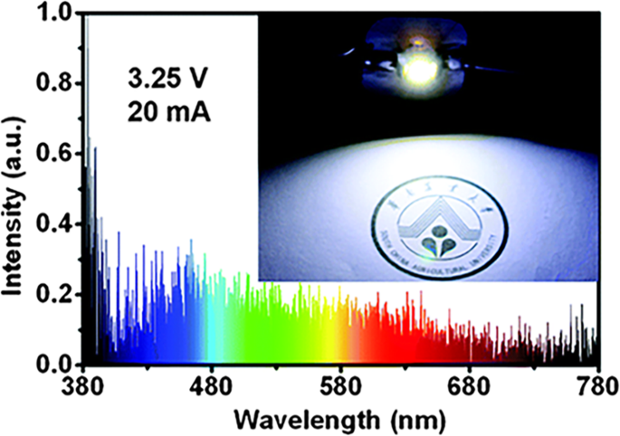
LSNs have great application value in drug delivery. Hongmin Chen synthesized luminescent mesoporous silica nanoparticles biofunctionalized by targeting motifs, which makes them applicable in drug delivery [47]. They first prepared APS-containing mesoporous silica particles, and subjected the products to calcination at 400 for 2 h. They synthesized mesoporous silica by the help of cetyltrimethyl ammonium bromide (CTAB). There were luminescent carbon dots in the silica matrix after calcination. The fluorescence intensity was at the maximum when the particles were excited at 380 nm. The target selectivity of FL-SiO2 was achieved by surface modification of RGD peptide with the help of APS. They also studied the RGD-FL-SiO2 loading and release of doxorubicin (Dox). After calcination, fluorescent mesoporous silica (FL-SiO2 ) can still load Dox effectively. The porous structure was not affected by calcination. They found that RGD-FL-SiO2 had good luminescent effect especially around the blood vessels of tumor in vivo imaging studies. 인테그린 αv β3 of the tumor was the key of the interactions. Although there are many excellent attempts to apply LSNs to medicine but less successful clinical tests in human beings means that there is still a long way to go for the real medicine applications.
Light-Emitting Devices
Due to their special emitting features, LSNs also play a vital role in light emission fields including the field emission- and liquid crystal-based display technologies [84]. WLEDs have received recent attention for their broad applications including general illumination and displays. Tunable color, high color purity, and luminescence efficiency are in line with the requirements of light-emitting diodes (LEDs) [85]. Quantum dot-based light-emitting diodes (QD-LEDs) have demonstrated recently, and may offer many advantages over conventional LED and organic light-emitting diodes (OLEDs) technologies in terms of color purity, stability, and production cost, while still achieving similar levels of efficiency. In order to improve the performances of polymer dots (Pdots) as WLED phosphors, Kaiwen Chang et al. [49] introduced some Pdots with different emission wavelength into the Stöber system to get encapsulated. The silica-encapsulated Pdots showed the same luminescence properties but markedly enhanced photostability.
To reduce the manufacturing complexity required for achieving full-color displays, it is more desirable to use a common device structure to achieve high efficiency for three primary colors (blue, green, and red). QDs have been widely used in display field because of its unique luminescent properties, such as high luminescent intensity, narrow emission spectra, and tunable emission. Chun Sun et al. [34] synthesized the perovskite QDs, CsPbBr3 , as the light-emitting core of WLEDS. Only the perovskite QDs are not enough for a LED device since photostability and stability are necessary for an optical device under long-time work and elevated temperature. There are anion-exchange reactions between different halide QD nanoparticles which would widen the narrow emission spectrum. QD/silica composite were fabricated in APS to avoid oxidation and decomposition. So they used APTES as the QDs’ capping agent and improved the silica coating process to avoid the decomposition of the QDs. Green and red QD/silica composites were synthesized and a WLED was obtained by the combination of the composites with a blue LED chip. The WLED had good performances with great air stability as depicted in Fig. 16.
The optical performances of the WLED:a the emission spectra, b the CIE color coordinates and the color triangle of WLED (red dashed line) with the NTSC TV standard (black dashed line), c the power efficiency, and d emission spectra after working for a while [34]
LSNs can keep good dispersion, brightness, and photostability of QDs. Hung-Chia Wang et al. [35] provided a new composite method for QDs and silica (Fig. 17). By mixing the QDs with mesoporous silica powder of which pore size was bigger than that of QDs in non-polar solution, mesoporous silica green PQD nanocomposite was obtained after washing and drying. The quantum dot showed better thermal stability and photostability after composited with silica. On the other hand, QDs are a typical kind of aggregation-caused quenching (ACQ) nanoparticles, which means that it is necessary to keep a good dispersion to get a good brightness and photostability. Kai Jiang et al. [86] synthesized carbon dots with red, green, and blue luminescence with phenylenediamines as precursors to enhance luminescence properties as solution and poly (vinylalcohol) (PVA) film. But it would exist quenching effect as solid-state CDs which was fatal for LED devices owing to aggregation and the result Förster resonance energy transfer (FRET). To avoid the dispersion and the resulting FRET phenomenon, Junli Wang et al. [36] embedded carbon dots into silica matrix (Fig. 18) by dispersing carbon dots into the N -(3-(trimethoxysilyl)propyl) ethylenediamine (KH-792) and heating to form a homogenous CD/silica film. A white LED was fabricated by drying the CD/silica solution on the inner wall. By the assistant of silica, CDs were well dispersed with an appropriate distance without quenching which improve the performance as powders. Figure 18 showed the emission spectra and performance in WLED. And the CIE coordinates (0.44, 0.42) and correlated color temperature (CCT) (2951 K) suggested that it was suitable for indoor illumination.
아 The formation progress of MP-CsPbBr3PQDs. ㄴ The luminescence intensity and the color triangle of WLED [35]
The performance of WLED showed as a the emission spectrum and b for CIE chromaticity and CCT [36]
Sensors
Luminescent silica showed the excellent performances on static luminescent materials, such as biolabeling and WLED phosphors. All these were based on their unique and stable optical properties. When it came to dynamic luminescent materials, LSNs also display the same wonder [9]. The luminescent sensors of pH [28], ions [87], and temperature [40] are following as representatives.
pH value have great influence on the luminescence intensity which inspires luminescent pH sensor. In the same principle as ref. [22], Atabaev et al. synthesized the same ratiometric pH sensor [28]. FITC was chosen as the pH-dependent luminescence dye and Y2 O3 :Eu
3+
as pH stable dye. With the Stöber coating of silica, Y2 O3 :Eu
3+
@SiO2 with FITC composite NPs were successfully synthesized. The change of pH was reflected by the ratio of fluorescence intensity (IFITC /나Y2O3:Eu3+ ). The standard dye led to a less influence of concentration and a more accurate result.
LSNs can also be used as ions sensors. Based on the changes of luminescence intensity with the measured physical quantity, LSNs have been applied to many sensor fields by the environment-dependent effect of the luminescence. Quenching effect is an effective detective tool to detect the changes of quenching factors such as ions and pH value with external quenching mechanisms such as FRET and photoinduced electron-transfer (PET) [9]. Sensors for metal ions are important fields whether in cells or open system. Won Cho et al. [37] synthesized europium (III) coordination polymer (EuCP) and found the specific quenching effect of Cu
2+
(Fig. 19). In view of this fact, they synthesized silica@EuCP microsphere which have the same sensitivity on Cu
2+
with less mass of europium. As an auxiliary material, silica can effectively reduce the amount of sensor materials. Both of them have their unique situations. Besides quenching effect, there are some different effects which can be used in the fields of sensors. 2,2-Dipicolylamine (DPA) and its derivatives have good affinity to heavy ions. And enhanced luminescence effect would happen after chelated with heavy ions. Yu Ding et al. [29] modified silica spheres with N ,N ′-bis (pyridine-2-ylmethyl)ethane-1,2-diamine (Fig. 20). The concentration of heavy ions (Cd
2+
Hg
2+
및 Pb
2+
) in samples can be determined by the change of fluorescence intensity. The test in real water samples and simulated biological samples confirmed the heavy metal ions-binding ability and the detection which has application prospects in the water monitoring and so on.
아 Confocal microscopy and OM (inset) images of silica@EuCP microspheres. ㄴ Luminescence spectra with different Cu (NO3 )2 in MeCN; luminescence intensity changes (c ) and photograph (d ) with different metal ion solutions (5 mM) [37]
The formation and sensing progress scheme of sensitive fluorescent sensor (FSCHP) [29]
Temperature sensors are also important applications of LSNs. Temperature is a basic variable in most science fields. The temperature dependence of radiative and non-radiative transition rates is the core content of temperature sensing which makes it possible for luminescence temperature sensing, with the contactless and large-scale advantages [9]. However, in order to be applied in practice, their stability is crucial as the environment of application is more complex than of that of experiment condition. Silica is an ideal matrix to improve their performance for application. Mirenda et al. [40] synthesized silica as the core and then TEOS was hydrolyzed with Ru (bpy)3 Cl2 to form the Ru (bpy)3 @SiO2 NP. The emission spectra of Ru (bpy)3 @SiO2 NPs (Fig. 21) showed that the intensity of Ru (bpy)3 @SiO2 NPs decreased linearly as the temperature rising as the result of the activated non-radiative
3
d-d state (20–60 °C, λexc = 463 nm). The polyethyleneimine (PEI)-modified glass with Ru (bpy)3 @SiO2 NPs showed the same trend as the NPs which proved that the potential as the temperature sensing. With cycling the temperature between 20 and 60 °C, the relative slope decreased until the seventh cycle which meant that it is necessary to condition to obtain the stable sensing materials. The influence of temperature on probes is complicated. So it is necessary to research the temperature-dependent luminescence of the probes to know how to apply it into temperature sensors. Temperature is a fundamental variable that governs diverse intracellular chemical and physical interactions in the life cycle of biological cells. The change of temperature reflects the level of cell metabolism. GdVO4 co-doped with Er
3+
(1 mol%) and Yb
3+
(1 mol%) has the potential to apply as the temperature sensor. To improve their performance as temperature sensor, Savchuk et al. [41] coated silica shell on the nanoparticles surface by Stöber method. The fluorescence intensity ratio (FIR) of Er, Yb:GdVO4 , 나520 /나550 , had a certain linear relationship with temperature in the range from 297 to 343 K after excitation at 980 nm. And the probes got enhanced thermal sensitivity, high thermal resolution and good stability in different solvents. And the result of the ex vivo experiment to monitor temperature evolution with the special sensor showed in Fig. 22 proved that Er, Yb:GdVO4 @SiO2 core-shell nanoparticles had a good thermal resolution as the temperature sensor in biomedical applications.
아 PL spectra of Ru (bpy)3 @SiO2 under different temperature. ㄴ The peak intensity changes as a function of temperature [40]
아나520 /나550 with different temperature for Er, Yb:GdVO4 and Er, Yb:GdVO4 @SiO2 . ㄴ The sketch map for the ex vivo temperature determination experiment. ㄷ The results of the temporal evolution of temperature for the Er, Yb:GdVO4 @SiO2 and a thermoresistor Pt-100 [41]
결론
In this article, LSNs with various functions demonstrate that silica is an ideal host material for luminescent dyes. The visualization of related parameters is the most special feature of luminescent dyes. Various luminescent materials have their own advantages but there are still some defects which limit their applications. Improved brightness, photostability, and thermal stability are the advantages of LSNs with the protection of silica. At the same time, it provides phosphors with a versatile platform which makes it possible to become multifunctional and specially modified. Excellent performance, adjustable adaptability, and potential versatility broaden the applications of fluorescent materials. LSNs have great potential in many unmentioned fields such as solar cells and photocatalysts. However, there is still a long way to apply LSNs to the actual species. Poor selectivity and low signal-to-noise ratio in complex conditions are factors that constrain LSNs for the practical applications which need to be further studied. Defined distances between phosphors and LSPR metal deserve more investigations to get the positive effect. Many new luminescent materials with excellent luminescence properties have been developed which means that it is necessary to improve the traditional synthetic methods to obtain LSNs. Silica is a traditional modified material but LSNs still have great potential for development.
약어
ACQ:
Aggregation-caused quenching
AIEgen:
Aggregation-induced emission luminogens
AMP:
Adenosine 5′-monophosphate
An18:
An aggregation-induced emission-based organic fluorogen derivatized from 9,10-distyrylanthracene with alkoxyl endgroup