Deinagkistrodon acutus 독으로 인한 급성 신장 손상에 대한 Phellodendri Chinensis Cortex Carbonisata에서 추출한 탄소 점의 보호 효과
초록
배경
새로운 나노 물질로서 탄소점(CD)은 생물 의학 응용 분야에서 엄청난 관심의 초점이 되어 왔습니다. 그러나 뱀 독에 의해 유발된 급성 신장 손상(AKI)을 억제하는 생물학적 활성에 대한 정보는 거의 없습니다.
방법
이 연구는 Phellodendri Chinensis Cortex(PCC)를 유일한 전구체로 사용하여 CD를 합성하는 녹색 1단계 열분해 공정의 개발과 Deinagkistrodon acutus(D. acutus) 독으로 인한 AKI가 처음으로 조사되었습니다. AKI 모델은 D를 주입하여 확립되었습니다. 귀두 6개 시점(1, 3, 12시간)에서 기능 장애, 염증 반응, 조직 손상 및 혈소판 감소증을 포함한 신장 이상에 대한 PCC Carbonisata-CD(PCCC-CD)의 잠재적인 보호 효과 및 쥐의 복강으로의 독 및 1, 2, 5 일)을 조사했습니다.
결과
이러한 결과는 PCCC-CD가 신장 기능 장애(혈청 크레아티닌(SCR), 혈액 요소 질소(BUN), 요 총 단백질(UTP) 및 미세단백뇨(MALB) 농도 감소) 및 화학 유인 물질(단핵구 주화성 단백질 1(MCP-1)), D. 귀두 독액. 독이 있는 쥐에 대한 PCCC-CD의 유익한 효과는 신장 조직학 및 혈소판 감소증의 변화와 유사했습니다.
결론
이러한 결과는 D에 의해 유도된 AKI에 대한 PCCC-CD의 놀라운 보호 효과를 입증했습니다. 귀두 venom은 CD의 생물의학적 응용을 확대할 뿐만 아니라 D에 의해 유도된 AKI에 대한 새로운 치료제 개발을 위한 잠재적인 표적을 제공합니다. 귀두 뱀에 물린 독.
소개
Deinagkistrodon acutus(D. acutus) , Viperidae 계통에 속하는, 중국에서 가장 위험한 육상 뱀 중 하나로 간주됩니다 [1]. D에 의한 Envenomation. 귀두 출혈, 혈소판 감소증 및 신장에 대한 직접적인 손상 가능성과 같은 일련의 증상과 관련이 있습니다[2, 3]. 급성 신장 손상(AKI)은 D에 의한 envenomation의 가장 심각한 전신 효과이자 일반적인 합병증입니다. 귀두 지속적인 신장 기능 장애와 높은 이환율로 직접 이어집니다[4, 5]. 현재 국소 치료는 해독제로 말 유래 과면역 안티베닌을 사용하는 것입니다. 그러나 그 효과는 특히 AKI의 발생에서 국소 조직 손상을 중화하는데 제한적이며 아나필락시스 및 발열 반응과 같은 몇 가지 불만족스러운 효과가 있습니다[6]. 또한, 안티베닌의 상대적으로 높은 비용과 열악한 안정성도 D에 물린 사람들의 불만족스러운 치료에 기여합니다. 귀두 , 특히 야생이나 시골 지역 [7,8,9]. 따라서, D의 치료를 위한 효과적이고 안전하며 저렴한 보완 의약품이 시급히 필요합니다. 귀두 독으로 인한 AKI.
10 nm 미만의 새로운 종류의 탄소 나노 물질인 탄소 점(CD)은 2004년 단일벽 탄소 나노튜브의 분리 및 정제에 의해 우연히 발견되었으며[10], 지난 10년 동안 그 놀라운 특성으로 인해 많은 관심을 받았습니다. 상당한 생체 적합성, 낮은 독성, 높은 수용성 및 풍부한 원료와 같은 새로운 특성[11,12,13]. CD의 출현은 주로 바이오이미징[14], 바이오의학[15], 약물 전달[16], 광촉매[17]를 포함한 다양한 "스마트" 나노시스템의 개발에 대한 연구에 기여했습니다.
고유한 생체 활성 잠재력을 가진 CD의 개발은 앞서 언급한 현저한 이점으로 인해 일부 질병의 효과적인 제어 또는 치료를 위한 새로운 세대의 약물 발견을 위한 많은 전략을 제공합니다. 세균성 각막염[18], 지혈[19], peroxidase-like[20], 항암, 항바이러스 및 항염증 활성[21]을 치료하는 항균과 같은 여러 생리활성이 보고되었습니다. 이러한 효과는 과학자들이 CD의 추가적인 제약 및 생의학 응용을 연구하는 데 관심을 갖게 되었습니다. 특히, Schizonepetae Spica Carbonisata [22]에서 유래한 CD의 D. 귀두 독으로 인한 출혈은 D에 의해 유도된 AKI에 대한 CD의 유익한 효과 조사에 대한 새로운 관점을 제공했습니다. 귀두 지금까지 잘 알려지지 않은 뱀에 물린 부분입니다.
PCC(Phellodendri Chinensis Cortex) Carbonisata(PCCC)-CD는 1단계 열분해 처리를 사용하여 PCC(1000년 이상 사용된 중국 전통 의학의 일종)를 직접 열분해하여 합성됩니다. PCCC-CD는 직경이 1.2~4.8nm 범위인 저독성 CD입니다. PCCC-CD는 현저한 지혈 효과가 있는 것으로 보고되어 CD의 생물 의학적 응용을 넓혔을 뿐만 아니라 PCCC의 지혈 물질 기반의 해명을 개척했습니다[23]. 참고로 PCCC는 숯을 가공하여 만든 한약재로 태평성경(太平聖藥)에 최초로 기록되어 있습니다. (978–992 AD, 중국). PCCC의 안전성 프로파일과 지혈, 항염증 등의 만족스러운 치료 효능은 중화인민공화국 약전에서 인정된 1000 년 이상의 지속적인 임상 적용을 장려했습니다. (PPRC, 2015). 그러나 PCCC-CD의 추가적인 기본 생물학적 활성에 대한 연구는 도전 과제였습니다. 특히, D에 의해 유도된 AKI의 억제에 대한 정보는 거의 없다. 귀두 독극물.
또한, 뱀에 물린 독극물은 신독성 성분을 통해 직접적으로 또는 면역 및 염증 매개체의 활성화 또는 조절을 통해 신장 생리학을 위험에 빠뜨릴 수 있다고 보고되었습니다[24]. 이러한 효과는 주로 생물의학적 매개변수[25](혈청 크레아티닌(SCR), 혈액 요소 질소(BUN), 요중 총 단백질(UTP) 및 미세알부민뇨(MALB) 농도), 염증성 사이토카인(인터루킨(IL)-1β), 항염증성 사이토카인(IL-10), 단핵구 주화성 단백질 1(MCP-1), 신장 조직학의 변화, 혈소판(PLT) 수. 본 연구에서 우리는 녹색의 1단계 열분해 방법을 사용하여 PCCC-CD를 합성하고 처음으로 D를 쥐의 복강 내 주사로 유도된 AKI의 이상 발달에 대한 보호 효과를 조사했습니다. 귀두 독.
자료 및 방법
화학물질
PCC 재료는 Beijing Qiancao Herbal Pieces Co., Ltd.(Beijing, China)에서 구입했으며 PCCC는 우리 연구실에서 준비했습니다. 분자량 1000 Da의 투석막은 Beijing Ruida Henghui Technology Development Co., Ltd.(Beijing, China)에서 구입했습니다. 세포 계수 키트(CCK)-8은 Dojindo Molecular Technologies, Inc.(Kumamoto, Japan)에서 구입했습니다. D의 동결건조된 뱀독. 귀두 실험용으로 An Ren Snake Farm(Yujiang County, Yingtan, Jiangxi, China)에서 제공했습니다. 다른 분석 등급의 화학 시약은 Sinopharm Chemical Reagents Beijing(Beijing, China)에서 구입했습니다. 마우스 MCP-1, IL-10 및 IL-1β 효소 결합 면역흡착 분석(ELISA) 키트는 Cloud-Clone Crop에서 구입했습니다. (중국 우한). 모든 실험은 탈이온수(DW)를 사용하여 수행되었습니다.
동물
동물 관리 및 연구 프로토콜은 베이징 중의과 대학 동물 실험 윤리 기관 위원회의 지원과 승인을 받았습니다. 수컷 Kunming 마우스(무게 30.0 ± 2.0 g)는 실험 동물 적합성 인증서와 함께 Beijing Vital River Laboratory Animal Technology Co., Ltd.에서 구입했으며 12시간 조명/ 자유로운 다크 사이클 음식과 물에 대한 접근.
베놈 솔루션 준비
동결건조된 독을 생리 식염수에 20분 동안 가볍게 혼합하면서 용해시키고(최종 농도:5 mg/mL) 필요할 때까지 -20 °C에서 보관했습니다.
PCCC-CD 준비
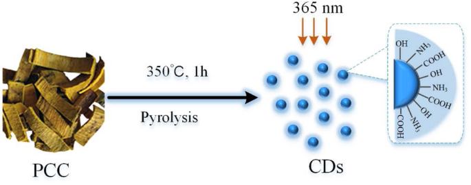
PCCC는 이전 방법에 따라 PCC를 단독 전구체로 사용하는 1단계 열분해 방법을 사용하여 제조되었습니다[23]. 간단히 말해서, PCC 샘플을 별도의 도자기 도가니에 넣고 예열된 노에서 350°C에서 1시간 동안 가열했습니다. 30°C로 냉각한 후, 얻어진 균일한 흑색 잔류물을 미세한 분말로 분쇄하고 1시간에 100°C의 물에 두 번 끓였습니다. 그런 다음 현탁액을 0.22 μm 셀룰로오스 아세테이트 막을 통해 사전 여과하여 용액을 수집했습니다. 비탄소계 불순물은 DW에 대해 72시간 동안 투석하여 제거하였다(유지 분자량:1000 Da). PCCC-CD 용액을 농축하고 사용할 때까지 4 °C에서 보관했습니다. 그림 1의 흐름도는 위의 과정을 보여줍니다.
<그림>
열분해 처리에 의한 Phellodendri Chinensis Cortex(PCC)의 탄소점(CD) 형성 과정의 그림
PCCC-CD의 특성
CD의 형태는 전자 에너지가 200 kV인 투과 전자 현미경(TEM, JEM-2100 electron, JEOL, Japan)을 사용하여 연구되었습니다. Tecnai G2 20 전자현미경(FEI, USA)을 사용하여 고해상도 TEM(HRTEM)을 사용하여 구조적 세부 사항을 조사했습니다. UV-Vis(CECIL, Cambridge, UK) 및 형광(F-4500, Tokyo, Japan) 분광기를 사용하여 자외선 가시광선(UV-Vis) 스펙트럼 및 형광 특성을 기록하고 측정했습니다. 푸리에 변환 적외선(FTIR) 분광법을 사용하여 400~4000 cm
− 1
스펙트럼 창에서 표면 작용기 정보를 분석했습니다. .
세포독성 분석
인간 L02 간세포 및 인간 배아 신장 293 T 세포주를 사용하여 CCK-8 분석을 사용하여 PCCC-CD의 잠재적 세포독성을 평가했습니다. L02 세포는 10% FBS(fetal bovine serum)가 포함된 Roswell Park Memorial Institute 1640(RPMI 1640) 배지에서 배양되었으며, 293 T 세포는 10% FBS를 포함하는 고 포도당이 포함된 Dulbecco의 변형 이글 배지(DMEM)에서 성장되었습니다. 두 세포주 모두 가습된 5% CO2에서 37 °C에서 배양되었습니다. .
L02 및 293 T 세포는 2.0 × 10
4
의 밀도로 시딩되었습니다. 96웰 플레이트에 세포/웰을 넣고 5% CO2의 가습 분위기에서 37 °C에서 배양 24 시간 동안. 그런 다음 두 세포 유형을 무혈청 배지에서 100μL의 서로 다른 농도(5000, 2500, 1250, 625, 156, 78, 39 및 19.5μg/mL)의 PCCC-CD 용액으로 처리하고 또 다른 24시간 동안 배양했습니다. 시간. 이어서, PCCC-CD를 함유하는 배지를 제거하고 세포를 인산완충식염수(PBS)로 2회 세척하였다. 세포독성은 CCK-8 시약 10μL를 첨가하고 37°C에서 4시간 동안 배양한 직후 450nm에서 플레이트를 판독하여 결정했습니다.
D. 귀두 독으로 인한 AKI 및 치료
D. 귀두 독으로 인한 AKI 모델
AKI 마우스 모델은 마우스에 D를 복강내 주사하여 확립되었습니다. 귀두 뱀 독. 마우스를 각각 36마리의 동물로 구성된 다음 5개 그룹으로 무작위로 나누었습니다:대조군; 모델; 및 고용량, 중간 및 저용량 PCCC-CD 치료 그룹. 모델 그룹은 D를 받았습니다. 귀두 1mg/kg(0.15mg/mL, 0.2mL) 체중의 독과 0.5mL 생리 식염수를 복강 내 투여한 반면, 고용량, 중간 용량 및 저용량 PCCC-CD 치료 그룹은 뱀 독과 PCCC의 동등한 양을 받았습니다. - 각각 8.0, 4.0, 2.0 mg/kg의 CD 추출물. 생리식염수(NS)만 복강내 주사한 마우스가 대조군으로 사용되었습니다. NS 투여 후 1, 3, 12시간 후에 각 그룹의 6마리의 마우스를 희생시켰다. 귀두 위에서 설명한 일정에 따라 독과 PCCC-CD를 제거하고 1일, 2일 및 5일에 희생된 마우스에 NS, D를 지속적으로 투여했습니다. 귀두 뱀 독과 PCCC-CD를 하루에 두 번.
UTP 및 MALB 농도 분석
각각의 처리제를 투여한 후, 대조군, 모델 및 PCCC-CDs-(4.0 mg/kg) 처리된 그룹의 동물을 배양 기간이 끝날 때까지(24시간) 소변 수집을 위해 즉시 적절한 대사 케이지에 넣었습니다. UTP 및 MALB의 농도는 자동 생화학 분석기를 사용하여 분석되었습니다.
신장 기능의 바이오마커 분석
신장 기능은 SCR 및 BUN 수준을 측정하여 평가되었습니다. 마우스를 안락사시키기 전에, 안와 뒤 신경총에서 혈액 샘플을 회수한 다음 4°C에서 최소 4시간 동안 플라스틱 튜브에 넣었습니다. 750×g에서 원심분리하여 혈청을 얻었다. 15분 동안 BUN 및 SCR 수준을 자동 생화학 분석기를 사용하여 분석했습니다.
염증성 사이토카인 수치 감지
마우스의 오른쪽 신장을 제거하고, 드라이아이스에서 급속 냉동하고 다음 절차에 사용할 때까지 -80 °C에 보관했습니다. 다른 그룹의 조직 샘플(100mg)을 얼음 위의 PBS로 균질화한 다음 750xg에서 원심분리했습니다. 15분 동안 제조업체의 지침에 따라 각각의 ELISA 키트를 사용하여 MCP-1, IL-10 및 IL-1β의 수준을 결정하기 위해 상등액을 수집했습니다.
신장 조직학
마우스 왼쪽 신장 조직 샘플을 4 °C에서 48시간 이상 동안 10% 중성 완충 포르말린에 고정하고, 탈수하고, 파라핀에 포매하고, 절편으로 절단한 다음, 헤마톡실린 및 에오신(H&E)으로 염색했습니다. 대조군, 모델 및 PCCC-CD 처리군 간의 형태적 변화를 비교하였다.
혈소판 감소 활성
PLT는 안와 뒤 신경총에서 채취한 쥐의 혈액에 대해 수행되었으며 자동 혈액 분석기(XS-800i, Sysmex Corporation Co., Ltd., Kobe, Japan)를 사용하여 검출되었습니다.
통계 분석
통계 분석은 사회 과학용 통계 패키지(SPSS, 버전 13.0)를 사용하여 수행되었습니다. 정규 분포 데이터와 동질 분산은 평균 ± 표준 편차(SD)로 표시됩니다. 일원 분산 분석(ANOVA)에 이어 최소 유의차(LSD) 테스트가 다중 비교에 사용되었습니다. 피 <0.05는 통계적으로 유의한 것으로 간주되었습니다.
<섹션 데이터-제목="결과">
결과
PCCC-CD의 특성
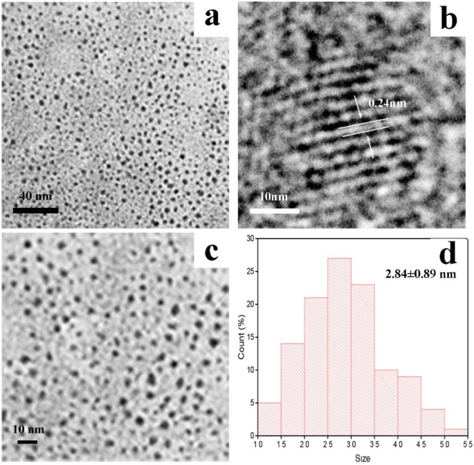
TEM은 PCCC-CD의 형태와 크기 분포를 직접 관찰하는 데 사용되었습니다(그림 2a, c, d). 준비된 CD는 구형이고 크기가 균일했으며 대부분은 뚜렷한 응집 없이 2.84 ± 0.89 nm였습니다. HRTEM 이미지는 CD의 격자 무늬(0.24 nm)를 보여주었으며, 이는 그림 2b와 같이 흑연 탄소에 해당합니다.
<그림>
아 및 c Phellodendri Cortex Carbonisatus 탄소 점(PCCC-CD)의 투과 전자 현미경(TEM) 이미지, b PCCC-CD의 고해상도 TEM 이미지, d PCCC-CD의 크기 분포
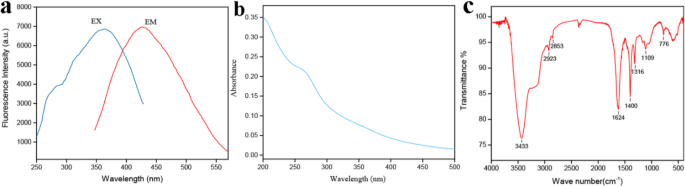
도 3a에 도시된 바와 같이, CD는 370nm 여기 후 445nm에서 최대 피크를 갖는 뚜렷한 청색 형광을 나타내었다. 따라서 UV-Vis 스펙트럼에 표시되는 PCCC-CD의 광학 정보는 CD 표면의 공액 유기 분자의 π-π* 전이에 해당하는 265 nm에서 강한 흡수 피크를 나타냅니다(그림 3b).
<그림>
Phellodendri Cortex Carbonisatus 탄소 점(PCCC-CD)의 광학적 특성. 아 형광 스펙트럼, b 자외선 가시 스펙트럼(UV-Vis) 및 c 푸리에 변환 적외선 스펙트럼(FTIR)
또한, 제형화된 CD의 작용기는 도 3c에 도시된 바와 같이 FTIR 스펙트럼을 기반으로 상세하게 식별되었다. 3433 cm
− 1
에서 피크 2923 cm
− 1
에서 C-H 신축 진동이 나타나는 동안 OH 및 NH 신축 진동의 흡수 밴드에 할당되었습니다. 및 2853 cm
− 1
. 또한 1624 cm
− 1
에서 흡수 피크 C=O의 존재를 나타냅니다. 1109 cm
− 1
에서 CO-C 결합이 관찰되었습니다. , 그리고 1400 cm
− 1
에서 피크 C-N 스트레칭에 기인합니다. 이러한 결과는 이전 보고서의 결과와 일치했습니다.
체외 세포독성
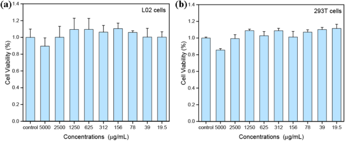
세포 독성을 평가하기 위해 L02 및 293 T 세포를 24시간 동안 다양한 농도의 PCCC-CD에 노출시키고 CCK-8 분석을 사용하여 생존력을 조사했습니다. 도 4a에 도시된 바와 같이, PCCC-CD로 처리된 L02 세포의 생존율은 5 mg/mL의 높은 농도에서도 대조군 세포와 비교하여> 85%였다. 유사하게, PCCC-CD는 최대 약 2500 μg/mL 농도에서 293 T 세포 성장에 영향을 미치지 않았습니다(그림 4b). 이러한 결과는 PCCC-CD가 낮은 세포 독성을 나타냄을 나타냅니다.
<그림>
(a의 CCK-8 분석에 의한 세포 생존력 ) L02 셀 및 (b ) 293 T 세포를 4 시간 동안 PCCC-CD와 함께 배양
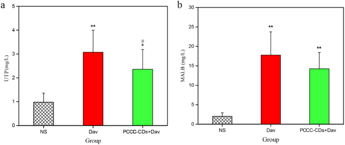
UTP 및 MALB 농도에 대한 PCCC-CD의 영향
대조군(0.98 ± 0.38 mg/L)의 UTP 수치와 비교하여 D에서 UTP 수치가 유의하게 증가했습니다. 귀두 독 처리(3.08 ± 0.92 mg/L, P <0.01) 및 PCCC-CD(2.36 ± 0.83 mg/L, P <0.05) 그룹(그림 5a). 또한, UTP 수치는 독 투여 후 수치와 비교하여 PCCC-CD 치료 후 감소했습니다(P <0.05). 또한, MALB의 수준은 D의 복강내 주사 후 24시간 후에 분명히 증가하였다. 귀두 독(17.78 ± 5.96 mg/L, P <0.01) 대조군과 비교(2.02 ± 0.91 mg/L). 대조적으로, PCCC-CD 처리 마우스는 MALB 수준에서 감소하는 경향을 보였다(14.25 ± 4.16 mg/L, Fig. 5b).
<그림>
Phellodendri Chinensis Cortex Carbonisata-탄소 점(PCCC-CD)이 소변의 총 단백질(UTP) 및 미세단백뇨(MALB)에 미치는 영향. 아 UTP 및 b 말브. 마우스를 생리 식염수(NS), D.acutus로 처리했습니다. 독(Dav, 1 mg/kg) 및 PCCC-CD(4 mg/kg) + Dav(1 mg/kg). 데이터는 각 그룹의 6마리 동물의 평균 ± SD로 표시됩니다. *삐 < 0.05 및 ** p < NS로 처리된 대조군과 비교하여 0.01.
#삐 < Dav 그룹과 비교하여 0.05
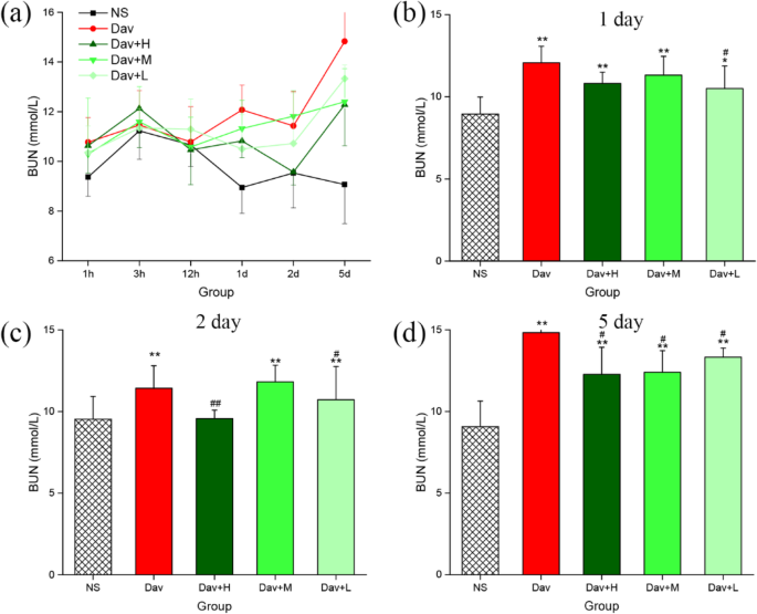
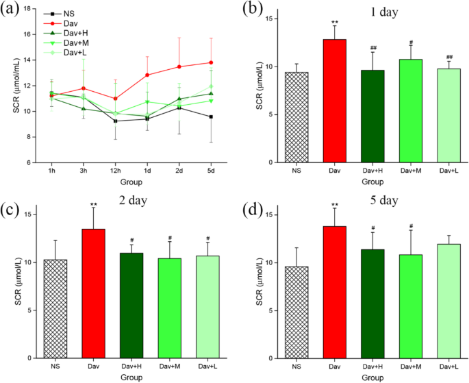
PCCC-CD는 D의 신장 기능 장애를 완화했습니다. 귀두 독으로 인한 AKI 마우스
BUN과 SCR의 수치를 측정하여 신장 기능을 평가하였으며, 이는 Fig. 6 및 7. D로 처리된 마우스. 귀두 독은 BUN 수치가 훨씬 더 높았습니다(12.07 ± 1.00 mmol/L [1일], P <0.01; 11.43 ± 1.37 mmol/L [2일], P <0.01; 14.83 ± 2.53 mmol/L [5일], P <0.01) 및 SCR(12.83 ± 1.43 μmol/L [1일], P <0.01; 13.48 ± 2.26 μmol/L[2일]; 피 <0.01. 13.80 ± 1.90 μmol/L [5일], P <0.01) NS 처리된 마우스보다 (BUN:8.95 ± 1.04[1일], 9.53 ± 1.40[2일] 및 9.07 ± 1.57[5일]mmol/L; SCR:8.95 ± 1.04[1일], SCR:9.40] , 10.27 ± 2.04[2일] 및 9.85 ± 1.99[5일] μmol/L) 1일부터 5일까지(그림 6a 및 7a). 참고로, 저용량 PCCC-CD 치료는 모델군과 비교하여 SCR 수준의 증가를 유의하게 억제했습니다(9.77 ± 0.79 μmol/mL, P <0.01) 및 BUN(10.50 ± 1.38 mmol/L, P <0.05), 높은 반면(9.62 ± 1.87 μmol/mL, P) <0.01) 및 중간(10.75 ± 1.48 μmol/mL, P <0.05) 용량은 D에 의해 유도된 SCR(그림 7b)의 증가를 억제했지만 BUN(그림 6b)의 증가를 억제하지 않았습니다. 귀두 고, 중, 저용량 PCCC-CD 처리군의 SCR 수치(그림 7c)는 감소하였다(10.97 ± 0.88, 10.42 ± 1.75, 10.68 ± 1.41). 각각; P <0.05) 2일차에는 모델군에 비해 BUN 수치가 높음(9.57 ± 0.52 mmol/L, P <0.01) 및 저용량(10.72 ± 2.04 mmol/L, P <0.05) PCCC-CD 그룹은 2일차에 중간 용량 그룹이 아닌 그룹(그림 6c). 6d 및 7d, 높은 수준(12.28 ± 1.65 mmol/L, P)에서 두 지수의 수준 모두에서 억제 효과가 관찰되었습니다. <0.01(BUN); 11.38 ± 1.80 μmol/mL, P <0.05(SCR), 중간(12.40 ± 1.33 mmol/L, P) <0.05(BUN); 10.83 ± 2.57 μmol/mL, P <0.05(SCR), 각각) PCCC-CD 용량. 또한, SCR에서 대조군과 PCCC-CD 처리군 간에 유의한 차이가 관찰되지 않았습니다.
<그림>
Phellodendri Chinensis Cortex Carbonisata - 탄소 점(PCCC-CD)이 혈액 요소 질소(BUN)에 미치는 영향. 아 BUN 농도는 1 h, 3 h, 12 h, 1 day, 2 day 및 5 day에 변화합니다. (b의 BUN 수준 ) 1 일(c ) 2 일(d) ) 5 일. 마우스를 생리식염수(NS), D.acutus로 처리했습니다. 독(Dav, 1 mg/kg) 및 PCCC-CDs + Dav(1 mg/kg)의 고(H), 중간(M) 및 저(L) 용량. 데이터는 각 그룹의 6마리 동물의 평균 ± SD로 표시됩니다. *삐 < 0.05 및 ** p < NS로 처리된 대조군과 비교하여 0.01.
#삐 < Dav 그룹과 비교하여 0.05
<그림>
혈청 크레아티닌(SCR)에 대한 Phellodendri Chinensis Cortex Carbonisata-탄소 점(PCCC-CD)의 효과. 아 SCR 농도는 1 h, 3 h, 12 h, 1 day, 2 days, 5 days로 변합니다. (b의 SCR 수준 ) 1 일(c ) 2 일(d) ) 5 일. 마우스를 생리 식염수(NS), D.acutus로 처리했습니다. 독(Dav, 1 mg/kg) 및 PCCC-CDs + Dav(1 mg/kg)의 고(H), 중간(M) 및 저(L) 용량. 데이터는 각 그룹의 6마리 동물의 평균 ± SD로 표시됩니다. *삐 < 0.05 및 ** p < 생리식염수(NS)로 처리한 대조군과 비교하여 0.01.
#삐 < Dav 그룹과 비교하여 0.05
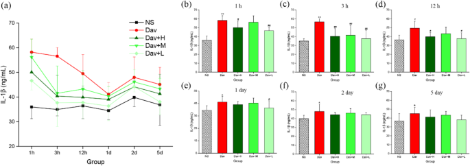
PCCC-CD 억제 사이토카인 분비
3가지 농도의 PCCC-CD가 D의 주사에 대한 반응으로 화학 유인 물질(MCP-1) 및 전염증성(IL-1β) 및 항염증성(IL-10) 사이토카인 생성에 미치는 영향. 귀두 독을 조사했다. 그림 8은 독을 주입하면 마우스 모델에서 방출되는 IL-1β가 증가함을 보여줍니다(1 h:58.19 ± 5.35 ng/mL, P <0.01; 3 h:56.57 ± 3.54 ng/mL, P <0.01; 12 h:49.48 ± 7.74 ng/mL, P <0.05; 1일차:41.09 ± 4.82 ng/mL, P <0.05; 2일차:47.96 ± 8.33 ng/mL, P <0.05; 5일차:45.11 ± 6.95 ng/mL, P <0.05) NS 처리된 마우스의 수준과 비교(1 h:35.96 ± 4.72 ng/mL, 3 h:34.94 ± 2.58 ng/mL, 12 h:36.42 . /mL, 2일:39.84 ± 3.71 ng/mL, 5일:36.82 ± 8.27 ng/mL). 그림 8b-d와 같이 D와 비교합니다. 귀두 독으로 유도된 그룹은 1-, 3- 및 12시간 동안 높은 노출(50.09 ± 7.68 ng/mL, P <0.05[1시간]; 40.36 ± 8.51 ng/mL, P <0.01[3시간]; 39.87 ± 4.64 ng/mL, P <0.05 [12시간]) 및 낮음(46.64 ± 3.83 ng/mL, P) <0.01[1시간]; 37.65 ± 9.61 ng/mL, P <0.01 [3 h]; 38.75 ± 6.64 ng/mL, P 각각 <0.05[12시간]) 용량 PCCC-CD는 IL-1β의 수준을 유의하게 감소시킵니다. 또한, 중간 용량 PCCC-CD는 IL-1β의 수준을 유의하게 감소시켰습니다(41.50 ± 11.08 ng/mL, P <0.01) 3 시간에 독을 처리한 쥐와 비교했지만 다른 시점에서는 그렇지 않았습니다.
<그림>
신장 조직의 IL-1β 수준에 대한 Phellodendri Chinensis Cortex Carbonisata-탄소 점(PCCC-CD)의 효과. 아 IL-1β 수치는 1 h, 3 h, 12 h, 1 day, 2 days, 5 days에 변화합니다. (b의 IL-1β 수준 ) 1 h, (c ) 3시간, (d ) 12시간, (e ) 1일, (f ) 2 일 및 (g ) 5 일. 마우스를 생리 식염수(NS), D.acutus로 처리했습니다. 독(Dav, 1 mg/kg) 및 PCCC-CDs + Dav(1 mg/kg)의 고(H), 중간(M) 및 저(L) 용량. 데이터는 각 그룹의 6마리 동물의 평균 ± SD로 표시됩니다. *p <0.05 및 ** p 생리식염수(NS)로 처리된 대조군과 비교하여 <0.01.
#삐 < Dav 그룹과 비교하여 0.05
항염증성 사이토카인 IL-10의 수준은 D에서 극적으로 증가했습니다. 귀두 다른 시점에서 독 치료군(23.27 ± 0.72 ng/mL, P <0.01; 22.03 ± 0.96 ng/mL, P <0.05; 21.76 ± 1.99 ng/mL, P <0.05; 26.31 ± 6.55 ng/mL, P <0.01; NS 처리군과 비교하여 각각 3 h, 12 h, 1일 및 2일). 대조적으로 고용량 및 중용량 PCCC-CD(각각 17.17 ± 4.04 및 17.25 ± 5.64 ng/mL, 둘 다 P <0.05) 3시간째에 독으로 인한 IL-10 분비를 억제한 반면, 저용량 PCCC-CD는 3시간, 12시간 및 1일째에 수준을 억제했습니다(17.17 ± .24, 17.83 ±3,17.83 ± 4.1 mL, 각각 모든 P <0.05). PCCC-CDs 처리군과 NS군 간에 유의한 차이는 관찰되지 않았다.
<그림>
신장 조직의 IL-10 수준에 대한 Phellodendri Chinensis Cortex Carbonisata-탄소 점(PCCC-CD)의 효과. 아 IL-10 수치는 1 h, 3 h, 12 h, 1 day, 2 days, 5 days에 변화합니다. (b의 IL-10 수준 ) 1 h, (c ) 3 h, (d ) 12 h, (e ) 1 일, (f ) 2 일 및 (g ) 5 일. 마우스를 생리 식염수(NS), D.acutus로 처리했습니다. 독(Dav, 1 mg/kg) 및 PCCC-CDs + Dav(1 mg/kg)의 고(H), 중간(M) 및 저(L) 용량. 데이터는 각 그룹의 6마리 동물의 평균 ± SD로 표시됩니다. *p <0.05 및 ** p 생리식염수(NS)로 처리된 대조군과 비교하여 <0.01.
#삐 < Dav 그룹과 비교하여 0.05
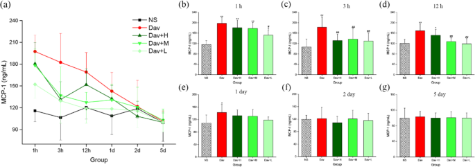
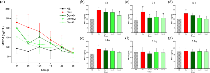
또한, Fig. 10은 D의 주입을 보여준다. 귀두 venom은 모델 그룹에서 MCP-1 방출을 유의하게 증가시켰습니다(1 h:197.45 ± 22.34 ng/mL, P <0.01; 3 h:182.42 ± 12.94 ng/mL, P <0.01; 12 h:169.20 ± 26.74 ng/mL, P <0.01; 24 h:142.81 ± 25.85 ng/mL, P <0.05)가 NS 처리 제어부 (1H 비교 :115.82 ± 14.80 NG / ㎖, 3 H :106.46 ± 13.76 NG / ㎖; 12 H :120.35 ± 15.75 NG / ㎖; 24 H :108.81 ± 25.60 NG / ML). 더 놀라운 사실은 독이 있는 쥐를 3회 PCCC-CD로 처리한 결과 MCP-1 수치의 증가가 억제되었다는 점입니다. 배지-(3 h:136.84 ± 39.94 ng/mL, P <0.01; 12 h:127.48 ± 13.75 ng/mL, P <0.01) 및 낮음-(1 h:152.13 ± 18.89 ng/mL, P <0.05; 3 h:129.54 ± 30.85 ng/mL, P <0.01; 12 h:118.75 ± 19.96 ng/mL, P <0.01) 용량 PCCC-CD는 1 h(178.20 ± 22.79 ng/mL)에서 중간 용량을 제외하고 1, 3 및 12 h에서 MCP-1 수준의 증가를 억제했습니다. 고용량 PCCC-CD 치료는 3 h에서만 MCP-1 수준에서 효과적으로 감소했습니다(131.42 ± 24.62 ng/mL, P <0.01).
<그림>
신장 조직의 MCP-1 수준에 대한 Phellodendri Chinensis Cortex Carbonisata 탄소 점(PCCC-CD)의 효과. 아 MCP-1 level changes in 1 h, 3 h, 12 h, 1 day, 2 days, and 5 days. MCP-1 levels in (b ) 1 h, (c ) 3 h, (d ) 12 h, (e ) 1 day, (f ) 2 days, and (g ) 5 days. Mice were treated with normal saline (NS), D.acutus venom (Dav, 1 mg/kg) and high (H), medium (M), and low (L) doses of PCCC-CDs + Dav (1 mg/kg). Data are represented as mean ± SD of six animals of each group. *p < 0.05 and ** p < 0.01 compared with control group treated with normal saline (NS).
#p < 0.05 compared with Dav group
In contrast, IL-1β, IL-10, and MCP-1 levels of the PCCC-CDs-treated groups at certain time points were not significantly different from those of the model group, and showed a decreasing tendency.
Effect of PCCC-CDs on the Inhibition of Thrombocytopenia
PLTs have a specific role in the pathogenesis of AKI; therefore, the PLT count was investigated and the results are shown in Fig. 11 [26]. Compared with the PLT values of the control group (1 h:[1228 ± 51] × 10
9
; 3 h:[1120 ± 36] × 10
9
; 12 h:[1245 ± 111] × 10
9
; day 1:[1177 ± 69] × 10
9
; day 2:[1195 ± 51] × 10
9
; and day 5:[1181 ± 46] × 10
9
), a drastic reduction occurred as early as 1 h after D. acutus venom administration, with a nadir occurring at 3 h. Subsequently, the PLT steadily increased up to day 5 (1 h:[354 ± 70] × 10
9
, P < 0.01; 3 h:[315 ± 77] × 10
9
, P < 0.01; 12 h:[435 ± 91] × 10
9
, P < 0.01; day 1:[663 ± 226] × 10
9
, P < 0.01; day 2:[941 ± 248] × 10
9
, P < 0.05; day 5:[1083 ± 89] × 10
9
). Of note, even at this time interval, the PLT values were significantly lower than those of control mice. In addition, administration of PCCC-CDs at a dose of 8 mg/kg markedly inhibited the venom-induced thrombocytopenia induced at 1 h ([435 ± 91] × 10
9
, P < 0.05), 3 h ([599 ± 290] × 10
9
, P < 0.05), 12 h ([929 ± 92] × 10
9
, P < 0.01), day 1 ([1028 ± 248] × 10
9
, P < 0.01), and day 2 ([1183 ± 89] × 10
9
, P <0.01). This inhibitory effect was also observed at a dose of 2 mg/kg PCCC-CDs at 3 h and day 2 and 4 mg/kg PCCC-CDs at day 2, in addition to increased PLT. Although there was no significant difference between the model and medium-dose groups, an increasing tendency was observed at other different time points.
Effects of Phellodendri Chinensis Cortex Carbonisata-carbon dots (PCCC-CDs) on platelet (PLT) counts in the blood. 아 PLT changes in 1 h, 3 h, 12 h, 1 day, 2 days, and 5 days. PLT in (b ) 1 h, (c ) 3 h, (d ) 12 h, (e ) 1 day, (f ) 2 days, and (g ) 5 days. Mice were treated with normal saline (NS), D.acutus venom (Dav, 1 mg/kg) and high (H), medium (M), and low (L) doses of PCCC-CDs + Dav (1 mg/kg). Data are represented as mean ± SD of six animals of each group. *p < 0.05 and ** p < 0.01 compared with control group treated with normal saline (NS).
#p < 0.05 compared with Dav group
Histopathological Observations
The renal injury in the venom group was histologically evaluated. As shown in Fig. 12a, in contrast to the NS-treated group, which showed normal glomeruli and tubular cellularity, marked changes were observed in the renal parenchyma of the D. acutus venom-treated group. These changes include marked haemorrhage, renal tubular dilation, and degeneration. Cotreatment with PCCC-CDs prevented D. acutus venom-induced renal damage, and the histopathological examination of the architecture of the renal tissues was almost normal with mild haemostasis, renal tubular dilation, and degeneration.
Effects of Phellodendri Chinensis Cortex Carbonisata-carbon dots (PCCC-CDs) on histopathological changes in kidney tissues in D.acutus venom (Dav)-induced AKI mice. After D.acutus venom challenged, kidney tissues from each experimental group were prepared for histological evaluation. Histological changes of kidney obtained from mice of different groups normal saline (NS), D.acutus venom (Dav, 1 mg/kg) and high (H), medium (M), and low (L) doses of PCCC-CDs + Dav (1 mg/kg) in (a ) 1 h, (b ) 3 h, (c ) 12 h, (d ) 1 day, (e ) 2 days, and (f ) 5 days
Discussion
As an emerging nanomaterial, CDs are beginning to occupy an important niche as innovative materials for next-generation nanomedicines. Compared to traditional heavy-metal-based quantum dots, CDs are good candidates for biomedical application because of their unique characteristics that have considerable potential advantages in the development of novel medicines with relatively low toxicity [27, 28].
The derived PCCC-CDs particles were quasi-spherical and well-dispersed in water, with abundant functional groups present on the surface. This observation is consistent with previously reported findings [23]. In addition, the as-prepared CDs showed low toxicity against L02 and 237 T cells, which indicated its suitability for biomedical applications.
The current study is the first, to the best of our knowledge, to demonstrate the remarkable bioactivities of PCCC-CDs on AKI induced by D. acutus snakebite. D. acutus is widely called “five pacer” or “hundred pacer” in Chinese folk medicine on account of the folkloric description that the people or animals bitten by D. acutus could not walk more than 100 steps. More than 90% of the population of D. acutus is found in China, and the frequency of critical conditions and even death related to the bite of this snake is higher than that by many other venomous snakes [29]. AKI is the most serious systemic effect and common complication, which leads to secondary renal ischemia and failure. An enhanced knowledge of relevant information on AKI induced by D. acutus envenomation would contribute to the development of novel therapeutic approaches. However, in contrast to the considerable knowledge available on the nephrotoxicity of snake venom in general [30, 31], information on the AKI induced by D. acutus venom is rare, which led us to investigate this potential medicine that is still in the early stages of development.
In this study, we established an AKI model by intraperitoneally injecting D. acutus venom into mice to assess the complex and multifactorial pathogenesis of venom-induced AKI. Furthermore, the model provided a tool for investigating the protective effects of PCCC-CDs against AKI induced by D. acutus venom.
Current experiments have shown the development of substantial AKI with distinct changes in inflammatory cytokines and serum and urinary biochemical index, as well histopathological evidence of renal injury after intraperitoneal injection of snake venom [31]. These findings indicated the possible factors that may mainly contribute to the venom toxicity are [32] (1) direct venom cytotoxicity against the kidney and (2) renal inflammatory reactions.
Specifically, renal insufficiency was confirmed approximately 24 h post venom injection based on oliguria associated with proteinuria and elevated serum biomarkers (SCR and BUN). We further affirmed the renal involvement in the D. acutus venom-treated group using biochemical analysis, which showed significantly elevated UTP and MALB levels compared with those of the control group. These findings indicated the presence of glomerular malfunction and tubular reabsorption in the venom-treated group [33], which were supported by evidence of histopathological change. In contrast, a significant reduction in the levels of UTP and MALB was observed in the envenomated medium-dose PCCC-CD-treated group. In addition, serum biochemical indicators (SCR and BUN) are other vital parameters used to determine the elevation of renal dysfunction in AKI and they remain clinical indicators in its diagnosis [34]. Injecting snake venom from day 1 to 5 also dramatically increased the levels of SCR and BUN, whereas treatment with PCCC-CDs reversed these effects, resulting in a faster recovery than that of the control group. More importantly, the distinct changes in the kidney tissue, marked haemorrhage, renal tubular dilation, and degeneration further indicated the direct impairment of the kidney by the venom. The attenuating effects of PCCC-CDs on the histopathological changes were demonstrated in this study. These results suggested that PCCC-CDs inhibited the AKI-induced abnormal manifestation of urinary and serum biochemical markers associated with kidney dysfunction as well as renal histological damage. Furthermore, these effects may be attributable to the amelioration of the direct nephrotoxicity of the D. acutus venom by the PCCC-CDs [35]. The protective effects of PCCC-CDs were evidenced by the inhibition of D. acutus venom-induced direct kidney damage.
An intense inflammatory response is a common feature induced by envenomation by venomous animals such as snakes and caterpillars [35,36,37]. The signs of systemic inflammation with mononuclear cell infiltration, neutrophilic leukocytosis, tubular epithelial cell degeneration, and necrosis have also been shown in kidney impairment induced by injecting snake venom. MCP-1 is a small molecule protein that plays a vital role in recruiting and activating leukocytes during inflammatory responses [38]. In addition, mononuclear phagocytes and lymphocytes may contribute to acute renal cell injury by different mechanisms such as the secretion of proinflammatory mediators, which may then induce resident renal cells to express chemokines [39]. The involvement of MCP-1, inflammatory cytokines (IL-1β) and anti-inflammatory cytokine (IL-10) in the inflammatory response in the pathogenesis of AKI in mice injected with D. acutus venom only was demonstrated in the present study. This observation indicated that the underlying mechanism of the D. acutus venom-induced AKI may be associated with the renal inflammatory response. The evidence that exposure to PCCC-CDs significantly reduced levels of IL-1β, IL-10, and MCP-1 suggests that CDs may exhibit renoprotection by inhibiting renal inflammatory reactions.
Furthermore, PLTs play a crucial role in acute haemostatic and inflammatory processes and are associated with diverse inflammatory pathologies [40, 41]. They are highly sensitive and respond quickly to biological changes when an organism is injured or bleeding, as the first cells to arrive at the site of acute injury to interact with endothelial cells and leukocytes [42]. PLTs are involved in the pathogenesis of AKI [43], and are considered a prognostic marker that is significantly associated with a worse outcome of AKI [44]. This study provided evidence that D. acutus venom conspicuously decreased the PLT count, which was consistent with the results of studies reporting that thrombocytopenia can be induced by snake venom [34, 45]. We observed that exposure to PCCC-CDs significantly elevated the PLT counts, which was consistent with the findings of a previous study [23].
The abnormalities of AKI induced by D. acutus venom were, to our knowledge, demonstrated for the first time in the current study and mainly included renal dysfunction associated with proteinuria, oliguria, elevated BUN and SCR levels, pathological kidney damage, inflammatory responses, and thrombocytopenia.
Remarkably, the PCCC-CDs demonstrated protective activity against D. acutus venom-induced AKI by inhibiting the associated impairments, as evidenced in this study for the first time. This study was a preliminary evaluation of the beneficial effects of PCCC-CDs on AKI induced by D. acutus venom, and further investigations of the underlying mechanism would be the focus of future studies.
Conclusion
The impressive protective effects of PCCC-CDs on D. acutus venom-induced AKI have been demonstrated in this study, for the first time, to the best of our knowledge. The AKI-related effects were mainly manifested as renal dysfunction, pathological kidney damage, inflammatory responses, and thrombocytopenia. These results indicated that PCCC-CDs have potential application prospects for use as a complementary medicine for the treatment of abnormalities induced by D. acutus venom-induced AKI. Furthermore, this provides a novel strategy for the study of active ingredients of traditional Chinese medicine formulations, and further broadens the biomedical applications of CDs.
데이터 및 자료의 가용성
All data generated or analysed during this study are included in this published article.