감염성 미생물의 효율적인 2광자 들뜬 광역학적 비활성화를 위한 수산기 의존성을 가진 수용성 풀러레놀
초록
수용성 풀러레놀 [C60 (OH)46 ] 높은 단일항 산소 양자 수율 및 효율적으로 생성된 활성 산소종을 나타냈다. 또한, 수용성 C60 (OH)46 노출된 히드록실기의 더 높은 조성을 갖는 것은 그러한 기의 더 낮은 조성을 갖는 것과 비교하여 우수한 2광자 안정성 및 특성을 가졌다. 따라서 준비된 풀러레놀은 효과적인 2광자 감광제가 될 수 있습니다. 수용성 C60 (OH)46 유리한 2광자 특성을 가졌다. 2광자 광역학 요법 동안 수용성 C60 (OH)46Escherichia coli에 대해 상당한 항균 활성을 보였습니다. 211.2 nJ pixel
−1
의 초저에너지 수준에서 800 스캔 및 760 nm의 광여기 파장으로.
소개
다양한 광감작제(PS) 분자가 지난 수십 년 동안 합성되었습니다[1]. 그러나 기존 PS의 임상 적용에는 몇 가지 문제가 있습니다. 대부분의 PS 분자는 소수성이며 수성 매질에서 쉽게 응집되어 양자 수율(QY)을 감소시킬 수 있습니다[2]. 또한, 응집된 PS는 단순히 정맥 주사할 수 없습니다. 죽은 조직에서 PS 분자의 선택적 축적도 건강한 세포의 손상을 방지하는 데 필요합니다. 이러한 문제로 인해 효과적인 PS 운반체를 개발하는 것은 PDT(광역학 요법)의 주요 과제로 남아 있습니다. 이에 따라 나노입자를 PS 운반체로 사용하는 것에 대한 관심이 높아지고 있다.
나노생명공학의 발전은 탄소 원자로만 구성된 새로운 종류의 나노구조[3,4,5,6,7,8,9,10,11], 즉 풀러렌 C60의 생물의학적 응용에 대한 관심을 자극했습니다. , 이것은 비독성이고 독특한 물리화학적 특성을 갖는 구상 분자(직경 0.72 nm)입니다. 친유성 C60의 작은 크기 분자는 생물학적 분자와의 입체적 호환성을 담당하고 막의 소수성 영역으로의 통합을 촉진합니다[12, 13]. 확장된 π 때문에 -분자 궤도의 공액 시스템, 풀러렌 C60 자외선 가시광선(UV-vis) 빛을 흡수하고 거의 100% 일중항 산소 QY(Φ)로 반응성 산소종(ROS)을 생성할 수 있습니다. Δ ). 또한, 풀러렌 C60의 물리화학적 특성은 ROS를 생성하고 PDT용 PS로 사용할 수 있습니다. 풀러렌은 또한 산화촉진제 효과를 유도할 수 있으며 이는 사용된 풀러렌, 조사된 세포 유형 및 실험 설정에 의해 결정될 수 있습니다[14,15,16,17]. C60 극성 용액에 대한 용해도가 극히 낮아 의학에서의 응용이 상당히 제한됩니다. 그러나 이중 결합이 있기 때문에 C60 수용성을 증가시키기 위해 화학 그룹을 사용하여 쉽게 수정할 수 있습니다. 따라서 수용성 C60 파생 상품은 신경 보호, 약물 및 유전자 전달, 광과민성 및 바이오센싱을 포함한 의료 응용 분야에 대한 기회를 증가시켰습니다.
다광자 레이저 현미경(2광자 레이저 현미경이라고도 함)은 얇은 래스터 스캔 평면 내에서만 형광을 여기시키기 위해 국소화된 "비선형" 여기의 사용을 수반합니다. 2광자 레이저 현미경은 다양한 이미징 연구에 사용되었습니다[18]. 일반적으로 근적외선(NIR) 레이저 여기와 결합되어 생체 이미징을 위한 고유한 최대 조직 투과율을 활용합니다. 이는 근적외선이 약간의 산란, 낮은 에너지 흡수, 최적의 조사 투과성 및 시편의 광표백 감소의 장점이 있기 때문입니다. NIR 레이저 여기와 2광자 레이저 현미경의 결합은 두꺼운 조직과 더 깊은 생물학적 표본의 형광 현미경에 선호되는 기술이 되었으며[19, 20] 다른 광여기 요법[21, 22]에 광범위하게 적용되었습니다. 게다가, 초저 에너지와 짧은 광여기 때문에 2광자 레이저 현미경은 PDT 수행에 대한 대안적인 접근 방식으로 간주됩니다[23]. 일부 PS는 독성이 있지만 [24, 25], 높은 ΦΔ PDT 수행에 우선순위를 둡니다. 높은 ΦΔ 이 값은 광 속성의 분자 활동을 평가하고 비선형 현미경 연구를 효율적으로 수행하기 위해 2광자 기술을 사용할 때 특히 바람직합니다. 이러한 값은 시편에 대한 입력 에너지 플럭스에 흡수된 에너지의 비율이 높기 때문에 바람직하며 시편에 대한 가능한 광손상을 최소화합니다[26]. 그러나 문헌에는 PDT에 2광자 특성을 가진 재료의 사용을 고려한 연구는 포함되어 있지 않습니다. 이러한 연구 공백을 메우기 위해 본 연구에서는 전자 공여 능력이 강하고 π가 큰 수용성 하이드록실화 풀러레놀을 적용했습니다. -전하 전달 효율을 증가시켜 2광자 특성을 향상시키는 공액 시스템. 구체적으로, 수용성 하이드록실화 C60 (OH)46 초저에너지 펨토초 레이저 조사와 2광자 여기(TPE, 여기 파장, 760 nm)에서 단 800회 스캔을 사용하여 효과적인 미생물 제거를 위한 2광자 PS로 유도 및 적용되었습니다. 초저에너지 펨토초 레이저 조사의 경우 에너지는 211.2 nJ pixel
−1
입니다. 출력은 2.112 mW(대물렌즈 후 레이저 출력 계산에 대해서는 "재료 및 방법" 섹션 및 그림 1a 참조, 여기서 x –예 초점 및 z - 레이저 시스템의 축 분해능은 각각 약 0.37538 및 0.90159 μm임); 또한 스캐닝 프로세스의 경우 총 유효 노출 시간은 약 3.2621 s, 스캔 속도는 4.0776 ms scan
-1
, 스캔 영역은 200 × 200 μm
2
(계산에 대한 자세한 내용은 "재료 및 방법" 섹션 참조). 수용성 하이드록실화 C60 (OH)46대장균을 거의 100% 제거했습니다. (대장균 , 그람 음성 박테리아 균주). 또한, 수용성 C60 (OH)46 더 높은 하이드록실 그룹 조성은 TPE 하에서 더 낮은 조성의 하이드록실 그룹에 비해 우수한 2광자 광특성을 나타냅니다. 따라서 파생된 수용성 C60 (OH)46 동시 PDT에서 악성 미생물을 제거하는 데 사용할 수 있는 상당한 잠재력이 있는 것으로 간주될 수 있습니다.
<그림>
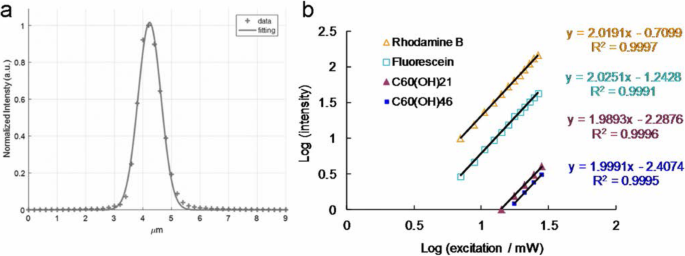
아z에 따르면 - 다른 위치, z에서 2차 고조파 발생 신호를 측정하는 데 사용되는 금 박막의 축 스캔 -레이저 시스템의 축 분해능(최대 반값의 전체 너비)은 약 0.90159 μm(가우스 함수를 사용한 피팅)입니다. ㄴ 재료 및 형광단의 여기 전력(로그)에 대한 TPL 강도 의존성; 704.0~2816.0 nJ pixel
−1
의 TPE 노출 로다민 B 및 플루오레세인의 경우, 1408.0 ~ 2816.0 nJ pixel
−1
수용성 C60용 (OH)21 풀러레놀, 1760.0 ~ 2816.0 nJ pixel
−1
수용성 C60용 (OH)46 풀레놀. 여기 파장, 760 nm. 전달 선량:OD600 0.05 of E. 대장균 및 3 μg mL
−1
재료. 데이터는 평균 ± SD(n =6)
자료 및 방법
수용성 풀레레놀, C60의 제조 및 특성화 (OH)21 및 C60 (OH)46 [27]
원시 풀러렌은 상업적으로 입수했으며(Sigma-Aldrich, St. Louis, MO, USA), C60 (OH)12 앞서 설명한 대로 전구체가 생성되었습니다. 먼저, 30% 과산화수소 용액(100 mL; Sigma-Aldrich, St. Louis, MO, USA)을 출발 물질에 0.5-1.0 g의 C60을 첨가했습니다. (OH)12 , 그리고 혼합물을 공기 하에 60℃에서 격렬하게 교반하였다. 냉각 후, 2-프로판올, 디에틸 에테르 및 헥산을 포함하는 용매 혼합물(각각 100-200mL; Sigma-Aldrich, St. Louis, MO, USA)을 용액에 첨가한 후 원심분리하고 따라내었습니다. 나머지 고체는 원심분리 및 경사분리 절차를 통해 200 mL의 디에틸 에테르로 두 번 세척되었습니다. 마지막으로 수용성 C60의 최종 제품 (OH)21 및 C60 (OH)46 잔류물을 실온에서 밤새 진공하에 각각 건조시켜 얻었다. 최종 제품의 무게는 열중량 분석을 통해 보정되었습니다. 최종 생성물의 형태는 C60에 대해 약 1.11 ± 0.03 nm 및 1.13 ± 0.04 nm의 해상도에서 고해상도 투과 전자 현미경(HR-TEM, JEOL 3010, Akishima, Tokyo, Japan)을 사용하여 관찰되었습니다. (OH)21 및 C60 (OH)46 , 각각. 동적 광산란(DLS, Malvern Nano-ZS90, Worcestershire, West Midlands, UK)도 재료의 크기를 결정하는 데 사용되었습니다. 준비된 재료의 노출된 작용기는 먼저 푸리에 변환 적외선(FTIR) 분광법(RX1, PerkinElmer, Waltham, MA, USA)을 통해 조사되었습니다. 재료의 UV-vis 분광법은 분광계(U-4100, Hitachi, Chiyoda-ku, Tokyo, Japan)를 사용하여 수행되었습니다. 풀러레놀의 표면 화학은 X선 광전자 분광기(XPS, PHI 5000 분광기(VersaProbe, Chanhassen, MN, USA))를 통해 조사하였다. 풀러레놀의 분자량은 FD(field desorption) 질량분석기(AccuTOF, GCx-plus, JEOL, Akishima, Tokyo, Japan)를 이용하여 측정하였으며, 그 결과 하이드록실기의 수는 21개와 46개로 확인되었으며, 각각.
박테리아 문화[28]
이. 대장균 , 우리 자신의 실험실에서 얻은 LB의 영양 한천 (리터당 :트립톤 10 g, 효모 추출물 5 g, 염화나트륨 8 g, 한천 15 g 및 pH 7.5로 조정)에서 재배되었습니다 (Sigma-Aldrich, St. Louis, MO, USA) 및 37 °C에서 배양합니다.
CFU(집락 형성 단위) 계산 방법을 사용한 생체 적합성 분석 [28]
이. 대장균 (OD600 ~ 0.05)에 물질(0–9 μg mL
−1
)을 첨가했습니다. ), 37 °C에서 3 시간 동안 배양했습니다(추가 파일 1:그림 S1). 배양 후, 혼합물을 원심분리하고 박테리아 펠릿을 희석했습니다(OD600 ~ 0.05). 10
−5
의 희석 비율 ~ 10
−8
그런 다음 배양된 박테리아에서 수행하고 한천 플레이트에 플레이팅했습니다. 플레이트를 밤새 인큐베이터(37 °C)에 보관합니다. 생존 박테리아의 수를 결정하고 CFU mL
-1
단위에 해당하는 백분율(%)로 표시했습니다. 배양 후. 데이터는 평균 ± SD(n =6).
ψΔ 측정 [29, 30]
이전 연구에 따르면 ψΔ 얻어 질 수있는. ψΔ 측정은 D2에서 수행되었습니다. 355 nm에서 O, meso 사용 -테트라(4-설포네이토페닐)포르핀 디하이드로클로라이드(TSPP; Sigma-Aldrich, St. Louis, MO, USA)를 참조로 사용(ψΔ =0.64).
형광 QY 측정 [31, 32]
조영제의 상대 광발광(PL) QY는 일반적으로 방출된 광자 대 흡수된 광자의 비율이며 다음과 같이 제공됩니다.
여기서 QY참조 =0.28은 기준으로서 디메틸 설폭사이드(DMSO; Sigma-Aldrich, St. Louis, MO, USA)에 용해된 Cy5.5의 QY, η ddH2의 굴절률 O =1.33(η참조 DMSO =1.48), 나 는 통합 형광 강도 및 A 여기 파장에서의 흡광도입니다. 1광자 여기(OPE) 또는 TPE는 동일한 QY를 산출합니다.
2광자 흡수(TPA) 및 이광자 발광(TPL) 측정을 위한 펨토초 레이저 광학 시스템 [23, 28, 33,34,35 ,36,37,38]
집에서 만든 펨토초 티타늄-사파이어(ti-sa) 레이저 광학 시스템(반복률 80 MHz, Tsunami, Spectra-Physics, Santa Clara, CA, USA)이 이전 연구에 따라 사용되었습니다.
TPA 측정
검류계 스캐너 속도 2 m ms
−1
, 여기 스펙트럼은 2.8 mW의 여기 전력으로 720–820 nm로 측정되었습니다[이는 대물렌즈 이전의 전력입니다. 대물렌즈 후(또는 샘플에서) 전력은 0.9856 mW 또는 98.56 nJ pixel
−1
입니다. ]. 따라서, 풀러레놀에 대한 여기 파장의 함수로서 상대 TPA 스펙트럼이 측정되었습니다.
TPL 스펙트럼 측정
물질은 760 nm의 여기 파장, 200 × 200 μm
2
의 스캔 영역에서 펨토초 레이저의 TPE에 노출되었습니다. , 주파수 10 kHz, 노출 시간 1.638 s/(scan, pixel) =100 μs, 128 × 128 pixels scan
−1
및 1562.5 × 1562.5 nm
2
의 픽셀 영역 . 초점 영역은 πd로 계산되었습니다.
2
/4, 여기서 d =0.61 λ/개구수(NA) )는 빔 웨이스트의 최대 절반에서 전체 너비입니다. 예를 들어, x에서 –예 760 nm 여기가 있는 축 초점 및 NA가 있는 × 40 오일 침지 대물렌즈 1.3의 d =0.61 × 800 nm/1.3 =375.38 nm =0.37538 μm, 그리고 z -축 분해능은 0.90159 μm로 측정되었습니다. 760 nm 여기의 경우, 개별 나노물질에 대한 스캔당 노출 시간은 (초점 영역/픽셀 영역) × 100 =4.0776 ms로 표현되며, 총 노출 시간 t =4.0776 ms × 스캔 횟수. A × 40 오일 침수 대물렌즈(NA 1.3) 신호를 수집하는 데 사용되었으며 스펙트럼 광도계의 감지 범위는 300–695 nm였습니다.
또한 레이저 출력 계산(mW 또는 nJ pixel
−1
) 샘플에 사용된 값은 다음과 같습니다. × 40 오일 침지 대물렌즈의 경우(NA 1.3), 파장 760 nm에서의 투과율은 이 광학 시스템에서 약 88%이고 레이저 파워는 파워 손실로 인해 원래 출력 파워의 40%만 출력에서 대물렌즈로 갔다. 결과적으로 목표(샘플에서) 이후에 계산된 에너지는 P입니다. 출력 (mW)*40%*88% =0.352 × P출력 (mW). 예를 들어, P출력 =2.8 mW, 대물렌즈(샘플에서) 후 계산된 에너지는 3.0 mW*40%*88% =0.9856 mW입니다. 10 kHz의 스캔 속도(각 펄스는 0.1 ms 픽셀 유지
−1
), 샘플에서 계산된 에너지(J pixel
−1
)는 P 정도였습니다. 출력 (mW)*40%*88%*0.1 ms =0.0352*P출력 (J 픽셀
−1
). 예를 들어, P출력 =2.8 mW, 에너지(J pixel
−1
) 샘플 =2.8 mW*40%*88%*0.1 ms =0.09856 μJ pixel
−1
=98.56 nJ 픽셀
−1
. 대물 렌즈(샘플에서)가 사용된 후 이 원고의 처리량을 표시했습니다.
TPE 절대 단면적 측정 [24, 36,37,38,39,40,41,42,43,44,45,46,47, 48]
TPE의 절대 단면적은 를 통해 발광 신호를 측정했습니다. 이전 연구에 따른 펨토초 레이저 광학 시스템. 플루오레세인과 로다민 B(Sigma-Aldrich, St. Louis, MO, USA)의 TPL을 확인해야 했습니다. 결과는 그림 1b에 나와 있으며 여기 전력 범위가 704 nJ pixel
-1
인 방출 강도의 의존성을 측정하여 얻은 것입니다. (7.04 mW) ~ 2816 nJ pixel
−1
(28.16 mW). TPE로부터의 발광을 결정하기 위해 여기 전력을 증가시키기 위해 플루오레세인에 대해 2.03 및 로다민 B에 대해 2.02의 지수와의 2차 의존성을 측정했습니다. 이전 연구에 따르면 플루오레세인과 로다민 B에 대한 TPE의 작용 단면적은 36.4 및 68.0 GM(1 GM =10
−50
cm
4
광자
−1
), 각각 760 nm 여기의 경우. 또한 Chris Xu 교수(Cornell University, NY, USA)가 친절하게 제공한 무료 웹사이트 http://www.drbio.cornell.edu/cross_sections.html을 참조했습니다. 플루오레세인과 로다민 B에 대한 TPE 작용 단면적은 각각 36.5와 66.1 GM으로 계산되었으며(표 1), 이는 Xu 교수의 연구실에서 얻은 것과 비교하여 5% 미만의 오류를 나타냅니다. 이 연구에서 로다민 B는 단면을 결정하기 위한 표준 참조로 선택되었으며 수용성 C60에 대한 TPE의 계산된 절대 단면적 (OH)21 및 C60 (OH)46 풀러레놀은 각각 약 1230.51 GM 및 1037.21 GM이었습니다. 샘플의 TPE 절대 단면적을 계산하기 위해 측정된 매개변수는 표 3에 나와 있습니다. 2광자 특성과 2광자 광역학 능력에서 재료에 대한 배치 간 변동은 관찰되지 않았습니다.
펨토초 레이저 광학 시스템(Fluorescence Lifetime Imaging Microscopy, FLIM용) [39, 45]
집에서 만든 펨토초 ti-sa 레이저 광학 시스템(반복 속도 80 MHz; Tsunami, Spectra-Physics, Santa Clara, CA, USA)이 이전 연구에 따라 사용되었습니다. 수명 데이터 및 매개변수는 TPE(Ex, 760 nm)에서 방출을 모니터링하면서 3중 지수 방정식 피팅을 사용하여 생성됩니다.
복사 및 비복사 붕괴율 계산 [46]
다양한 환경에서 형광 염료의 발광 특성을 조사할 때 PL QY와 수명은 모두 주요 매개변수입니다. QY(Q )는 다음과 같이 표현할 수 있습니다.
$$ Q=\frac{\varGamma }{\varGamma +k} $$ (2)
여기서 Γ 는 복사 감쇠율이며 k 는 비방사성 붕괴율입니다. 형광 수명은 일반적으로 여기 상태의 전자가 바닥 상태로 붕괴하는 데 필요한 평균 시간으로 정의됩니다. TPL 수명 τ 또한 붕괴율과 관련될 수 있으며 다음과 같이 설명됩니다.
$$ \tau =\frac{1}{\var감마 +k} $$ (3)
다음 식. (2) 및 (3), 복사 및 비복사 붕괴율을 계산할 수 있습니다.
광자를 흡수하면 형광 분자의 약하게 결합된 전자 중 하나인 형광단이 더 높은 에너지 준위로 승격됩니다. 그러면 형광단은 여기 상태에 있습니다. A* . 이 상태는 준안정입니다. 따라서 형광단은 안정적인 바닥 상태인 A로 돌아갑니다. . 형광 광자를 방출하여 복사적으로 그렇게 할 수 있습니다. hν
$$ A\ast ->A+ h\nu $$
또는 여기 상태 에너지를 열로 소산하여 비방사적으로:
$$ A\ast ->A+\mathrm{heat} $$
여기 상태의 인구 감소는 사용 가능한 탈 여기 경로에 따라 다릅니다. 형광은 첫 번째 전자적으로 여기된 단일항 상태인 S의 가장 낮은 진동 에너지 준위의 복사 비활성화입니다. 1 , 전자 접지 상태로 돌아가기, S0 . 단일항 상태는 스핀 플립 없이 약하게 결합된 전자에 의해 채워질 수 있는 에너지 준위입니다. 흡수 및 방출 과정은 Aleksander Jablonski의 이름을 딴 에너지 준위 도표로 설명됩니다.
형광 수명, τ , 형광단이 전자적으로 여기된 상태로 남아 있는 평균 시간 S1 흥분 후. τ 모든 여기 상태 인구 감소 프로세스에 대한 속도 매개변수 합계의 역으로 정의됩니다. Eq. (3) 여기서 비방사율 상수 k 내부 변환에 대한 비율 상수의 합 kic 그리고 삼중항 상태 k로 교차하는 시스템 간 속도 상수 isck =kic + 카isc . 형광 방출은 항상 S의 가장 낮은 진동 수준에서 발생합니다. 1 , 형광단이 여기 경로에 대한 기억이 없음을 나타내는 Kasha의 규칙으로 알려진 규칙; 예를 들어, OPE와 TPE는 동일한 형광 스펙트럼, QY 및 수명을 산출합니다.
레이저 노출 후 박테리아 생존율 측정 [28]
CFU 계산 방법
박테리아(OD600 ~ 0.05)에 물질(3 또는 6 μg mL
−1
)을 첨가했습니다. ) 어둠 속에서 37 °C에서 3 시간 동안 배양함으로써. 배양 후, 혼합물을 원심분리하고 박테리아 펠릿을 희석했습니다(OD600 ~ 0.05) 및 211.2 nJ pixel
−1
의 TPE 전력에 노출 800회 스캔(총 유효 노출 시간의 약 3.2621 s, Ex, 760 nm). 그런 다음 10
−5
의 희석 비율 ~ 10
−8
그런 다음 배양된 박테리아에서 수행하고 한천 플레이트에 플레이팅했습니다. 플레이트를 밤새 인큐베이터(37 °C)에 보관합니다. 생존 박테리아의 수를 결정하고 CFU mL
-1
단위에 해당하는 백분율(%)로 표시했습니다. 배양 후. 데이터는 평균 ± SD(n =6).
라이브/데드 키트
박테리아(OD600 ~ 0.05)에 물질(3 또는 6 μg mL
−1
)을 첨가했습니다. ) 어둠 속에서 37 °C에서 3 시간 동안 배양함으로써. 배양 후, 혼합물을 원심분리하고 박테리아 펠릿을 희석했습니다(OD600 ~ 0.05) 및 211.2 nJ pixel
−1
의 TPE 전력에 노출 800회 스캔(총 유효 노출 시간의 약 3.2621 s, Ex, 760 nm). 그런 다음, 펠릿을 LIVE(SYTO 9, 녹색 형광으로 표시됨)/DEAD(요오드화프로피디움 지시에 따라 PI, 적색 형광으로 표시됨) 키트(Thermo Fisher Scientific, Waltham, MA, USA). 박테리아의 생존력은 항균 테스트를 위해 정량화되었으며 거의 모든 나노 물질 처리 박테리아가 처리 후 죽은 것으로 나타났습니다. PDT에서 물질의 효율적인 항균 효과를 결정하기 위해 CFU 계수 방법을 통해 유사한 생존력을 정량화했습니다. 데이터는 평균 ± SD(n =6).
ROS 감지 [23, 29, 34, 35, 49,50,51,52,53,54,55]
단일 산소(
1
O2 )
(a) 재료(3 또는 6 μg mL
−1
) 박테리아(OD600)로 처리되었습니다. ~ 0.05), 그 후 어둠 속에서 37 °C에서 3 시간 동안 배양했습니다. 이어서, 혼합물을 TPE 광여기(211.2 nJ pixel
-1
, 800 스캔; Ex, 760 nm) 그리고 마지막으로 SOSG(Singlet Oxygen Sensor Green) 시약(1 μM; Thermo Fisher Scientific, Waltham, MA, USA)과 혼합했습니다(Ex/Em:488/525 nm). 측정을 위해 형광 분광계를 사용했습니다. ROS 중화를 위해 혼합물에 항산화제 α 30 ppm을 혼합했습니다. -tocopherol/methyl linoleate (Sigma-Aldrich, St. Louis, MO, USA) 어둠 속에서 동일한 처리로 TPE 광여기에 노출. (b) 재료(3 또는 6 μg mL
−1
) 박테리아(OD600)로 처리되었습니다. ~ 0.05), 그 후 어둠 속에서 37 °C에서 3 시간 동안 배양했습니다. 이어서, 혼합물을 TPE 광여기(211.2 nJ pixel
-1
, 800 스캔; Ex, 760 nm) 및 최종적으로 10 μM의 트랜스-1-(2'-메톡시비닐)피렌(t -MVP, Thermo Fisher Scientific, Waltham, MA, USA)/0.10 M SDS(Sigma-Aldrich, St. Louis, MO, USA)(Ex/Em:352/465 nm). ROS 중화를 위해 혼합물에 항산화제 α 30 ppm을 혼합했습니다. -토코페롤/메틸 리놀리에이트(Sigma-Aldrich, St. Louis, MO, USA) 어둠 속에서. t의 반응 - MVP
1
O2 1-피렌카르복스알데히드로 분해되는 동안 형광을 내는 디옥세탄 중간체를 생성합니다. 또한, 이 고선택성 형광 프로브는 하이드록실 라디칼, 슈퍼옥사이드 또는 과산화수소와 같은 다른 활성 산소 종과 반응하지 않습니다. 측정을 위해 형광 분광계를 사용했습니다. ROS 중화는 이전에 설명한 처리와 동일하게 수행되었습니다.
과산화물 라디칼 음이온(O2
.−
)
(a) 재료(3 또는 6 μg mL
−1
) 박테리아(OD600)로 처리되었습니다. ~ 0.05), 그 후 어둠 속에서 37 °C에서 3 시간 동안 배양했습니다. 이어서, 혼합물을 TPE 광여기(211.2 nJ pixel
-1
, 800 스캔; Ex, 760 nm) 및 최종적으로 2,3-비스(2-메톡시-4-니트로-5-술포페닐)-2H-테트라졸륨-5-카르복사닐리드(XTT, 0.45 mM; Sigma-Aldrich, St. Louis, MO , 미국). 이 자료의 목적은 O2와 상호작용하는 것입니다. . -
XTT-formazan을 생성하여 강한 흡수(파장 470 nm)를 생성합니다. 이 흡수를 모니터링하기 위해 UV-vis 분광계가 사용되었습니다. ROS 중화를 위해 혼합물에 항산화제 α 30 ppm을 혼합했습니다. -tocopherol/methyl linoleate (Sigma-Aldrich, St. Louis, MO, USA) 어둠 속에서 동일한 처리로 TPE 광여기에 노출. (b) 재료(3 또는 6 μg mL
−1
) 박테리아(OD600)로 처리되었습니다. ~ 0.05), 그 후 어둠 속에서 37 °C에서 3 시간 동안 배양했습니다. 이어서, 혼합물을 TPE 광여기(211.2 nJ pixel
-1
, 800 스캔; Ex, 760 nm) 최종적으로 50 mM bicarbonate buffer(pH 8.60) 및 glutathione(γ)과 혼합 -l-글루타밀-1-시스테이닐-글리신, GSH, Sigma-Aldrich, St. Louis, MO, USA)/0.80 mM 중탄산염 완충액(O2에 대한 Ellman의 분석 . -
발각). 이후 선행 연구의 절차에 따라 다음과 같은 실험을 수행하였다. GSH 손실(%)은 샘플과 음성 대조군 간의 흡광도 차이를 음성 대조군의 흡광도로 나눈 값으로 계산되었습니다. 생성된 O2의 신호 . -
이전 계산에서 설명한 대로 구했습니다. 데이터는 평균 ± SD(n =6).
흡수 분석[35]
이. 대장균 (OD600 ~ 0.05)를 3 μg mL
−1
와 함께 인큐베이션했습니다. 재료. 3 μg mL
−1
의 흡광도 재료는 UV-vis 분광법(Abs, 약 203 nm)에 의해 기록되었습니다. 재료는 E와 혼합되었습니다. 대장균 (OD600 ~ 0.05) 1시간부터 10시간까지 각각 37 °C에서 원심분리(1200 rpm)하여 과잉 물질을 제거하고 상층액을 유지하고 흡광도를 측정합니다. 수집된 상층액과 원래 물질 간의 흡광도 차이를 추정하여 각 시점에서의 흡수율을 산출했습니다. 데이터는 평균 ± SD(n =6).
통계 분석 [56]
통계적 유의성은 분산 분석에 의한 것이었다. 피 값은 모든 처리에 대해 통계적으로 유의한 것으로 간주되었습니다.
결과 및 토론
수용성 풀러레놀의 특성
수용성 C60 (OH)46 (fullerenol)은 원형 및 단분산으로 결정되었으며 이전 연구에 따라 합성되었습니다 [27]. 풀러레놀의 평균 측면 크기는 저배율(그림 2a)과 HR-TEM 이미지(그림 2b)를 사용하여 측정한 바와 같이 약 1.13 ± 0.04 nm였습니다. 또한, 풀러레놀은 d -풀러레놀 {1\( \overline{1} \)00} 격자 무늬의 간격. 그러나 이러한 입자는 pH 7.0의 수용액에서 수소 결합을 통해 응집체를 형성할 수 있습니다. 형성된 응집체의 평균 크기는 DLS 분석에 의해 밝혀진 바와 같이 약 130 nm였습니다. 또한, 응집체는 pH 7.0 수용액, 1x 인산염 완충 식염수 및 배양 배지와 같은 다양한 생리적 환경에서 3 개월 동안 매우 안정적으로 유지되었습니다(추가 파일 1:표 S1). 풀러레놀의 UV-vis 흡수 스펙트럼에서 흡광도 피크는 약 216 및 309 nm에서 관찰되었으며 이러한 피크는 π –π * 방향족 C=C 결합의 전이 및 n –π * 각각 C=O 숄더의 전환. π -수성 분산액에서 일반적으로 관찰되는 바와 같이 풀러레놀의 전자 전이는 산소를 함유하여(그림 2c), 풀러레놀의 존재를 확인합니다. 준비된 재료의 특성을 확인하기 위해 FTIR, XPS 및 질량 분석기를 사용하여 추가 특성화를 수행했습니다. FTIR을 사용하여 준비된 재료의 노출된 작용기를 분석했습니다. 분석 결과 다음과 같은 특성 재료 밴드가 나타났습니다. 약 1109 cm
−1
에서 C–O 신축 밴드 (밴드 1), 약 1271 cm
−1
에서 페놀릭 C–OH 스트레칭 밴드 (밴드 2), 약 1422 cm
−1
에서 3차 알코올 C=O 스트레칭 밴드 (밴드 3), 약 1674 cm
−1
에서 C=C 신축 밴드 (밴드 4), 약 1721 cm
−1
에서 C=O 스트레칭 밴드 (밴드 5), 약 3318 cm
-1
에서 CH 분자간 수소 결합 및 카르복실레이트 O-H 스트레칭 밴드 (밴드 6). 또한 CO2 밴드 간섭이 관찰되었습니다. 이 밴드는 노출된 하이드록실 및 카보닐 그룹과 방향족 C=C 결합을 나타냅니다(그림 2d). 일반적으로 탄소 원자를 지배적으로 포함하는 풀러레놀의 표면 화학을 조사하기 위해 XPS를 수행했습니다. 풀러레놀의 deconvoluted C(1s) 스펙트럼은 비산소화 고리(C–C/C=C, 286.1 eV), C–O 결합(286.9 eV) 및 C=O 결합(288.0 eV)을 나타냅니다. 또한, O(1s)/C(1s) 비율은 약 35.8%였습니다(그림 2e). 풀러레놀의 분자량은 또한 FD 질량 분석기를 사용하여 결정되었습니다(추가 파일 1:그림 S2). the number of hydroxyl groups (C–OH) was confirmed to be 46, which was consistent with the atomic ratios and bonding compositions of the fullerenol summarized in Fig. 2. These characterization results confirm the successful synthesis of fullerenol.
Functional characterization of the synthesized water-soluble C60 (OH)46 fullerenol. 아 Low-magnified TEM image and b HR-TEM image of a water-soluble fullerenol illustrating the materials {1\( \overline{1} \)00} lattice planes and the mean size of 1.11 ± 0.03 nm with a d -spacing of 0.213 nm. ㄷ UV–vis and d FTIR spectra of nanomaterial. 이 Deconvoluted C(1s) XPS spectra and fitted peaks obtained using Gaussian function:nonoxygenated ring (C–C/C=C), C–O bond, and C=O bond, respectively. The atomic ratio and bonding composition of fullerenol are shown as summarized in the table. The O(1s)/C(1s) atomic ratio is 35.8%
ROS Generation of Water-Soluble Fullerenol Under TPE
A PS absorbs and transfers light energy to other nonabsorbing molecules to generate ROS, which kill targeted cells, damage tumor vasculature, and activate an antitumor immune response. PSs have a particular arrangement of electrons in their molecular orbitals. Similar to nearly all molecules, at ground (singlet) state, PSs have couples of electrons with opposite spins in low-energy molecular orbitals. The absorption of light at an appropriate wavelength lifts an electron to a high-energy orbital without changing its spin. This is a short-lived (nanoseconds) excited singlet (S1 ) state, and the PS can lose its energy and return to the ground state by emitting light (fluorescence) or heat. Alternatively, intersystem crossing, wherein the spin of the excited electron is inverted, can occur in the S1 상태. This electron spin inversion is responsible for the relatively long life (lasting microseconds) of the excited triplet (T1 ) state. Radiative triplet-to-singlet transitions are inhibited because they require a change in electron spin, which is a slow process. From the T1 state, the PS can return to the ground state by emitting light (phosphorescence) or transferring energy to another molecule. It can also lose energy through internal conversion or radiationless transitions when colliding with other molecules. The longer the life of the PS in the T1 state is, the higher are its chances of colliding with another molecule, resulting in ROS production [57,58,59]. The photosensitization of water-soluble fullerenols results in their transition to a long-lived T1 state and subsequent energy or electron transfer to molecular oxygen, yielding ROS such as
1
O2 및 O2. -
, which have major roles in PDT. Therefore,
1
O2 및 O2. -
produced by water-soluble C60 (OH)46 must be detected directly using laser irradiation. To detect
1
O2 및 O2. -
formation during PDT, in this study, PDT was initiated by combining excited the triplet water-soluble C60 (OH)46 , oxygen, and light configured to a suitable wavelength and energy as well as by introducing SOSG, t -MVP, XTT, and GSH reagents [33, 34, 49,50,51]. To exploit the potential bactericidal capability of the materials, a wavelength of approximately 760 nm was determined to be the most efficient for deriving the relative maximum TPA ratio of the water-soluble C60 (OH)46 under TPE (Fig. 3a); this is attributable to the interband transitions involved [52]. This wavelength was used in subsequent experiments in this study. The water-soluble C60 (OH)46 was photoexcited through TPE at a power of 211.2 nJ pixel
−1
with 800 scans (Ex, 760 nm; total effective exposure time, ~ 3.2621 s) and delivered dose of 3 or 6 μg mL
−1
(Additional file 1:Table S2). Furthermore, to confirm the involvement of ROS in the PDT effects of the water-soluble C60 (OH)46 , α -tocopherol was used for ROS neutralization [49, 53]. The quantity of generated ROS was reduced after the addition of α -tocopherol, but the observed bacterial viability increased as expected. Additionally, the quantity of generated ROS depended on the delivered dose. To prevent
1
O2 및 O2. -
production possibly engendered by inadvertent exposure of water-soluble C60 (OH)46 to white light—which could have compromised the experiments in this study [60]—subsequent PDT experiments were conducted in the dark. This study focused on the quantities of generated
1
O2 및 O2. -
. The water-soluble C60 (OH)46 exhibited considerable antibacterial effects, demonstrating its potential for application in PDT. Notably, after the same experiment, the water-soluble C60 (OH)21 (Additional file 1:Figs. S3, S4; Fig. 3a) was less effective in forming
1
O2 및 O2. -
when compared with the water-soluble C60 (OH)46 (Additional file 1:Table S2). The water-soluble C60 (OH)46 generated more
1
O2 및 O2. -
than did the water-soluble C60 (OH)21; additionally, the water-soluble C60 (OH)46 and water-soluble C60 (OH)21 had ΦΔ values of approximately 0.93 and 0.85, respectively (for reference, ΦΔ =0.64 is the QY of TSPP dissolved in D2 O [29, 30]).
아 Relative TPA spectra of the material. TPE as a function of the wavelength (720–820 nm) at 98.56 nJ pixel
−1
that was used to monitor the signals. Delivered dose, 3 μg mL
−1
water-soluble C60 (OH)46 or C60 (OH)21 fullerenol. The number of surviving b material-treated bacteria was determined by CFU counting assay and is expressed as the percentage (%) for c bacteria that corresponds to the unit of CFU mL
−1
. Delivered dose, OD600 ~ 0.05 of E. 대장균 and 0–9 μg mL
−1
water-soluble fullerenol. d Measurement of phosphorescence spectra at 1270 nm for material. Delivered dose, 3 μg mL
−1
water-soluble fullerenol. Data are means ± SD (n =6)
Antimicrobial Ability Determination Using TPE
Before the execution of antimicrobial experiments, the toxicity of water-soluble fullerenols must be examined to exclude factors that could contribute to bacterial elimination and confound experimental results. In addition, to prevent possible ROS production engendered by the inadvertent exposure of experimental materials to white light, which could confound experimental results [35], PDT experiments must be conducted in the dark. This study applied Gram-negative E. 대장균 as the experimental template. A CFU counting assay was conducted to determine the number of surviving bacteria (expressed herein as a percentage, corresponding to CFU mL
−1
). The bacteria were treated with two types of the prepared water-soluble fullerenols (dose range, 0 to 9 μg mL
−1
) and incubated in the dark for 3 h at 37 °C to determine absorbance at 600 nm (OD600 ~ 0.05; Additional file 1:Fig. S1). The growth levels of the bacteria treated with the water-soluble fullerenols were first monitored by measuring absorbance at 600 nm. The initial absorbance was 0.05 OD600 , and the absorbance associated with both materials reached approximately 0.37 over time. Accordingly, neither material inhibited bacterial proferation. Moreover, the materials engendered a nearly 0 log10 reduction in the number of surviving bacteria (Fig. 3b), corresponding to a viability of approximately 100% (Fig. 3c). Accordingly, the materials were determined to exhibit excellent biocompatibility with the bacteria. Consequently, the materials subjected to 3 h of incubation in the dark at 37 °C were used to conduct experiments. Although the water-soluble fullerenol could generate ROS, interactions between materials and reagents (i.e., SOSG, t -MVP, XTT, and GSH) may result in false-positive ROS signals, thereby confounding PDT results [52]. Therefore, to exclude this possibility, bacteria were introduced and treated with materials in the present study. The amount of ROS generated from the photoexcited material-treated E. 대장균 was observed. Table 2 presents the observed amount of ROS, revealing a similar trend to that in Tables S2–S3 (Additional file 1:materials alone and material-treated-Gram-positive Bacillus subtilis (B. subtilis )); these results were consistent with the
1
O2 phosphorescence signal emitted from the materials at 1270 nm (Fig. 3d). PDT against E. 대장균 was performed using irradiation with a low dose of energy (211.2 nJ pixel
−1
with 800 scans, total effective exposure time ~ 3.2621 s; Ex, 760 nm). The effects PDT on the viability of E. 대장균 treated with two-photon photoexcited materials were then determined (Fig. 4). No bactericidal effects were observed on bacteria alone (with or without laser exposure) or on the panel of material-treated bacteria without laser treatment (Fig. 4a). After TPE, bacterial viability was relatively low; specifically, the viability observed for the panel that was treated with the water-soluble C60 (OH)21 was nearly 15%, corresponding to an approximately 0.823 log10 reduction (Fig. 4b). By contrast, the bacterial viability observed for the panel treated with the water-soluble C60 (OH)46 was approximately 0 (100% elimination efficiency, corresponding to a ~ 7.736 log10 reduction). When the dose was increased, complete bactericidal effects were observed for both materials (Fig. 4c, d). However, antimicrobial effects did not differ by bacteria type (Gram-negative E. coli or Gram-positive B. 자막 ) after photoexcitation (Additional file 1:Fig. S5). In addition, regarding the fullerenols that eliminated bacteria, a higher composition of hydroxyl groups increased bactericidal capability when compared with a lower composition under identical treatment conditions.
Viability (%) was quantified according to the determined viable count of material-treated bacteria through a CFU assay conducted using short excitation with a TPE power of 211.2 nJ pixel
−1
with 800 scans (approximately 3.2621 s of total effective exposure time; Ex, 760 nm) to deliver a dose of a , b 3 or c , d 6 μg mL
−1
. Delivered dose, OD600 0.05 of E. 대장균 . Data are presented as means ± SD (n =6). For C60 (OH)46 - and C60 (OH)21 -treated E. 대장균 with photoexcitation, a피 <0.001 and p =0.662, b피 <0.001 and p =0.658, c피 <0.001 and p <0.001, and d피 <0.001 and p <0.001. *p value obtained by Student’s t test
Observation of Water-Soluble Fullerenol-Treated E. 대장균 Using TEM and Investigation of Two-Photon Properties
To observe the disruption of material-treated bacteria after photoexcitation, the water-soluble C60 (OH)46 with high PDT efficiency was selected, and bacteria were imaged using TEM. Bare E. 대장균 (Fig. 5a) were incubated with the water-soluble fullerenol for 3 h, resulting in the substantial adsorption of materials on the bacterial surfaces. Nevertheless, no unusual morphologies were observed, indicating normal live bacterial morphology (Fig. 5b). Uptake assay results revealed the adsorption of materials onto the bacterial surface, with the corresponding burst rate being approximately 85% within the first 3 h of incubation (Fig. 5c); the rate reached saturation from the 3rd to the 10th hour. Therefore, the materials were adsorbed and formed an external barrier on the bacterial surface. However, the E. 대장균 exhibited a distorted appearance and severe morphological changes over 3 days of incubation (Fig. 5d), resulting in a 0.940 log10 reduction that corresponded to a nearly 18% viability (Fig.5f; Additional file 1:Fig. S6). Material absorption and coating on the bacterial surface suppressed the absorption of nutrients essential for microbial growth and engendered changes in membrane (wall) permeability, thereby inducing internal osmotic imbalances and inhibiting microbial growth. In other words, the water-soluble fullerenol had antibacterial (bacteriostatic or bactericidal) effects after 3 days of incubation. Furthermore, the photoexcited material-treated bacteria, particularly E. 대장균 , exhibited unique morphologies with severe damage after 3 h of incubation (Fig. 5e, f; Additional file 1:Fig. S6). No heat-generated bubbles formed on the bacterial surface incurred damage, indicating that the water-soluble fullerenol did not have photothermal-mediated heat properties after photoexcitation (Additional file 1:Fig. S7). 의 생존 가능성 E. 대장균 was also determined through fluorescence and quantification (Fig. 6). The green fluorescence indicative of living bacteria in Fig. 6a reveals that the bacteria exposed to laser treatment alone were largely undamaged, which is consistent with the results presented in Fig. 5a. Dead bacteria were detectable after treatment with the materials and laser exposure (red fluorescence in Fig. 6b), a finding that is also consistent with that in Fig. 5e. Bacterial viability was quantified for further antimicrobial testing. Nearly complete elimination of the material-treated bacteria (Fig. 6c) was observed. Viability was also quantified using a CFU assay (Figs. 4a, b and 5f, and Additional file 1:Fig. S6) to demonstrate the antibacterial efficiency of the water-soluble C60 (OH)46 in PDT. According to the results in Figs. 4, 5, and 6; Table 2; and Table S2 (Additional file 1), E. 대장균 treated with the water-soluble C60 (OH)46 was susceptible to photoexcitation, leading to a higher death rate, increased ROS generation, and more severe morphological collapse compared with E. 대장균 treated with the water-soluble C60 (OH)21 . In general, the absolute cross section for TPE makes fluorophores efficient for nonlinear microscopic studies because the ratio of the energy absorbed to the input energy flux to a specimen is high, thereby minimizing possible photodamage to specimens [39, 40]. When two-photon techniques are used to image molecular activities in living biological preparations and turbid tissues, a favorable cross section is desirable [61]. In the present study, the absolute cross section for TPE calculated for the water-soluble C60 (OH)46 was approximately 1037 GM (Goeppert-Mayer unit, with 1 GM =10
−50
cm
4
s photon
−1
) at a 760-nm excitation wavelength (fluorescein was the standard reference for the cross section [39, 40]; Fig. 1b and Tables 1 and 3); the absolute cross section calculated for the water-soluble C60 (OH)21 was approximately 1230 GM, which is similar to values obtained in relevant studies [62, 63]. These absolute cross sections could facilitate the two-photon process. Moreover, the fluorescence of the water-soluble C60 (OH)46 was illuminated through a two-photon process (Fig. 1b). The relative fluorescence QY was approximately 0.02 (the QY of Cy5.5 in dimethyl sulfoxide [31] served as a reference:QYref =0.28); similarly, the absolute QY [64] was approximately 0.01, and the same QYs were derived for one-photon excitation and TPE [31]. By contrast, the water-soluble C60 (OH)21 had lower relative and absolute QYs (0.06 and 0.05, respectively). In addition, this study investigated the lifetime of the fullerenols. The effects of radiative and nonradiative decay rates on QY and lifetime were calculated. The average lifetime of the water-soluble C60 (OH)46 was approximately 7.797 ns, as calculated from observed lifetimes of 0.149, 1.775, and 19.679 ns; the average lifetime of the water-soluble C60 (OH)21 was approximately 5.251 ns (Fig. 7 and Table 4). Therefore, the ratio of radiative to nonradiative decay rates of the water-soluble C60 (OH)46 was approximately0.020 (derived from rates of approximately 2.565 × 10
6
s
−1
to 1.257 × 10
8
s
−1
), whereas that of the water-soluble C60 (OH)21 was approximately 0.064 (approximately 1.143 × 10
7
s
−1
and 1.790 × 10
8
s
−1
; Additional file 1:Table S4). This finding is attributable to the existence of a hydroxyl group on the surface of the water-soluble fullerenol, which induced the nonradiative recombination of electron–hole pairs, leading to the inhibition of intrinsic state emission. However, hydroxylgroups at the edge of the water-soluble fullerenol may have a high occupied molecular orbital. This can be attributed to the strong orbital interaction between hydroxyl groups, which could thus increase the efficiency of intersystem crossing (rather than fluorescence generation) and generate numerous nanomaterial triplets with a high composition of hydroxyl groups; therefore, this would result in a high ΦΔ value for the water-soluble fullerenol and induce the fullerenol to react with oxygen according to the Jablonski diagram [65]. Consequently, two-photon PDT can be effectively performed using ultralow energy in an extremely short time, thereby providing an alternative approach to killing malignant species.
TEM 이미지. 아 Showing bare bacteria without any treatment. Bacteria treated with material for b 3 h and d 3 days of incubation. 이 The photoexcited material-treated bacteria (3 h of incubation) with a TPE power of 211.2 nJ pixel
−1
with 800 scans (approximately 3.2621 s of total effective exposure time; Ex, 760 nm). ㄷ Uptake assay of bacteria and material at 37 °C. 에 Viability (%) was quantified following the determined viable count of material-treated bacteria via CFU assay by short excitation with the same treatment. Delivered dose OD600 ~ 0.05 of E. 대장균 and 3 μg mL
−1
water-soluble fullerenol C60 (OH)46 . Data are means ± SD (n =6)
Images obtained after laser photoexcitation exposure (211.2 nJ pixel
−1
) with 800 scans (approximately 3.2621 s of total effective exposure time; Ex, 760 nm) of a , b material-treated bacteria. The Live/Dead kit was used to stain bacteria before images were obtained. Scale bar, 50 μm. ㄷ Viability (%) determination results. Delivered dose, OD600 ~ 0.05 of E. 대장균 and 3 μg mL
−1
water-soluble fullerenol C60 (OH)46 . For the percentages alive and dead, p <0.001. *p value obtained using Student’s t test. Data are presented as mean ± SD (n =6)
Time-resolved room-temperature PL decay profiles of material (98.56 nJ pixel
−1
). Excitation wavelength, 760 nm. Delivered dose, OD600 ~ 0.05 of E. 대장균 and 3 μg mL
−1
재료. Data are presented as means ± SD (n =6)
Conclusions
This study revealed that a water-soluble fullerenol material with a higher composition of hydroxyl groups had superior photoproperties to those of a fullerenol material with a lower composition of hydroxyl groups; the superior photoproperties can be attributed to the reduced laser exposure and materials used for treatment. Furthermore, the water-soluble fullerenol with a higher composition of hydroxyl groups exhibited high TPA, a favorable absolute cross section for TPE, and high two-photon stability. Therefore, this fullerenol has potential as a two-photon PS in two-photon PDT coupled with TPE. This property is probably due to the presence of a hydroxyl group on the surface of the water-soluble fullerenol, which caused the nonradiative recombination of electron–hole pairs, leading to the inhibition of intrinsic state emission. Moreover, hydroxyl groups at the edge of the water-soluble fullerenol may have a high occupied molecular orbital; this may be ascribed to the strong orbital interaction between the hydroxyl groups, thereby increasing intersystem crossing (rather than fluorescence generation) efficiency and generating numerous material triplets with a high composition of hydroxyl groups. Therefore, the water-soluble fullerenol would have a high ΦΔ value and react with oxygen according to the Jablonski diagram. Consequently, two-photon PDT can be effectively performed using ultralow energy in an extremely short time. Accordingly, this efficient alternative approach to managing malignant species presents possibilities for future clinical applications.