대퇴골두 골괴사증(ONFH)에서 microRNA-410(miR-410)의 기능적 역할에 대해서는 알려진 바가 거의 없습니다. 따라서 본 연구의 목적은 Wnt-11을 표적으로 하는 miR-410을 조사하여 ONFH의 예방에서 골형성 및 파골세포 기전을 조절하는 것이었습니다.
방법
15개의 ONFH 샘플과 15개의 정상 샘플이 수집되었습니다. 임상 검체에서 대퇴골두, 조골세포, 파골세포의 병리학적 변화가 관찰되었다. ONFH의 쥐 모델에는 agomir-miR-410, Wnt-11-siRNA 또는 oe-Wnt-11이 주입되었습니다. MiR-410; Wnt-11; 조골세포 관련 인자 알칼리성 포스파타제(ALP), 뼈 감마-카르복시글루타메이트 단백질(BGLAP) 및 Collα1 발현; 파골세포 관련 인자인 acid phosphatase 5(ACP5), cathepsin K(CTSK), MMP9, Bcl-2 및 Bax 발현을 RT-qPCR 및 웨스턴 블롯 분석으로 테스트했습니다. 혈청 내 파골세포 기능 지수 NTX-1 및 CTX-1과 함께 골형성 기능 지수 ALP 및 OCN을 ELISA로 테스트했습니다.
결과
MiR-410, ALP, BGLAP 및 Collα1은 임상 샘플의 ONFH 조직에서 강화된 Wnt-11, ACP5, CTSK 및 MMP9뿐만 아니라 분해되었습니다. miR-410을 상향 조절하고 Wnt-11을 하향 조절하여 쥐의 골밀도(BMD)와 BV/TV를 향상시켰고, 대퇴골간, 대퇴골두 및 척주의 BMD 수준을 높였으며, 또한 쥐의 혈청 칼슘과 인 수준을 높였습니다. , 골 세포의 세포 사멸은 억제되었지만 OCN, ALP, BGLAP 및 Collα1 발현은 증가하고 쥐에서 ACP5, CTSK, NTX-1, CTX-1 및 MMP9 발현은 감소했습니다.
결론
이 연구는 miR-410을 상향 조절하거나 Wnt-11을 하향 조절하면 조골 세포를 증가시키고 파골 세포를 감소시켜 ONFH의 발생을 완화한다고 제안했습니다. 따라서 miR-410은 ONFH 치료의 잠재적 표적이 될 수 있습니다.
소개
대퇴골두 골괴사증(ONFH)은 고관절에 영향을 미치는 가장 친숙한 질병 중 하나로 심한 통증이나 관절 장애를 유발하며 주로 중년층에서 발생한다[1]. 중국에 812만 명의 환자가 있는 성인 ONFH의 치료는 여전히 외과의사에게 어려운 과제입니다[2]. 고지혈증, 자가면역질환, 응고장애, 알코올중독, 코르티슨과민증과 같은 많은 위험인자들이 ONFH의 발생과 관련이 있다[3]. 외과적 치료는 자가 골수와 같이 보조제를 포함하거나 포함하지 않는 핵심 감압을 포함하는 반면, 고관절 전치환술(THR)은 관절 유지로 치료되지 않는 노인 또는 진행성 골괴사증에 대해 유지합니다[4]. 그러나 ONFH는 THR이 자주 필요한 환자에서 불만족스러운 예후를 보입니다[5]. 따라서 ONFH의 치료를 위한 정확한 치료 목표를 찾는 것이 시급합니다.
MicroRNA(miRNA)는 세포 기능 및 유전자 발현의 주요 조절자로서 내피 항상성에 막대한 기능을 발휘하며 새로운 치료법이 될 수 있습니다[6]. 연구에 따르면 miR-410은 여러 인간의 악성 암 유형에서 비정상적으로 발현되는 것으로 밝혀졌으며 자궁내막암, 폐암, 골수종 및 유방암에서 종양 억제제로 사용될 수 있다고 보고되었습니다[7,8,9,10]. 또 다른 연구에서는 PTEN/AKT/mTOR 신호 전달 경로를 억제하여 6-하이드록시도파민에 의해 유도된 파킨슨병 세포 모델에 대한 miR-410의 신경 보호 효과가 있음을 밝혔습니다[11]. 또한 miR-410은 FKBP5 및 AKT 신호 전달 경로를 표적으로 삼아 급성 림프구성 백혈병 어린이의 악성 생물학적 행동을 조절하는 것으로 밝혀졌습니다[12]. 또한 췌장암에서 1형 지오텐신 II를 표적으로 하는 miR-410의 억제 효과를 입증한 논문이 있습니다[13]. Wnt 단백질은 시스테인이 풍부한 분비 지당단백질의 일종으로 발달과 질병에 막대한 기능을 발휘한다[14]. Wnt-11은 Wnt 신호 전달 경로에 속하며 발암에 중추적인 역할을 하는 양성 조절자입니다[15]. 자궁경부암에서 편평세포암 항원과 Wnt11 발현의 임상적 의의를 보고한 연구가 있다[16]. 또한, TGF-β1 및 Wnt11 시너지 신호가 Rho 키나제-액틴-MRTF-A 신호전달에 의해 평활근에서 sm-α-액틴의 발현을 유도한다는 것이 제시되었다[17]. 위의 문헌에 기초하여 본 연구의 목적은 ONFH의 예방에 있어서 Wnt-11을 조절하는 miR-410의 역할을 조사하는 것이며, Wnt-11을 표적으로 하는 miR-410이 골형성 및 파골세포를 조절할 수 있다는 가설을 제안합니다. ONFH의 예방 메커니즘.
자료 및 방법
윤리 성명서
이 연구는 수도의과대학 북경시지탄병원 기관검토위원회의 승인 하에 수행되었으며 헬싱키 선언의 신조를 따랐습니다. 참가자들은 이 연구에 참여하기 위해 서면 동의서를 제공했습니다. 모든 동물 실험은 국립 보건원의 실험 동물 관리 및 사용 지침과 일치했습니다. 이 프로토콜은 수도 의과대학 베이징 스지탄 병원 동물 실험 윤리 위원회에서 승인했습니다.
연구 주제
2017년 1월부터 2018년 9월까지 수도의과대학 북경시지탄병원 정형외과에서 치료를 받은 환자 30명을 선정했다. 이 중 ONFH 환자 15명이 THR 수술을 받았고, 환자의 중앙 연령은 50.6 ± 4.3 세였고, 체질량은 57.0 ± 5.6 kg이었고 남자 7명, 여자 8명이었다(ONFH군). 다른 15명의 대퇴골 경부 골절 환자가 THR 수술을 받았고, 중앙 연령은 59.6 ± 3.3 세였고, 체질량은 50.0 ± 5.6 kg이었고 남자는 9명, 여자는 6명이었다(대조군). 두 그룹 사이에 성별, 연령 및 체중에는 뚜렷한 차이가 없었습니다(P> 0.05)로 비교할 수 있었습니다. 연골하 괴사 부위의 조직을 대퇴골두의 세로선을 따라 끌로 채취하여 -80 °C에 보관하였다. 각 그룹은 병리학 및 분자생물학으로 각각 조사되었습니다.
헤마톡실린-에오신(HE) 염색
시료는 4% 파라포름알데히드로 고정하고 10% 에틸렌디아민테트라아세트산으로 탈회하였으며, 탈회 용액은 주 1회 교체하였다. 뼈 샘플의 색상을 관찰하고 석회화 정도를 측정했습니다. 완전한 탈회 후, 샘플을 파라핀에 포매하고 4 μm 두께로 슬라이스하였다. 소성된 부분을 xylene I 및 xylene II에 차례로 10분 동안 담그고 탈납된 부분을 절대 알코올 I, 절대 알코올 II, 95% 알코올, 80% 알코올 및 70% 알코올에 각각 2분 동안 침지시켰다. 그런 다음 섹션을 hematoxylin으로 3분 동안 염색하고 1% 염산 알코올로 2분 동안 분화시켰다. 다음으로 절편을 50%, 70%, 80% 알코올에 차례로 2분 담그고 에오신에 5분 담그었다. 다음으로 절편을 95% 알코올, 무수알코올 I, 무수알코올 II에 3분 동안 침지시킨 후, 자일렌 I 및 자일렌 II에 차례로 5분 동안 침지시켰다. 마지막으로 단면을 중성 고무로 밀봉하고 현미경으로 검사했습니다.
알칼리성 인산분해효소(ALP) 염색
각 샘플에서 하나의 섹션을 선택하고 60 °C에서 60분 동안 굽습니다. 파라핀 절편은 크실렌 I 및 크실렌 II에 의해 각각 15분 동안 탈납되었으며, 무수 알코올 I, 무수 알코올 II, 95% 에탄올, 90% 에탄올, 80% 에탄올 및 75% 에탄올에 차례로 5분 동안 침지되었으며, 3 증류수로 2분 3회 세척하였다. ALP 염색 키트(NanJing JianCheng Bioengineering Institute, Nanjing, China)에서 준비한 일부 기질 액체로 섹션을 떨어뜨려 기질 액체가 샘플을 완전히 덮도록 한 다음 15분 동안 빛을 피하고 37°C에서 부화했습니다. 여분의 염료 용액은 버리고 즉시 발색제 A를 5분 동안 떨어뜨리고 3차 증류수로 30초 동안 세척하였다. 그런 다음 절편을 발색제 B로 30초 동안 염색하고 시약으로 30초 동안 대조염색했습니다. 사진은 200배 광학현미경으로 촬영하였으며, 조골세포의 수는 현미경 영상분석시스템(Image-Proplus 6.0)을 통해 계산하였다.
타르타르산염 저항성 산성 인산분해효소(TRAP) 염색
각 샘플에 대해 하나의 섹션을 선택하고 60 °C에서 60 분 동안 로스팅했습니다. 파라핀 섹션은 크실렌 I 및 크실렌 II에 의해 15분 동안 탈납되었고, 무수 알코올 I, 무수 알코올 II, 95% 에탄올, 90% 에탄올, 80% 에탄올 및 75% 에탄올에 차례로 5분 동안 침지되었습니다. 섹션은 30 s 동안 준비된 고정 용액으로 고정되었습니다. 절편을 염색 랙에 삽입하고 새로 준비된 TRAP 염색 용액(Sigma, St. Louis, MO, USA)이 담긴 어두운 상자에 넣고 염색 용액을 절편으로 완전히 덮은 다음 절편을 부화시켰다. 1 시간 동안 37 °C 수조 냄비. 다음으로, 섹션을 2분 동안 헤마톡실린으로 대조염색하고 건조시켰다. TRAP 염색은 시간이 지남에 따라 부패하기 때문에 섹션을 밀봉하지 않고 현미경으로 직접 검사합니다. 사진은 200배 광학현미경으로 촬영하였으며, 파골세포의 수는 현미경 영상분석시스템(Image-Proplus 6.0)으로 계수하였다.
면역조직화학 염색
석회질 제거 후 절편을 파라핀 왁스에 묻혀 4 μm 두께로 썬다. 섹션은 기존의 자일렌으로 탈랍되고 구배 알코올로 탈수되고 3% H2로 부화되었습니다. O2 (Sigma-Aldrich Chemical Company, St Louis, MO, USA)에서 7 °C에서 30분 동안 가열한 다음 0.01 M 시트르산 완충액으로 95 °C에서 20분 동안 끓였습니다. 절편을 정상 양 혈청 작동액으로 10분 동안 차단하고 1차 항체 Wnt-11(1:100, Invitrogen, Carlsbad, California, USA)과 4°C에서 밤새 혼합, 이차 항체(즉석 사용 가능한 이차 항체 키트(PV-6000), ZSGB-Bio, Beijing, China) 20 분, 양고추냉이 과산화효소 표지 스트렙토마이세스 오브알부민 작동 유체(SA/HRP, Beijing ComWin Biotech Co. Ltd, Beijing, China) 2분 섹션은 diaminobenzidine (DAB) (Sigma-Aldrich Chemical Company, St Louis, MO, USA)에 의해 개발되었고 hematoxylin (Shanghai Bogoo Biological Technology Co., Ltd., Shanghai, China)으로 대조 염색 된 다음 밀봉되었습니다. 인산염 완충 식염수(PBS)는 음성 대조군(NC)으로 1차 항체를 대체했습니다. 광학현미경하에서 양성세포는 세포질에 갈색반응물이 존재하고 강한양성발현은 짙은 갈색을 띤 노란색, 약한양성발현은 옅은 갈색을 띠고 음성발현은 착색이 없었다. 세포의 염색은 광학현미경으로 관찰하였고, 각 절편에 대해 5개의 고배율 필드(400×)를 관찰하였고, 필드당 100개의 세포를 계수하였다. 평균값은 각 구간의 관찰 결과였다. 양성 세포의 비율과 분포에 따라 음성(-), 단세포 염색, 양성 세포 5% 미만; 약한 양성(+), 산란 또는 작은 세포 덩어리 염색, 양성 세포의 수는 5-24%였습니다. 양성(++), 플레이크 또는 클러스터 세포 염색, 양성 세포의 수는 25-50%; 및 강력 양성(+++), 확산 세포 염색, 양성 세포의 수가 50% 이상이었다. 면역조직화학의 결과는 두 사람이 독립적으로 이중맹검 점수로 평가하였다.
역전사 정량적 중합효소 연쇄 반응(RT-qPCR)
전체 RNA는 Trizol 추출 키트(Invitrogen, Carlsbad, California, USA)에 의해 조직 샘플에서 추출되었습니다. RNA의 농도와 순도를 측정했습니다. 프라이머는 Takara Bio Inc.(Otsu, Shiga, Japan)에서 고안하고 합성했습니다(추가 파일 1:표 S1). 그런 다음 PrimeScript RT 키트(Takara Biotechnology Ltd., Dalian, China)를 사용하여 RNA를 cDNA로 역전사했습니다. 반응 용액은 SYBR® Premix Ex Taq
TM
지침을 참조하여 형광 정량적 PCR에 사용되었습니다. II 키트(Takara Biotechnology Ltd., Dalian, China). 형광 정량적 PCR은 ABI PRISM® 7300 시스템(Applied Biosystems, Massachusetts, USA)에서 실행되었습니다. U6은 miR-410의 로딩 대조군이었고, 글리세르알데히드 포스페이트 탈수소효소(GAPDH)는 Wnt-11, ALP, 뼈 감마-카르복시글루타메이트 단백질(BGLAP), Collα1, 타르타르산 저항성 산성 인산 가수분해효소 5(ACP5), 카텝신의 내부 매개변수였습니다. K(CTSK), 매트릭스 메탈로펩티다제(MMP)9, Bcl-2 및 Bax. 타겟 유전자의 상대적 전사 수준은 2
-△△Ct
로 계산되었습니다. 방법 [18].
서부 얼룩 분석
전체 단백질은 대퇴골두 조직에서 추출되었습니다. 각 샘플의 단백질 농도를 측정하고 샘플 로딩을 탈이온수로 조정하여 크기가 일정한지 확인했습니다. 나트륨 도데실 설페이트 분리 겔 및 스페이서 겔(10%)을 제조하였다. ice bath와 centrifugation 후 같은 양의 microsampler로 전기영동을 하여 시료를 분리한 후 gel 상의 단백질을 nitrocellulose membrane으로 옮겼다. 니트로셀룰로오스 막을 4 °C에서 밤새 5% 탈지분유로 밀봉하고 1차 항체 Wnt-11(1:150, Santa Cruz Biotechnology, Santa Cruz, CA, USA), ALP(1:100, Sigma, St Louis, MO, USA), BGLAP, Collα1 및 MMP9(1:1000), ACP5(1:100), CTSK(1:500, Abcam, Cambridge, MA, USA), Bcl-2 및 Bax(1:500, Proteintech, Chicago, USA) 및 밤새 부화했습니다. 그리고 샘플을 HRP로 표지된 이차 항체 IgG(1:1000, Wuhan Boster Biological Technology Co., Ltd., Hubei, China)로 떨어뜨리고 1시간 동안 배양하였다. 멤브레인을 향상된 화학발광 반응 용액(Pierce, Rockford, IL, USA)에 1분 동안 담가 두었습니다. 액체를 제거한 후, 샘플을 식품 보존 필름으로 덮었다. 현상 및 고정 후 결과를 관찰하였다. GAPDH는 내부 매개변수이고 단백질 임프린트 이미지는 ImageJ2x 소프트웨어로 분석되었습니다.
ONFH의 쥐 모델 구축
체중이 300 g에서 350 g 사이인 90마리의 수컷 Sprague-Dawley(SD) 쥐(Shanghai SLAC Laboratory Animal Co., Ltd., Shanghai, China)가 선택되었습니다. 환경은 18–25 °C, 습도는 45–70%로 설정되었습니다. 쥐는 소음을 피하기 위해 별도의 케이지에서 키웠습니다. 적응 섭식 1주일 후 후속 실험을 진행했습니다.
모델은 외상성 ONFH에 의해 확립되었습니다. 모델링 방법은 SD 랫트를 복강으로 마취시킨 후 제모 및 피부 준비를 하였다. 쥐의 피부를 자르고 조직을 분리하고 대퇴골두 노출 후 둥근 인대를 잘랐다. 쥐의 대퇴골 경부의 골막을 긁어내고 대퇴골두 주변의 혈액 공급을 파괴했습니다. 관절낭과 근육, 피부를 겹겹이 봉합하고 다시 살균하였다.
동물 그룹화
70마리의 성공적인 모델링된 SD 쥐를 7개의 그룹으로 배포했으며 각 그룹에는 10마리의 쥐가 있었습니다. ONFH 그룹; agomir-NC군(0.5 mL의 miR-410 agonist NC(200 nM, Guangzhou RiboBio Co., Ltd., Guangdong, China)은 ONFH의 성공적인 확립 1주일 후 고관절 주위에 주사), agomir-miR-410 군 (0.5 mL의 miR-410 agonist(200 nM, Guangzhou RiboBio Co., Ltd., Guangdong, China)는 ONFH의 성공적인 확립 1주일 후 고관절 주위에 주입되었다), siRNA-NC 그룹(0.4 mL의 NC의 NC는 Wnt -11 벡터(1 × 10
9
포함 플라크 형성 단위(PFU)(Shanghai GenePharma Co. Ltd., Shanghai, China)는 ONFH의 성공적인 확립 1주일 후 고관절 주위에 주입되었습니다. 1 × 10
9
PFU)(Shanghai GenePharma Co. Ltd., Shanghai, China)는 ONFH의 성공적인 확립 1주일 후 고관절 주위에 주사되었다. 및 0.4 mL 상향조절된 Wnt-11 벡터의 NC(1 × 10
9
포함 PFU)(Shanghai GenePharma Co. Ltd., Shanghai, China)는 ONFH의 성공적인 확립 1주일 후 고관절 주위에 주사되었고, agomir-miR-410 + OE-Wnt-11 군(miR-410 agonist 0.5 mL) 및 0.4 mL 상향조절된 Wnt-11 벡터(1 × 10
9
포함) PFU)(Shanghai GenePharma Co. Ltd., Shanghai, China)는 ONFH의 성공적인 설립 1주일 후 고관절 주위에 주사되었다. 한편, 정상군(복강에 식염수만 주입, 쥐 10마리)을 대조군으로 하였다. ONFH 4 주 후, 마이크로 CT, 골 측정 분석, X-선 관찰, 골밀도 측정, 혈청 칼슘 및 인 수준 측정을 수행했습니다.
Micro-CT 테스트 및 Osteonometrics 분석
쥐의 오른쪽 대퇴골두를 마이크로 CT 기계(미국 매사추세츠주 제너럴 일렉트릭(GE) Company)에 놓고 무작위로 장착된 표준 팬텀도 스캔하였다. 스캐닝 매개변수는 다음과 같습니다:분해능 27 μm × 27 μm × 27 μm, 주사 전류 450 mA, 주사 전압 80 kV, 단일 주사 시간 88 min. 쥐의 대퇴골두의 미세구조를 자세히 관찰하였다. 사양에 비추어 보정한 후, 재구성을 위해 3개의 입방체 관심 영역(ROI)을 서로 다른 쥐 그룹에서 무작위로 선택했습니다(0.5 × 0.5 × 0.5 cm
3
). 이미지 처리는 기계 소프트웨어 GE Microview로 수행되었으며 ROI는 뼈 계측으로 재구성 및 분석되었습니다. 골밀도(BMD) 및 골용적분율(BV/TV)의 매개변수를 선택했습니다.
X선 관찰, BMD 측정, 혈청 칼슘 및 인 수준 측정
수술 4주 후, 각 그룹의 쥐에게 10% 클로랄 수화물을 복부를 통해 주사했습니다. 마취 후 랫트를 앙와위 자세로 고정하고 사지를 고정한 후 X선 사진으로 검사하였다. 대퇴골두의 형태와 밀도의 변화가 관찰되었다. 이중 에너지 X선 골밀도 측정기로 좌측 대퇴골, 대퇴골두, 척추의 골밀도를 측정하였다. 수술 4주 후 각 그룹의 랫트는 채혈 전 12시간 동안 금식하였다. 혈액 샘플(5 mL)은 아침에 심장을 통해 수집되어 진공 청소된 응고 튜브에 넣었습니다. 혈청을 15분 동안 3000 r/min 원심분리에 의해 분리하였다. 혈청 칼슘 및 인 수치는 자동 생화학 분석기로 측정되었습니다.
전자 현미경 관찰
쥐 뼈 조직 덩어리를 3.5% glutaraldehyde로 고정하고 5% 염산 용액과 1% osmic acid로 탈회한 후 gradient acetone으로 탈수하고 uranium acetate와 citric acid로 이중염색하고 마지막으로 Epon-61로 포매하였다. 반박형 단면을 준비한 후 투과전자현미경으로 초박형 단면을 관찰하였다.
TdT 매개 dUTP-비오틴 닉 말단 라벨링(TUNEL) 분석
파라핀이 포함된 섹션은 기존의 탈랍 및 탈수로 전처리되었습니다. 절편을 펩신(0.25~0.5% 염산 용액)으로 25분 동안 부화시킨 다음 50 μL TUNEL 반응 혼합 용액을 적하하고 젖은 상자에서 60분 동안 부화시킨 다음 50μL agent-peroxidase를 떨어뜨리고 a에서 부화시켰다. 30 분 동안 젖은 상자. 섹션이 착색되고 현미경으로 관찰되었는지 여부에 관계없이 섹션을 DAB 시약으로 현상했습니다. 절편에 물을 첨가하여 발달을 멈추고 헤마톡실린으로 2분 동안 염색하였다. 그런 다음 절편을 95% 에탄올 I-II, 무수 에탄올 I-II에 각각 3-5분, 크실렌 I-II에 3-5분 동안 담그고 중성 검으로 밀봉했습니다. 결과는 광학현미경으로 분석했습니다.
효소 결합 면역흡착 분석(ELISA)
마취 후 대퇴동맥의 혈액을 채취하고 1시간 휴식 후 원심분리하여 혈청 샘플을 채취하였다. ELISA 키트(Shanghai enzyme-linked Biotechnology Co., Ltd., Shanghai, China)의 사양에 따라 다양한 농도 표준(100 μL)이 있는 7개의 표준 웰을 차례로 추가하고 100 μL의 빈 웰을 추가했습니다. 표준 희석 용액을 사용하고 나머지 웰에 100 μL 샘플을 추가하여 테스트했습니다. 효소 플레이트를 필름으로 코팅하고 1 시간 동안 방치한 후 액체를 버렸다. 용액(100 μL)을 모든 웰에 첨가하고 효소 플레이트를 1 시간 동안 필름으로 덮었다. 세척 후 100μL B 용액을 모든 웰에 첨가하고 효소 플레이트를 필름으로 30분 동안 덮었다. 그런 다음 테트라메틸벤지딘 기질 용액(90 μL)을 첨가하고 효소 플레이트를 필름으로 10-20분 동안 덮고 50 μL 종결 용액을 모든 웰에 첨가했습니다. 각 well의 optical density(OD) 값을 microplate reader로 측정하여 샘플의 실제 농도를 계산하였습니다.
이중 루시퍼라제 리포터 유전자 분석
miR-410과 Wnt-11 3' untranslated region(UTR)의 결합 부위와 표적 관계는 생물정보학 소프트웨어 http://www.targetscan.org를 사용하여 예측되었습니다. miR-410 결합 부위를 포함하는 Wnt-11 3'UTR 프로모터 영역 서열을 구성하여 pMIR-REPORT
TM
에 삽입했습니다. Wnt-11 3'UTR 야생형 플라스미드(Wnt-11-WT)를 제형화하기 위한 루시페라제 벡터 플라스미드(Ambion, Company, Austin, TX, USA). 그리고 플라스미드와 돌연변이 결합 부위를 기반으로 Wnt-11 3'UTR 돌연변이 플라스미드(Wnt-11-MUT)를 제작하였다. 구입한 플라스미드 추출 키트(Promega, Madison, Wisconsin, USA)의 단계를 따르고 대수 세포를 96웰 플레이트에 접종했습니다. 세포 컨플루언스(cell confluence)가 약 70%일 때, Lipofectamine 2000이 형질감염을 위해 채택되었습니다. Wnt-11-WT 및 Wnt-11-MUT를 각각 NC 및 miR-410 모방체(Shanghai GenePharma Co. Ltd., Shanghai, China)와 혼합한 다음 293 T 세포에 공동 형질감염시켰다. 형질감염 48시간 후 세포를 모아 용해하고 루시퍼라제 검출 키트(BioVision, San Francisco, CA, USA)와 Glomax 20/20 luminometer(Promega, Madison, Wisconsin, USA)를 사용하여 루시퍼라제 활성을 테스트했습니다.
통계 분석
모든 데이터는 SPSS 21.0 소프트웨어(IBM Corp. Armonk, NY, USA)로 처리되었습니다. 측정 데이터는 평균 ± 표준 편차로 전달되었습니다. 일원 분산 분석(ANOVA)은 다중 그룹 비교를 위해 수행되었으며 t 두 그룹 비교 및 Fisher의 최소 유의차에 대한 검정 t 테스트(LSD-t)는 ANOVA 후에 활용되었습니다. 피 값 <0.05는 통계적으로 유의한 차이를 나타냅니다.
<섹션 데이터-제목="결과">
결과
임상 샘플의 ONFH 조직의 병리학적 변화
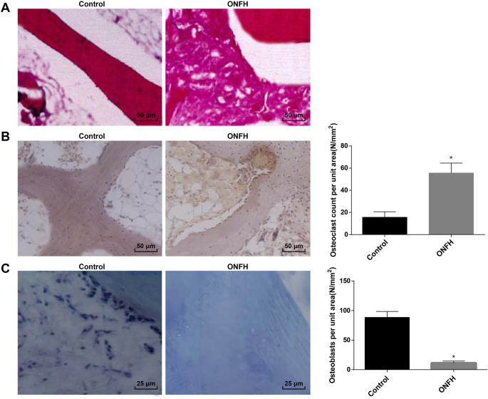
대퇴골 경부 골절군(대조군)과 ONFH군에서 대퇴골두의 병리학적 변화를 관찰하기 위해 H염색법을 채택하였다. 그 결과 대조군에서 골소주 밀도가 잘 분포되어 있고 구조가 온전한 것으로 보고되었다. ONFH군에서는 골소뇌에 빈 골소공이 많았고 골세포는 감소하였고 골소주는 지속적으로 변화하였다. 그 밖에도 골소뇌 주변에 다른 많은 조직 과형성이 있었다(Fig. 1a).
<그림>
임상 샘플의 ONFH 조직의 병리학 적 변화. 아 각 그룹의 H 염색 결과(200 ×). ㄴ TRAP 염색 결과 및 각 그룹의 TRAP 양성 세포 수(200 ×). ㄷ 각 그룹에서 ALP 염색 및 ALP 양성 세포 수의 결과(400 ×). *피 <0.05 대 대조군
TRAP 염색은 TRAP가 주로 파골세포에서 발견되었으며 종종 아마란스에서 성숙한 파골세포를 식별하는 데 사용되는 것으로 나타났습니다. 대조군과 비교하여 ONFH군에서 TRAP 양성 파골세포의 수가 증가하였고, 파골세포의 형태가 다양하고, 세포가 크고 다핵성으로 나타났다. 파골세포와 인접한 골소뇌에서는 골흡수를 보이는 골결손이 뚜렷이 관찰되었다(Fig. 1b).
ALP 염색 결과 ALP는 골기질 성숙 석회화 조절에 관여하는 조골세포 분화 및 성숙의 표지 효소임을 알 수 있었다. 따라서 ALP 발현은 일반적으로 조골 세포를 식별하는 데 사용되었습니다. 갈색 또는 커피 입자는 성숙한 조골 세포의 세포질에서 볼 수 있습니다. 대조군에 비해 ONFH군에서 ALP 양성 조골세포의 수가 감소하였다(Fig. 1c).
MiR-410, ALP, BGLAP 및 Collα1이 분해되고 Wnt-11, ACP5, CTSK 및 MMP9가 임상 샘플의 ONFH 조직에서 강화됨
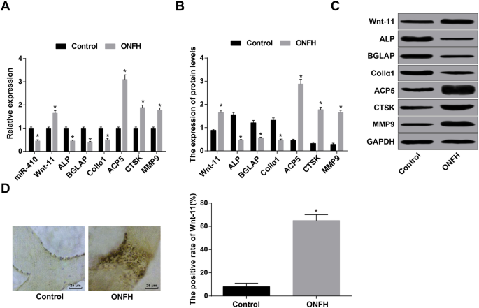
웨스턴 블롯 분석 및 RT-qPCR은 ONFH 그룹의 miR-410 발현이 대조군에 비해 감소한 반면 Wnt-11 발현은 증가함을 보여주었습니다. 조골세포 관련 인자 ALP, BGLAP 및 Collα1의 발현이 저하됨; 파골세포 관련 인자 ACP5, CTSK 및 MMP9 발현이 상승했습니다(모든 P <0.05) (그림 2a-c).
<그림>
MiR-410, ALP, BGLAP 및 Collα1은 임상 샘플의 ONFH 조직에서 감소하고 Wnt-11, ACP5, CTSK 및 MMP9는 증가했습니다. 아 RT-qPCR에 의한 miR-410, Wnt-11, 조골세포 및 파골세포 관련 유전자의 검출. ㄴ 웨스턴 블롯 분석으로 검출된 Wnt-11 및 조골세포 및 파골세포 관련 단백질. ㄷ Wnt-11과 조골세포 및 파골세포 관련 단백질의 단백질 밴드. d Wnt11 발현(400 ×) 및 면역조직화학염색에 의해 검출된 Wnt11 단백질 양성 비율의 비교. *피 <0.05 대 대조군
Wnt-11 발현을 면역조직화학법으로 검사한 결과 Wnt-11 단백질은 주로 세포질에서 발현되었고 양성 발현은 기본적으로 갈색을 띤 노란색 또는 갈색이었다. 대조군과 대조적으로 ONFH군에서 Wnt-11 단백질 발현의 양성률이 높아졌다(P <0.05) (그림 2d).
상향 조절된 miR-410 및 하향 조절된 Wnt-11 쥐의 BMD 및 BV/TV 상승
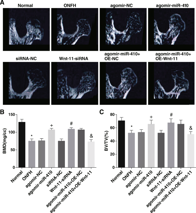
Micro-CT는 정상군에서 대퇴골두의 모양이 둥글고, 목의 두께가 균일하며, 골소주의 구조가 연속적이고 고르게 분포되어 있음을 시사하였다. ONFH군, agomir-NC군, siRNA-NC군, agomir-miR-410 + OE-Wnt-11군 쥐의 대퇴골두가 점차 무너지고 골흡수영역이 점차 나타나 대퇴골의 목이 가늘어졌다. , 뼈 소주가 부서지고 연속성이 파괴되었습니다. agomir-miR-410군, Wnt-11-siRNA군, agomir-miR-410 + OE-NC군에서 쥐의 모습은 그대로 유지되었고, 허탈도 일어나지 않았고, 명백한 골흡수영역도 나타나지 않았고, 골소주 정상적으로 배열되었고 구조가 완성되었습니다(그림 3a).
<그림>
높게 발현된 miR-410과 제대로 발현되지 않은 Wnt-11은 쥐의 BMD와 BV/TV를 증가시킵니다. 아 각 그룹의 쥐에 대한 Micro-CT 결과. ㄴ 골밀도 분석 결과. ㄷ BV/TV 분석 결과. *피 <0.05 대 정상 그룹.
+피 <0.05 vs. agomir-NC 그룹.
#피 <0.05 대 siRNA-NC 그룹.
&피 <0.05 vs. agomir-miR-410 + OE-NC 그룹
뼈 측정 결과에 따르면 정상 그룹과 대조적으로 ONFH 그룹의 BMD 및 BV/TV는 우울했습니다(둘 모두 P <0.05). agomir-NC 그룹과 siRNA-NC 그룹과 관련하여 agomir-miR-410 그룹과 Wnt-11-siRNA 그룹에서 BMD와 BV/TV가 상승했습니다(모두 P <0.05). agomir-miR-410 + OE-NC 그룹과 비교하여, agomir-miR-410 + OE-Wnt-11 그룹에서 BMD와 BV/TV가 떨어졌습니다(둘 다 P <0.05) (그림 3b, c).
miR-410의 과발현 및 Wnt-11의 열악한 발현은 대퇴골간, 대퇴골두 및 척주의 BMD 수준을 높이고 혈청 칼슘을 증가시킵니다. 및 쥐의 인 수준
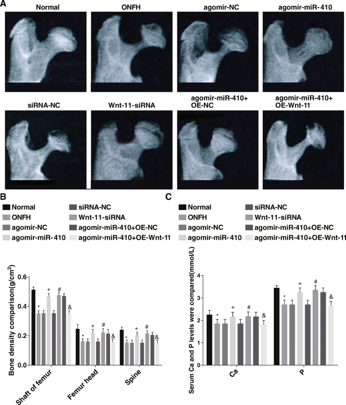
X-ray상 정상군에서 대퇴골두의 모양이 둥글고 대퇴골두 밀도가 균일하며 구조가 완전함을 관찰하였다. ONFH군, agomir-NC군, siRNA-NC군, agomir-miR-410 + OE-Wnt11군에서는 쥐의 대퇴골두 모양이 얇아지고 골흡수영역이 나타났다. agomir-miR-410 그룹, Wnt11-siRNA 그룹, agomir-miR-410 그룹 + OE-NC 그룹에서 생김새가 고르고 대퇴골두의 두께가 완전했습니다(그림 4a). <그림>
상향조절된 miR-410 및 하향조절된 Wnt-11은 대퇴골간, 대퇴골두 및 척주의 골밀도 수준을 증가시키고 또한 쥐의 혈청 칼슘 및 인 수준을 증가시킨다. 아 동물 X선 관찰 결과. ㄴ Comparison of the bone mineral density of the femoral shaft, femoral head, and spine in each group. ㄷ Comparison of the serum calcium and phosphorus levels in each group. *피 <0.05 vs. the normal group.
+피 <0.05 vs. the agomir-NC group.
#피 <0.05 vs. the siRNA-NC group.
&피 <0.05 vs. the agomir-miR-410 + OE-NC group
BMD and serum calcium and phosphorus level determination reported that compared to the normal group, the BMD of the femoral shaft, femoral head, and spine, as well as the serum calcium and phosphorus levels, fell in the ONFH group (all P <0.05). In contrast with the agomir-NC group and siRNA-NC group, the BMD of the femoral shaft, femoral head, and spine, as well as serum calcium and phosphorus levels, ascended in the agomir-miR-410 group and Wnt-11-siRNA group (all P <0.05). In relation to the agomir-miR-410 + OE-NC group, the BMD of the femoral shaft, femoral head, and spine, as well as the serum calcium and phosphorus levels in the agomir-miR-410 + OE-Wnt-11 group, was abated (all P <0.05) (Fig. 4b, c).
Silencing Wnt-11 and Upregulating miR-410 Alleviate the Pathological Changes of Rat Tissues and Restrain Apoptosis of Osteocytes
The results of HE staining showed that the bone trabeculae were neat and clear and arranged regularly and tightly. Bone cells filled the bone lacunae, and the calcified zone was well connected to the subcartilage bone trabeculae in the normal group. In the ONFH group, agomir-NC group, siRNA-NC group, and agomir-miR-410 + OE-Wnt-11 group, the bone trabecula was sparse; thinned, and even broken; the structure was disordered; and some fragments appeared. Some osteocytes in the bone lacuna were necrotic and a large number of bone lacuna was in emptiness that no bone cells filled in, and obvious proliferation of granulation tissue was observed in the necrosis bone trabecular space, which was wrapped around the necrotic bone trabeculae. In the agomir-miR-410 group, the Wnt-11-siRNA group, and the agomir-miR-410 + OE-NC group, the appearance of the rats remained intact, no obvious bone resorption area appeared and the bone trabeculae were arranged normally and the structure was complete (Fig. 5a).
Poor expression of Wnt-11 and overexpression of miR-410 alleviate pathological changes of rats tissues and restrain apoptosis of osteocytes. 아 Morphological observation of the femoral head of rats in each group (200 ×). ㄴ Ultrastructural observation of bone cells in rats of each group (8000 ×). ㄷ The results of TUNEL staining (200 ×). d Detection of apoptosis rate by TUNEL assay. 이 Detection of the Bax and Bcl-2 mRNA expression by RT-qPCR. 에 , 지 Detection of the Bax and Bcl-2 protein expression by western blot analysis. *피 <0.05 vs. the normal group.
+피 <0.05 vs. the agomir-NC group.
#피 <0.05 vs. the siRNA-NC group.
&피 <0.05 vs. the agomir-miR-410 + OE-NC group
Electron microscopic observation observed that the shape of the bone cells in the normal group was consistent with the lacunae, and there was a small gap between the cell and the lacunae wall. The organelles were abundant, the cells in the lacunae were normal, the nucleus was egg-shaped, the nuclear membrane was intact, the chromatin did not agglutinate, and the cytoplasmic pseudo-foot stretched to the peripheral bone small tube and was connected with the adjacent bone cells. Osteoblasts were located on the surface of the bone trabecula, the sample was long, and there were many organelles. In the ONFH group, agomir-NC group, siRNA-NC group, and agomir-miR-410 + OE-Wnt-11 group, a large number of lipid deposits were found in the cytoplasm of osteoblasts, the gap between capsule and lacunae well was further widened, edema bright bands appeared between the nuclear membrane and cytoplasm, nuclear margination showed with compression, nuclear membrane was intact, mitochondria in cytoplasm was swollen, and the endoplasmic reticulum and Gore apparatus disappeared. In the agomir-miR-410 group, Wnt-11-siRNA group, and agomir-miR-410 + OE-NC group, the morphology of rat osteoblasts was normal, chromatin aggregation was found in the nucleus of a small number of cells, no obvious lipid droplets were found in the cytoplasm, and the nuclear membrane was intact (Fig. 5b).
The results of TUNEL staining demonstrated that compared to the normal group, the apoptosis rate of osteocytes in the ONFH group was raised (P <0.05). In contrast with the agomir-NC group and the siRNA-NC group, the apoptosis rate of osteocytes abated in the agomir-miR-410 group and the Wnt-11-siRNA group (both P <0.05). In relation to the agomir-miR-410 + OE-NC group, the apoptosis rate of osteocytes enhanced in the agomir-miR-410 + OE-Wnt-11 group (P <0.05) (Fig. 5c, d).
Western blot analysis and RT-qPCR reported that the Bcl-2 expression reduced and Bax expression raised in the ONFH group compared to the normal group (both P <0.05). By comparison with the agomir-NC group and the siRNA-NC group, Bcl-2 expression ascended and Bax expression descended in the agomir-miR-410 group and the Wnt-11-siRNA group (all P <0.05). In contrast with the agomir-miR-410 + OE-NC group, the Bcl-2 expression declined and Bax expression appended in the agomir-miR-410 + OE-Wnt-11 group (both P <0.05) (Fig. 5e–g).
Overexpression of miR-410 and Low Expression of Wnt-11 Increase the Number of Osteoblasts and Decrease the Number of Osteoclasts
TRAP staining revealed that in the normal group, the morphology of osteoclasts with positive TRAP staining was different, most of them were shuttle or fusiform, few large osteoclasts were polygons, and most of them were distributed around the bone cerebellum. In the ONFH group, agomir-NC group, siRNA-NC group, and agomir-miR-410 + OE-Wnt-11 group, TRAP-positive cells were ascended, the morphology of cells was diverse in large polygons and polykaryotes, bone resorption was found in the bone trabeculae adjacent to osteoclasts, and typical resorption lacunae were formed. In the agomir-miR-410 group, Wnt-11-siRNA group, and agomir-miR-410 + OE-NC group, the number of cells positive for TRAP staining was reduced, the cells were in a long strip shape and the morphology was more regular, the polygonous polynuclear osteoclasts were rare, and the bone structure of adjacent bone trabeculae was relatively complete (Fig. 6a).
Overexpression of miR-410 and low expression of Wnt-11 increase the number of osteoblasts and decrease the number of osteoclasts. 아 Results of TRAP staining in osteoclasts of rats (200 ×). ㄴ Count of positive cells in TRAP staining. ㄷ Results of ALP staining in osteoblasts of rats in each group (200 ×). d Count of positive cells determined by ALP staining. *피 <0.05 vs. the normal group.
+피 <0.05 vs. the agomir-NC group.
#피 <0.05 vs. the siRNA-NC group.
&피 <0.05 vs. the agomir-miR-410 + OE-NC group
ALP staining presented that in the normal group, in osteoblasts, which were positive for ALP staining, the morphology was small and round. The cells were distributed in aggregation, most of which were located in the bone trabecular space of bone marrow cavity and on the surface of some bone trabeculae. In the ONFH group, agomir-NC group, siRNA-NC group, and agomir-miR-410 + OE-Wnt-11 group, the number of osteoblasts positive for ALP staining was dispersedly distributed in the trabecular space of the bone marrow cavity. In the agomir-miR-410 group, Wnt-11-siRNA group, and agomir-miR-410 + OE-NC group, the number of osteoblast-positive cells enhanced, and they were distributed in the trabecular space of the bone marrow cavity and the surface of the part of the bone trabecular (Fig. 6c).
By comparison with the normal group, the number of osteoblasts per unit area was suppressed and the number of osteoclasts was heightened in the ONFH group (both P <0.05). In relation to the agomir-NC group and the siRNA-NC group, the number of osteoblasts per unit area was heightened and the number of osteoclasts was declined in the agomir-miR-410 group and Wnt-11-siRNA group (all P <0.05). In contrast to the agomir-miR-410 + OE-NC group, the number of osteoblasts per unit area was declined and the number of osteoclasts was raised in the agomir-miR-410 + OE-Wnt-11 group (both P <0.05) (Fig. 6b, d).
Highly Expressed miR-410 and Lowly Expressed Wnt-11 Elevate the Expression of Osteocalcin (OCN), ALP, BGLAP, and Collα1, as well as Abate C- and N-terminal Telopeptides of Type I collagen (NTX-1, CTX-1), ACP5, CTSK, and MMP9 expression in ONFH Rats
The results of ELISA revealed that in relation to the normal group, the level of osteogenic function index ALP and OCN degraded while the levels of osteoclast function index NTX-1 and CTX-1 heightened in the ONFH group (all P <0.05). In contrast with the agomir-NC group and the siRNA-NC group, ALP and OCN ascended and NTX-1 and CTX-1 descended in the agomir-miR-410 group and the Wnt-11-siRNA group (all P <0.05). By comparison with the agomir-miR-410 + OE-NC group, ALP and OCN dropped and NTX-1 and CTX-1 appended in the agomir-miR-410 + OE-Wnt-11 group (all P <0.05) (Fig. 7a).
Highly expressed miR-410 and lowly expressed Wnt-11 elevate the expression of OCN, ALP, BGLAP, and Collα1 as well as abate NTX-1, CTX-1, ACP5, CTSK, and MMP9 expression in ONFH rats. 아 Detection of osteogenesis and osteoclast function indices in the serum of rats in each group by ELISA. ㄴ Detection of osteoblast- and osteoclast-related genes by RT-qPCR. ㄷ , d Detection of osteoblast- and osteoclast-related genes protein expression by western blot analysis. *피 <0.05 vs. the normal group.
+피 <0.05 vs. the agomir-NC group.
#피 <0.05 vs. the siRNA-NC group.
&피 <0.05 vs. the agomir-miR-410 + OE-NC group
The results of western blot analysis and RT-qPCR revealed that in relation to the normal group, the expression of osteoblast-related factors ALP, BGLAP, and Collα1 degraded, and osteoclast-related factors ACP5, CTSK, and MMP9 expression ascended (all P <0.05). In contrast with the agomir-NC group and the siRNA-NC group, the expression of ALP, BGLAP, and Collα1 elevated, and ACP5, CTSK, and MMP9 expression abated in the agomir-miR-410 group and the Wnt-11-siRNA group (all P <0.05). By comparison with the agomir-miR-410 + OE-NC group, the expression of ALP, BGLAP, and Collα1 depressed, and ACP5, CTSK, and MMP9 expression heightened in the agomir-miR-410 + OE-Wnt-11 group (all P <0.05) (Fig. 7b–d).
Elevated Wnt-11 and Decreased miR-410 are Found in ONFH Tissues of Rats as well as Wnt-11 is the Target Gene of miR-410
The targeting relationship between miR-410 and Wnt-11 gene was analyzed by an online analysis software. It was displayed that a specific binding region existed between Wnt-11 gene sequence and miR-410 sequence, implying that Wnt-11 was the target gene of miR-410 (Fig. 8a). Luciferase activity assay was utilized to verify this relationship (Fig. 8b). The results presented that compared to the NC group, the luciferase activity depressed in the miR-410 mimics group (P <0.05), but there was no significant difference in the luciferase activity of MUT 3′UTR (P> 0.05), indicating that miR-410 could specifically bind to Wnt-11.
Increased Wnt-11 and decreased miR-410 are found in ONFH tissues as well as Wnt-11 is the target gene of miR-410. 아 Prediction of binding sites of miR-410 on Wnt-11 3′UTR. ㄴ Luciferase activity detection for verifying the relationship between miR-410 and Wnt-11. ㄷ Detection of miR-410 and Wnt-11 expression by RT-qPCR. d , e Wnt-11 protein expression determined by western blot analysis. 에 Expression of Wnt-11 in femoral head of rats in each group detected by immunohistochemistry (200 ×). 지 Comparison of the positive rate of Wnt-11 protein expression in each group. *피 <0.05 vs. the normal group.
+피 <0.05 vs. the agomir-NC group.
#피 <0.05 vs. the siRNA-NC group.
&피 <0.05 vs. the agomir-miR-410 + OE-NC group
The results of western blot analysis and RT-qPCR revealed that in relation to the normal group, miR-410 expression declined and Wnt-11 expression raised in the ONFH group (both P <0.05). By comparison with the agomir-NC group, miR-410 raised and Wnt-11 reduced in the agomir-miR-410 group (both P <0.05). In contrast with the siRNA-NC group, Wnt-11 declined in the Wnt-11-siRNA group (P <0.05). Wnt-11 enhanced in the agomir-miR-410 + OE-Wnt-11 group relative to that in the agomir-miR-410 + OE-NC group (P <0.05) (Fig. 8c–e).
>Wnt-11 expression was verified by immunohistochemistry. Wnt-11 protein was mainly expressed in the cytoplasm, and the positive expression was mainly brownish yellow or brown. Compared to the normal group, the positive rate of Wnt-11 protein expression in the ONFH group was raised (P <0.05). By comparison with the agomir-NC group and the siRNA-NC group, the positive rate of the Wnt-11 protein expression descended in the agomir-miR-410 group and Wnt-11-siRNA group (both P <0.05). In relation to the agomir-miR-410 + OE-NC group, the positive rate of Wnt-11 protein expression enhanced in the agomir-miR-410 + OE-Wnt-11 group (P <0.05) (Fig. 8f, g).
Discussion
ONFH, a kind of bone destruction disease, is caused by blood supply failure and coagulation and fibrinolysis system disorder and finally causes the femoral head to collapse [19]. A previous study has discussed miRNA expression in bone marrow mesenchymal stem cells induced by hormone in mice with ONFH [20]. Also, a recent study has provided a proof that the expression profile of miRNA of bone marrow-derived mesenchymal stem cells in osteogenesis related to steroid-induced ONFH [21]. Furthermore, it was revealed the Li-nHA/GM/rhEPO stents can elevate both Wnt and HIF-1/VEGF pathways and promote osteogenesis and angiogenesis, which is beneficial to the repair of ONFH induced by glucocorticoids [22]. Based on these facts, the study aimed to explore the effects of miR-410 in the prevention of ONFH by targeting Wnt-11.
Our study has provided substantial evidence in relation to the notion that miR-410, ALP, BGLAP, and Collα1 degraded and Wnt-11, ACP5, CTSK, and MMP9 enhanced in ONFH tissues. A recent study has promoted that the expression of miR-410 in osteosarcoma is declined, and the anti-tumor effect is shown [23]. Another study has presented miR-410 expression was reduced in human estrogen receptor-positive tissues of breast cancer [24]. It is reported that secreted factor Wnt-11 expression is ascended in several types of cancer, containing colorectal cancer, where it advances the migration and invasion of cancer cells [25]. Similarly, a previous study has proved that Wnt-11 expression is heightened in hormone-independent prostate cancer [26]. ALP is a useful index for the state diagnosis and clinical prognosis of the disease [27]. OCN (bone gamma-carboxyglutamate protein; BGLAP) is a widely conserved molecule related to mineralization of bone matrix [28]. ACP5 is necessary for osteoclast differentiation and bone resorption, and it promotes cell movement by regulating adhesion kinase phosphorylation [29]. CTSK is a critical protease in charge of osteopontin, degrading type I collagen and other bone matrix proteins [30]. MMPs can degrade and modify most of the components of extracellular matrix and basement membrane and push forward an immense influence on cancer invasion and metastasis [31]. Also, our study revealed that Wnt-11 is the target gene of miR-410. Similarly, Zhang et al. found that miR-410 targeted the inferred binding site in the Wnt3a 3′-UTR to modulate the Wnt signaling pathway [32].
In addition, it was revealed that upregulated miR-410 and downregulated Wnt-11 increased BMD and BV/TV of ONFH rats. It has been suggested previously that a decrease of spinal BMD and an increase of urinary DPD/Cr ratio in non-traumatic ONFH patients [33]. Another study has verified that BMD of the femoral head and lumbar vertebrae in the ONFH group were degraded relative to that in the control group [34]. Additionally, imaging analysis revealed muscone can restore BMD and BV/TV ratio of the necrotic femoral head, while histologic examination further confirmed the protective effect of muscone on alcohol-induced ONFH [35]. A result emerged from our data that highly expressed miR-410 and lowly expressed Wnt-11 restrained apoptosis of osteocytes. A study has demonstrated that silencing miR-410 can induce cell proliferation and reduce the apoptosis of human umbilical vein endothelial cells induced by oxidized low-density lipoprotein [36]. It is reported that the descended miR-410 expression and ascended SOCS3 expression could reduce the expression of anti-apoptosis factor Bcl-2 and promoted the apoptosis of cells [37]. In addition, in androgen deficient LNCaP cells, the downregulation of Wnt-11 can prevent neuroendocrine-like differentiation and lead to apoptosis of prostate cancer cells [26]. The study also showed that the downregulation of Wnt-11 and upregulation of miR-410 enhanced ALP, BGLAP, and Collα1 expression and reduced ACP5, CTSK, and MMP9 expression. A study has indicated that Wnt-11 expression heightened in cervical cancer cells may result in activation and phosphorylation of JNK-1 by activating Wnt/Jnk pathway and boosts the proliferation and migration/invasion of tumor cells [15]. It has been suggested that Wnt-11 gene silencing in colorectal cancer cell lines reduced the invasive ability of cells [25]. Furthermore, the overexpression of miR-410 can restrain the invasion, migration, proliferation, and epithelial mesenchymal transformation of osteosarcoma cells via directly targeting TRIM44 [23]. Furthermore, a previous study stated that upregulated miR-410 inhibited the growth of cholangiocarcinoma in a xenograft mouse model by inducing apoptosis [38].
Conclusion
In summary, our investigation revealed that miR-410 was lowly expressed and Wnt-11 was highly expressed in ONFH, and upregulating miR-410 or downregulating Wnt-11 increased osteoblasts and reduced osteoclasts to alleviate ONFH. However, clinical researches might be further carried out to detect the efficacy of miR-410 and Wnt-11 for the treatment of ONFH.