항-Epcam 앱타머(Syl3c)-독소루비신의 표적 전달을 위한 기능화된 리포솜:C26 결장암을 보유한 마우스의 시험관 내 및 생체 내 항종양 연구
초록
이 연구에서 우리는 항-EpCAM 앱타머-결합 DSPE-mPEG2000의 사후 삽입을 통해 항-EpCAM(상피 세포 접착 분자) 앱타머와 함께 표면 기능화된 PEG화 나노리포좀 독소루비신(DOX)을 가지고 있습니다. Caelyx®(ED-립)로. 제형의 크기, 전하, 방출 프로파일, 및 세포독성 및 세포 흡수를 측정하였다. ED-립의 특성화는 제타 전위의 감소와 함께 크기 및 PDI의 약간 증가를 보여주었으며 이는 삽입 후 효율적으로 수행되었음을 나타냅니다. 유세포 분석 및 형광 현미경 검사 결과에 따르면 ED-lip은 Caelyx®와 비교하여 C26 세포주에서 세포 흡수율을 향상시켰습니다. ED-lip은 또한 표적 리간드로서 항-EpCAM 앱타머의 효능을 나타내는 Caelyx®보다 더 많은 세포독성 효과를 가졌습니다. C26 종양이 있는 마우스에서 제형의 약동학 및 조직 생체 분포는 동물 모델에서 Caelyx®와 비교하여 ED-lip이 DOX의 분포 프로파일에 영향을 미치지 않는다는 것을 보여주었습니다. 또한 ED-lip은 Caelyx®에 비해 DOX의 종양 축적을 효과적으로 개선하고 동물의 생존을 촉진했습니다. 이러한 결과는 항-EpCAM 앱타머를 사용한 Caelyx®의 기능화가 암 치료에 유망하며 추가 조사가 필요함을 시사합니다.
소개
100~200nm 크기의 나노 약물 전달 시스템(NDDS)은 EPR(Enhanced Permeability and Retention) 효과를 통해 종양 미세 환경에 수동적으로 축적됩니다. 이것은 느슨한 내피 라이닝과 약한 림프 배수를 통해 발생합니다. 그러나 최근 데이터에 따르면 투여된 약물의 1% 미만만이 종양 부위에 도달할 수 있습니다[1]. 종양의 조밀한 세포외 기질(ECM)에 침투하는 능력의 부족, 방출된 약물의 순환으로의 복귀 및 종양의 이질성이 이러한 실패의 원인이 됩니다[2]. 내인성 및 외인성 자극을 사용하여 NDDS의 종양 축적을 개선하기 위해 다양한 전략이 사용되었습니다[3]. 이러한 NDDS는 빛과 같은 외인성 자극에 반응할 수 있으며 종양 영상화에 사용할 수 있습니다[4]. 항암제로 사용할 수 있는 다양한 무기 나노물질이 있다[5, 6]. 그러나 무기 나노 물질의 경우 독성 및 환경 안전성에 주의를 기울여야 합니다[7,8,9,10,11].
능동 표적 전달은 NDDS가 치료제를 종양에 보다 효율적으로 전달하고 비표적 조직에 대한 노출을 최소화하는 데 도움이 되는 중요한 접근 방식입니다[12, 13]. 표적 전달을 위한 이상적인 표적 제제는 특정 세포 또는 조직 성분에 의해 상향 조절되는 세포 표면 단백질 또는 수용체에 친화성을 갖는 분자입니다[14].
상피 세포 부착 분자(EpCAM)는 활성 표적화를 위한 후보 리간드로 간주되는 막횡단 당단백질입니다. 최근 연구 결과에 따르면 EpCAM은 정상적으로 낮은 발현의 건강한 상피 세포를 가지고 있는 반면 암세포에서는 그 발현이 더 높은 수준(최대 1000배)으로 나타납니다[15,16,17]. 암이 발생하는 동안 EpCAM의 발현 패턴은 정상 상피의 기저 및 기저외측 막에서 종양 상피 세포의 정점 표면으로 변경됩니다[18]. 이러한 차별적인 발현은 EpCAM을 약물의 치료 지수를 향상시킬 수 있는 약물 전달을 위한 매우 흥미로운 리간드로 만듭니다[19].
EpCAM은 암 줄기 세포(CSC) 또는 종양 개시 세포(TIC) 마커로 입증되며, 이는 암에서의 발현이 불량한 예후와 관련이 있습니다[20]. CSC 또는 TIC는 동일한 유형의 더 많은 세포를 생성할 수 있는 자가 재생 능력이 있는 세포로, 종양 발달 및 전이에 중요한 역할을 합니다[21]. EpCAM의 과발현은 다양한 고형 종양의 CSC에서 보고되었습니다[22]. 최근 압타머는 광범위한 연구에서 많은 관심을 받았고 NDDS에서 표적 리간드로 사용될 수 있는 잠재적으로 강력한 분자로 부상하고 있다[23, 24]. 압타머는 세포 표면 수용체와 같은 표적에 친화성을 갖는 2차 및 3차 구조를 갖는 DNA 또는 RNA 기반 올리고뉴클레오티드 서열이다[23, 24]. 압타머는 또한 비면역성이며 화학적 및 열적 안정성과 함께 저분자량(8-25kDa)을 가지고 있다는 점과 같이 여러 이점이 있습니다. 또한 합성 및 화학적 변형이 저렴하고 확장 가능합니다[25]. 항-EpCAM-특이적 앱타머를 통한 NDDS의 선택적 표적화는 화학요법제를 종양 미세환경으로 전달하기 위한 효과적인 표적화 옵션으로 간주될 수 있다[19, 26]. 이와 관련하여 다양한 연구에서 항-EpCAM 앱타머 기능화된 나노캐리어가 종양 세포에 대한 항암제의 전달을 효과적으로 개선할 수 있음을 보여주었습니다[15, 27, 28].
이 연구의 목표는 NDDS의 모델로 독소루비신(DOX)(ED-lip)이 로딩된 항-EpCAM DNA 앱타머(SYL3C)-PEG화 나노리포좀을 개발하는 것입니다. 이러한 기능화는 앱타머의 아민 그룹과 DSPE-mPEG2000의 카르복실 그룹 간의 EDC/NHS 커플링 화학에 의해 수행되었습니다. 크기, 제타 전위, 독소루비신 캡슐화의 백분율, 방출 프로필 및 세포독성을 특징으로 하는 ED-립. 그런 다음, 우리는 이러한 ED-립이 C26 결장암 종양이 있는 마우스에서 표적화를 통해 시험관 내 세포 흡수를 개선하고 DOX를 종양에 전달할 수 있는지 여부를 평가했습니다.
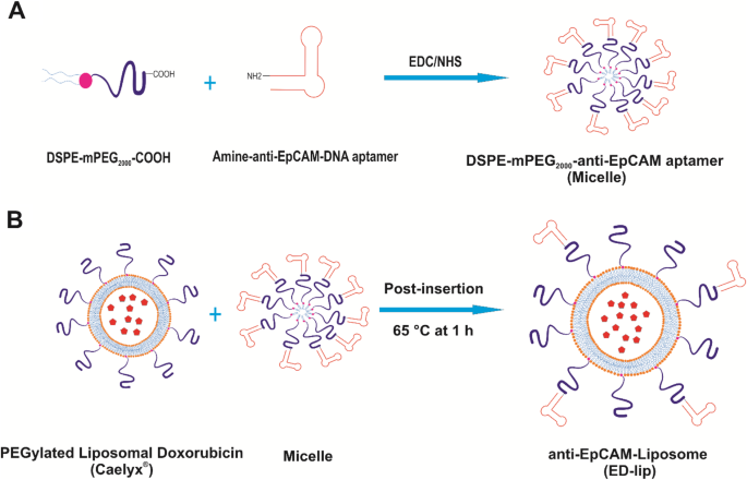
<그림>
항-EpCAM 기능화된 Caelyx®(ED-립)의 제조. 아 DSPE-mPEG2000에 Anti-EpCAM 앱타머 연결 DSPE-mPEG2000의 카르복실기(-COOH)에 항-EpCAM 앱타머의 1차 아민(-NH2)의 공유 결합을 통해 . ㄴ 항-EpCAM 기능화 Caelyx®(ED-lip)의 제조를 위한 삽입 후 방법
결과 및 토론
Caelyx®, PEG화 리포솜 독소루비신은 가장 널리 사용되는 화학요법제 중 하나이며 난소암, AIDS 관련 카포시 육종 및 다발성 골수종의 치료를 위해 표시된 최초의 FDA 승인 나노입자입니다. Caelyx®는 EPR 효과를 통해 수동적으로 종양 부위에 침투했습니다[29]. Caelyx®는 DOX의 약동학 및 반감기를 크게 개선했습니다. 그러나 Caelyx®의 주요 한계는 세포 흡수가 불충분하고 종양 부위에서 약물의 낮은 방출 속도입니다[29]. 여기에서 우리는 SYL3C 앱타머를 표적 리간드로 사용하여 리포솜 독소루비신(ED-lip)을 기능화하여 암세포 표면의 EpCAM 분자를 표적화하여 활성 표적화 과정을 통해 DOX를 특정 표적 부위에 전달할 수 있습니다.
DSPE-mPEG의 활용2000 압타머로
현재 연구에서 우리는 DSPE-mPEG2000의 활성 카르복실기에 대한 아민 작용화된 항-EpCAM 앱타머의 접합을 위해 EDC/NHS 커플링 화학을 사용했습니다. -쿠. EDC/NHS 커플링 화학과 아미드 결합의 형성을 사용하는 이 커플링 반응의 장점은 안정성과 앱타머 간의 비특이적 상호작용을 줄이는 것입니다[30]. 압타머는 1차 아민 또는 티올기로 변형되고 각각 말레이미드의 카르복실 또는 피롤기를 활성화하기 위해 공유 결합될 수 있다[31]. 티올기로 변형된 압타머는 DSPE-PEG2000의 말레이미드 작용기에 접합되었습니다. . 그런 다음 DSPE-PEG2000 -압타머는 리포솜 구조에 후삽입되어 리포솜의 외부 표면을 장식합니다[32]. 말레이미드 티올 화학의 한 가지 중요한 제한 사항은 보관하는 동안 앱타머의 티올기가 산화에 의해 영향을 받아 두 개의 티올로 변형된 앱타머 사이에 이황화 결합(S-S)이 형성될 수 있다는 것입니다. 이러한 이량체 앱타머는 DSPE-PEG2000의 말레이미드 작용기와의 접합 반응에 참여할 수 없습니다. [30]. 따라서 EDC/NHS 반응을 사용하면 제품 수율이 증가하고 삽입 후 방법이 향상됩니다.
Aptamer는 합성 및 규모 확장의 용이성, 낮은 전신 독성 및 면역원성의 결여를 포함하여 항체에 비해 몇 가지 이점이 있습니다[33]. 여기서, 지질에 앱타머 접합 후, 항-EpCAM 앱타머로 장식된 Caelyx®(ED-립)를 만들기 위해 사후 삽입 방법이 모집되었습니다. 일반적으로 삽입 후 기술은 리포솜 표면에 앱타머를 부착하는 간단하고 효과적인 방법이며 리포솜으로의 더 높은 비율의 앱타머 결합을 제공합니다[34].
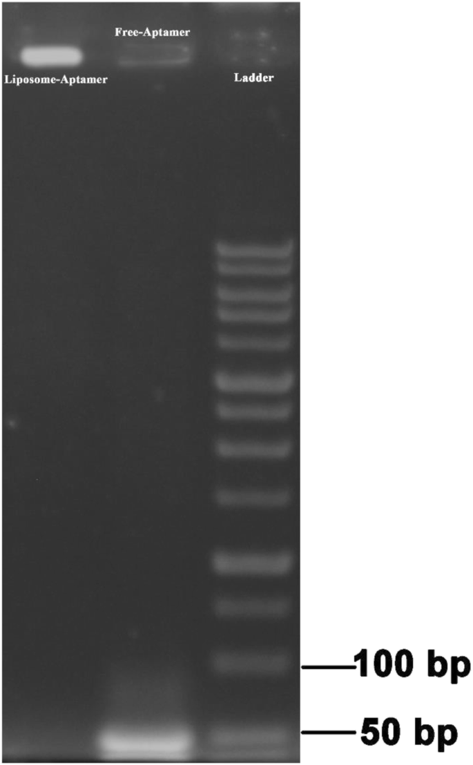
우리는 리포솜에 항-EpCAM 앱타머를 삽입한 후 평가하기 위해 겔 전기영동 이동성 이동 분석을 사용했습니다. 그림 2에서 보는 바와 같이 음전하를 띠는 압타머는 겔 내에서 이동하였고 ED-lip 제제의 경우 ED-lip이 웰 라인에 갇혀 젤을 통해 이동할 수 없었기 때문에 ED-lip 제제에 대한 대응 밴드가 없는 동안 그 밴드가 관찰되었다. 이러한 결과는 압타머가 결합된 미셀이 리포솜 표면에 성공적으로 사후 삽입되었음을 나타냅니다.
<그림>
ED-립 제형의 아가로스 겔 전기영동. 샘플을 아가로스 겔에 로드합니다. UV 빛은 젤을 시각화했습니다. 사다리, 자유 압타머 및 PL 접합 압타머에 해당하는 웰이 표시됩니다. 리포솜-앱타머에 해당 밴드가 없음은 삽입 후 확인을 나타냄
ED-lip의 물리화학적 특성
Caelyx® 및 ED-lip의 물리화학적 특성은 표 1에 나와 있습니다. 준비된 제제의 크기와 전하량은 항-EpCAM 앱타머를 사용한 Caelyx®의 변형이 입자 크기에 큰 영향을 미치지 않는 것으로 나타났습니다(p> 0.05). 압타머(Caelyx®) 삽입 전 리포솜 크기는 PDI가 0.11인 약 96 nm였으며, 삽입 후(ED-lip) 리포솜 크기가 부분적으로 117 nm로 증가했으며 PDI가 0.14로 종양. 이전 연구의 결과는 또한 표적 리간드의 통합이 리포솜의 크기와 PDI를 증가시킨다는 것을 나타냈습니다[35, 36]. 또한 ED-lip의 제타 전위(-19.25)는 Caelyx®(-12)보다 더 음이 되었습니다. RNA-aptamer를 리포솜에 결합하면 리포솜의 제타 전위가 감소하는 것으로 나타났습니다[37]. ED-lip의 크기 증가와 음의 제타 전위는 리포솜 표면에 접합된 앱타머가 성공적으로 삽입되었다는 증거가 될 수 있습니다[38]. 이러한 결과는 Caelyx® 표면에 앱타머가 부착되어 입자 크기가 약간 증가하고 앱타머 기능화된 Caelyx®에서 더 음의 제타 전위가 발생한다는 우리의 이전 연구와 일치합니다[38, 39]. 그러나 더 나은 크기와 PDI로 더 효율적인 삽입 후 리포솜에 도달하려면 배양 시간과 온도 측면에서 삽입 후의 효능을 테스트해야 합니다. Caelyx®와 ED-lip의 캡슐화 효율은 100%였습니다(표 1 참조).
리포솜 표면에 사후 삽입된 앱타머의 수는 [6]에 설명된 대로 결정되었습니다. 포스페이트 분석에 의해 결정된 리포솜 제형의 인지질 함량의 총량은 14mM이었다. 평균 크기가 100 nm인 리포솜의 평균 지질 분자 수는 8×10
4
이므로 각 밀리리터의 리포솜 수는 거의 10
14
입니다. [38]. 압타머의 분자량은 g/mol이었다. DSPE-mPEG2000의 수 -압타머는 인산염 분자의 몰이 접합된 분자의 몰에 해당하는 인산염 분석 방법에 따라 결정되었습니다. 이러한 데이터를 기반으로 하여 분취액 1ml당 앱타머 분자의 수는 10
15
입니다. .
DOX 출시 프로필
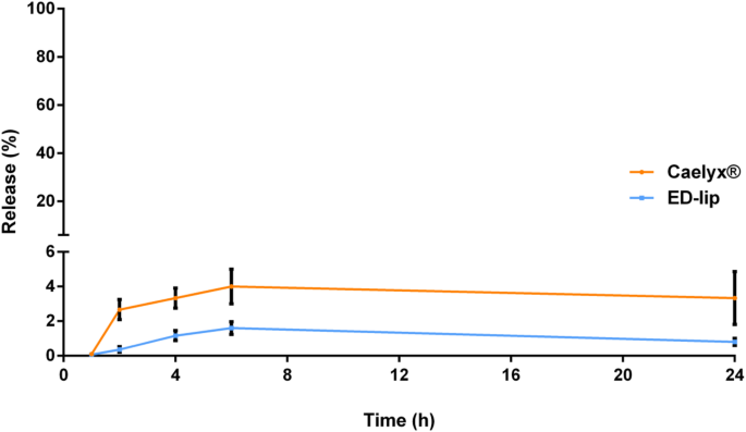
Caelyx®의 외부 표면에 앱타머 결합 미셀을 삽입하면 DOX의 방출 프로파일에 영향을 미칠 수 있습니다. 따라서 우리는 50% FBS와 5% 덱스트로스 내 Caelyx®와 비교하여 DOX 형태 ED-립의 방출을 평가했습니다. 이 매질은 혈장 내 제형의 방출 거동을 모방할 수 있습니다[40]. 그림 3은 연구 24시간 동안 Caelyx® 및 ED-립 제형의 DOX 방출에 유의미한 차이가 없으며 무시할 수 있는 양의 DOX만 방출되었음을 보여줍니다. 이것은 리포솜 표면에 앱타머를 삽입하는 것이 DOX의 막 안정성과 방출 프로필에 영향을 미치지 않는다는 것을 나타내는 이전 연구와 일치합니다[38, 39]. 이는 주로 pH 구배 기반 원격 로딩 방법을 사용하여 제형화된 Caelyx® 제형의 안정성 때문입니다[41].
<그림>
연구를 출시합니다. 연구 24시간 동안 덱스트로스에 50% FBS가 존재하는 37°C에서 Caelyx® 및 ED-lip의 DOX 함량 누출 프로필. 평균 ± 표준 편차(SEM)로 표시되는 데이터(n =3)
형광현미경에 의한 세포 상호작용 및 세포 흡수
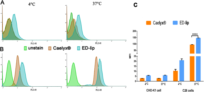
리포솜 제형의 세포 상호 작용 및 세포 흡수는 4 °C 및 37 °C에서 평가되었으며 그림 4에 나와 있습니다. ED-lip의 표적화 효능 평가는 평균 형광 강도(MFI) 간에 차이가 없음을 나타냅니다. 4 °C와 37 °C에서 Caelyx®와 ED-lip으로 처리한 CHO-K1 세포의 모습(그림 4a, c). 그러나 데이터는 표적화된 ED-lip이 4° 및 37°C에서 Caelyx®와 비교하여 C26 세포에 의해 상당히 더 높은 흡수를 가짐을 입증했으며(그림 4b, d) 37°C에서 통계적으로 유의했습니다(p <0.0001). ED-립은 Caelyx®와 비교하여 상당한 흡수율을 보였습니다(p <0.001). 이러한 결과는 항-EpCAM 앱타머로 인한 ED-lip 강화된 표적 특이성이 CHO-K1 세포와 비교하여 C26 세포주에 대한 친화도가 더 높다는 것을 나타내었다. 유리 DOX는 지질 이중층을 자유롭게 통과하여 세포로 들어가므로 제형 중 세포 흡수율이 가장 높기 때문에 세포 독성이 가장 높습니다. Caelyx®의 경우 PEGylation은 세포내이입 속도를 제한하고 세포독성을 감소시켰습니다. 그러나 리포솜(ED-lip) 표면에 항-EpCAM 앱타머가 존재하면 제형이 세포로 내재화되는 속도가 향상되고 Caelyx®와 비교하여 세포 독성이 증가합니다[38]. 형광 현미경의 데이터는 37 °C에서 C26 세포주에서 ED-lip과 유리 DOX의 세포 흡수 사이의 차이가 중요하지 않음을 보여주었습니다(그림 5). 그러나 ED-lip과 DOX 모두 C26 세포 흡수에서 Caelyx®와 통계적으로 유의한 차이를 나타내는 표 2에 묘사된 내재화된 DOX의 강도를 기반으로 한 스케일링(p <0.001). C26 세포는 표면에서 낮은 수준의 EpCAM을 발현하지만[42], 이 연구의 데이터는 항-EpCAM 앱타머의 존재가 리포솜의 내재화 과정 속도를 향상시킬 수 있음을 시사했습니다[43].
<그림>
4°C 및 37°C에서 평가된 리포솜 제형의 세포 상호작용 및 세포 흡수. 아 4°C 및 37°C에서 제형의 CHO-K1 세포 상호작용. ㄴ 4 °C 및 37 °C에서 C26 세포와 제형의 상호 작용. ㄷ 그래프는 CHO-K1 및 C26 세포에 대한 제형의 평균 MFI를 입증했습니다. 평균 ± 표준 편차(SEM)로 표시되는 데이터(n =3). ****p <0.0001
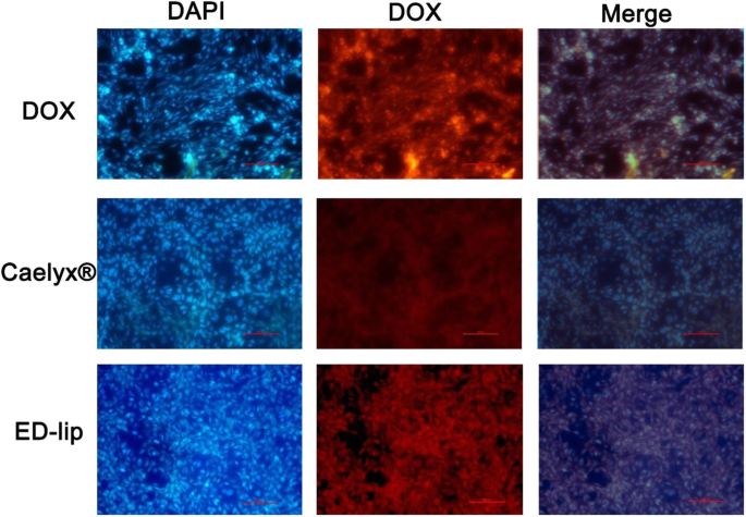
<그림>
형광현미경. 형광 현미경으로 시각화한 C26 세포주에서 DOX의 세포 내재화 결과. DAPI로 염색된 세포. free DOX와 ED-lip은 Caelyx®에 비해 DOX 내재화 수준이 더 높습니다. × 200 배율로 검사한 세포
세포독성 연구
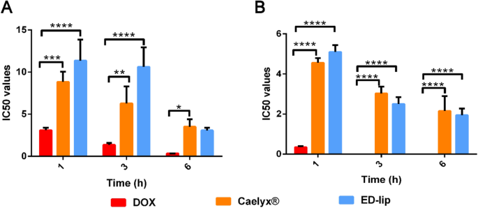
유리 DOX, Caelyx® 및 ED-lip 제제의 세포독성 효과는 그림 6에 나와 있습니다. 1, 3, 6시간 동안 세포를 처리하고 다음 72시간 동안 배양하는 데 사용되는 다양한 농도의 제제가 표시됩니다. 데이터는 모든 제형이 시간 및 용량 의존적 방식으로 세포에 영향을 미친다는 것을 입증했습니다. ED-lip 제형으로 처리된 C26 세포의 생존율은 Caelyx® 처리된 세포와 비교하여 감소했습니다. EpCAM 음성 세포인 CHO-K1 세포는 C26 세포에 비해 ED-lip에 대한 반응이 낮기 때문에 항-EpCAM 앱타머는 표적 세포에 Caelyx®의 특이적 전달을 증가시킨 것으로 보입니다. 이러한 결과는 C26 세포에 의한 ED-lip의 특정 세포 흡수를 확인할 수 있습니다. 이러한 결과는 표적 세포에 약물을 선택적으로 전달하고 오프 타겟을 피함으로써 약물의 부작용을 줄이기 위해 특정 표적 제제와 함께 표적 약물 전달 사용의 중요성을 강조했습니다[44]. 이전에 보고된 바와 같이, 앱타머 및 항체와 같은 특정 표적 리간드를 사용한 Caelyx®의 활성 표적화는 활성 종양 표적화 및 표적 세포에 대한 특정 약물 전달을 증가시켜 차례로 DOX의 치료 효능을 향상시킵니다[35, 39].
<그림>
다양한 노출 시간 후 CHO 세포 및 C26 세포에 대한 ED-lip, Caelyx® 및 유리 독소루비신의 시험관 내 세포독성 효과(IC50). μg/ml ± 표준편차(SEM)로 표시되는 데이터(n =3). *p <0.05, **p <0.01, ***p <0.001, ****p <0.0001
생체분포 및 약동학
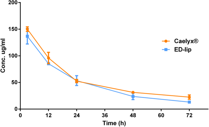
항-EpCAM 앱타머가 DOX의 생체내 분포에 어떤 영향을 미치는지 평가하기 위해, 우리는 피하 C26 결장암 종양이 있는 마우스에 10 mg/kg의 ED-lip 및 Caelyx®를 주사했습니다. Caelyx®와 ED-lip 주사 후 3, 12, 24, 48, 72시간 후의 혈장 DOX 농도는 그림 7에 나와 있습니다. 결과는 두 그룹의 혈장 DOX 농도 거동이 유사했으며 두 제형 간에 유의한 차이가 없었습니다. 표 3에 나타난 바와 같이, 리포솜 표면에 대한 항-EpCAM 앱타머의 접합은 순환 반감기를 39.3시간에서 34.2시간으로, MRT를 47.6시간에서 42.9시간으로 약간 감소시켰다(표 3 참조). 약동학적 매개변수는 리포솜에 대한 항-EpCAM 앱타머의 접합이 t½을 약간 감소시키는 것으로 나타났습니다. 이전 보고서와 일치하는 MRT는 리포솜 표면에 대한 앱타머의 접합이 리포솜의 제거를 가속화한다는 것을 보여주었습니다[38]. 단핵 식세포 시스템(MPS)에 의한 단백질 흡착 및 이에 따른 제거는 리간드-접합 나노입자의 혈액 제거를 가속화하는 원인이 될 수 있습니다[45].
<그림>
DOX의 플라즈마 수준. 주입 후 3, 12, 24, 48 및 72시간에 혈액 내 DOX 양의 시간에 대한 농도 결과. 평균 ± 표준 편차(SEM)로 표시되는 데이터(n =3)
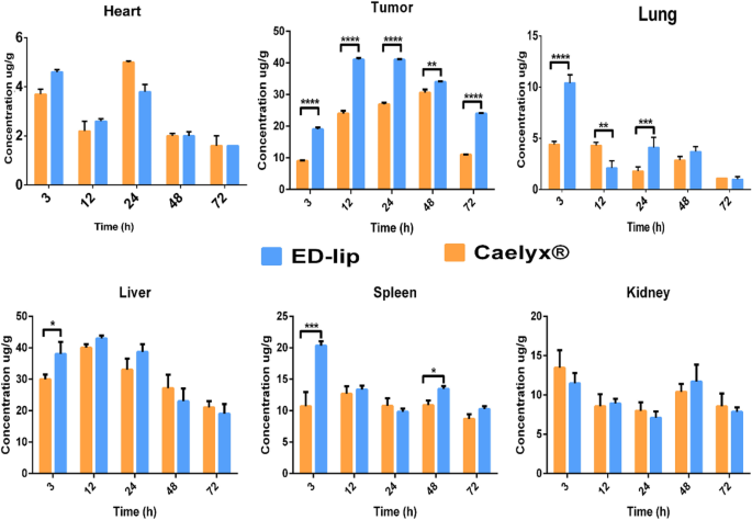
그림 8에서 볼 수 있듯이, Caelyx®와 ED-lip을 투여받은 그룹에서 주요 적출 기관의 DOX 농도를 비교했습니다. 유리 DOX의 가장 중요한 부작용은 Caelyx®가 이러한 부작용의 위험을 상당히 감소시키는 심장 독성입니다[46]. 간, 폐 및 비장에서 ED-lip의 생체 분포는 3시간에 Caelyx®보다 상당히 높습니다. 새는 혈관의 존재와 삽입 후 ED-립의 크기 및 PDI 증가는 초기에 이러한 조직에 ED-립이 더 많이 축적되는 이유일 수 있습니다. 나노입자의 크기가 150 nm로 증가하면 나노입자의 간, 폐 및 비장 축적이 향상되는 것으로 나타났습니다[47]. 한편, 생체 분포 연구 결과는 종양에 나노 입자가 축적되는 EPR 메커니즘을 명확하게 보여줍니다. 그림 8은 ED-lip과 Caelyx®가 종양 부위에 점차적으로 축적되어 약 12시간에 최대에 도달하고 24시간까지 안정 상태를 유지한 다음 48시간과 72시간에 점차적으로 감소한다는 것을 분명히 보여줍니다. 3, 12, 24, 48, 72시간의 모든 시점에서 흥미롭습니다. 종양에서 ED-lip의 축적은 Caelyx®보다 훨씬 더 많으며, 이는 항-EpCAM 앱타머를 사용한 활성 표적화의 효능 때문일 수 있습니다. 이러한 결과는 리포솜 표면에 대한 앱타머의 부착이 신장의 DOX 분포에 영향을 미치지 않았음을 나타냅니다. 따라서 항-EpCAM 앱타머는 종양 혈관 내피 세포에서 EpCAM 분자의 과발현으로 인한 것일 수 있는 리포솜의 종양 특이적 침투를 효과적으로 촉진하는 것으로 보입니다[48]. 이전에는 항-EpCAM 앱타머가 이종이식 종양에서 종양 침투를 향상시킬 수 있다고 표시되었습니다[49]. CSC 또는 TIC는 또한 항-EpCAM 치료의 표적이다. EpCAM을 표적으로 하기 위해 표적 리간드로 항-EpCAM 앱타머를 투여하면 CSC를 표적으로 하는 유망한 효과가 나타났습니다[22, 50]. 여기서 ED-lip의 효율적인 항종양 효과 중 일부는 CSC의 성공적인 표적화 때문일 수 있음을 시사할 수 있습니다.
<그림>
조직 생체 분포. 심장, 종양, 간, 폐 비장 및 신장에서의 DOX 생체 분포 결과. 농도(μg/g)는 각 기관에 대해 주사 후 3, 12, 24, 48 및 72시간에 보고되었습니다. 평균 ± 표준 편차(SEM)로 표시되는 데이터(n =3). *p <0.05, **p <0.01, ***p <0.001, ****p <0.0001
체내 항종양 활동
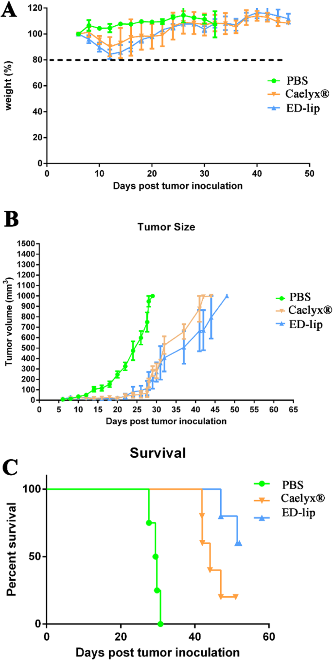
ED-lip의 치료 효능은 C26 결장암 종양 모델에서 평가되었습니다. 종양 크기, 체중 및 생존은 거의 2개월 동안 모니터링되었으며 결과는 그림 9 및 표 4에 요약되어 있습니다. 데이터는 ED-lip이 Caelyx®뿐만 아니라 마우스 체중에 명백한 영향을 미치지 않음을 나타냅니다(그림 9a 참조). ). 도 9b에 도시된 바와 같이, Caelyx®와 ED-lip의 정맥 주사 후, 종양 성장 속도는 주사 후 30일까지 효율적으로 억제되었으며, 리포솜 그룹에서 유의한 차이가 없었다. 주사 후 30일 후, 종양 성장 속도는 가속화되었지만 약물 투여 그룹의 성장 속도는 PBS 투여 그룹보다 여전히 더 느렸습니다. Caelyx® 그룹과 ED-lip 간의 차이는 주입 후 30일 동안 유의하지 않았습니다. 생존 결과는 Kaplan-Meier 플롯으로 표시됩니다. 그림 9c는 ED-lip이 PBS 또는 Caelyx®와 비교하여 생존 곡선을 개선함을 보여줍니다. 생존 연구의 주요 지표는 표 4에 요약되어 있습니다. ED-lip 그룹의 3마리 마우스의 종양은 완전히 치유되었으므로 이 그룹에 대한 MST는 정의되지 않았습니다. ED-lip 치료는 TTE를 41.1일에서 49.7일로 증가시켰고 3마리의 마우스에서 종양의 완전한 제거로 인해 정의되지 않은 MST로 90.27% TGD로 효과적인 항종양 활성을 보였습니다(표 4 참조).
<그림>
C26 결장암이 있는 암컷 BALB/c 마우스에서 제형의 생체내 치료 효능. 마우스에 단일 용량의 제형(10 mg/kg)을 IV 주사하였다. 아 각 실험 그룹에서 BALB/c의 각 중량 백분율 프로필을 나타냅니다. ㄴ BALB/c 마우스의 종양 크기 추적을 나타냅니다. ㄷ BALB/c에 대한 생존 그래프를 보여줍니다. 평균 ± SD로 표시되는 데이터(n =5)
종양 크기 데이터는 ED-립이 종양 성장을 극적으로 억제할 수 있음을 입증했습니다. 생존 분석 결과 ED-lip 치료는 MST와 TTE를 증가시키는 것으로 나타났습니다. ED-lip을 받은 그룹은 Caelyx®에 비해 TGD%가 더 높았고 더 효과적이었습니다. 우리의 발견은 ED-lip 처리군의 종양 조직에서 높은 수준의 DOX 농도와 일치합니다. 따라서, 압타머-결합 리포솜 DOX는 침투를 개선하고 결과적으로 종양 부위의 약물 축적을 향상시키며, 이는 차례로 Caelyx®의 효능을 증가시키고 생존 데이터에서 더 높은 TGD%로 이어집니다. 종합하면, 이러한 발견은 항-EpCAM 앱타머가 약물 전달을 위한 중요한 표적화제로 작용할 수 있음을 나타냅니다.
결론
여기에서 우리는 삽입 후(ED-lip)를 통해 항-EpCAM(SYLC3) 앱타머로 표면 기능화된 Caelyx®를 가지고 있습니다. 유세포 분석 및 형광 현미경 검사는 C26 세포에서 높은 수준의 DOX 흡수를 보여 aptamer가 ED-lip의 내재화 과정 속도를 향상시킬 수 있음을 나타냅니다. 약동학 데이터는 DSPE-mPEG-EpCAM 삽입 후 Caelyx®와 비교하여 DOX의 약동학을 변경하지 않았음을 나타냅니다. 그러나 조직 생체 분포는 주입 후 72시간 후에도 Caelyx®와 비교하여 ED-lip의 더 많은 종양 축적을 보여주었습니다. 우리는 ED-lip이 C26 종양이 있는 마우스에서 개선된 치료 효과를 가지고 있음을 입증했습니다. ED-lip으로 치료한 마우스에서 개선된 생존 매개변수는 EpCAM 표적 DOX 리포솜이 암 치료를 위한 유망한 약물 전달 운반체임을 시사하며 추가 조사가 필요합니다.
재료 및 방법
자료
5'-Amine-anti-EpCAM DNA 앱타머(5'-CACTACAGAGGTTGCGTCTGTCCCACGTGTCATGGGGGGTTGGCCTG-3')(SYL3C)는 BIONEER(biotechnology company, Daejeon, South Korea)에서 구입했습니다. DSPE-mPEG2000 -COOH는 Avanti Polar Lipids(Alabaster, AL)에서 구입했습니다. Dowex®, 1-에틸-3-(3-디메틸아미노프로필) 카르보디이미드(EDC), N-히드록시숙신이미드(NHS), 페니실린 스트렙토마이신 및 DAPI가 포함된 Fluoroshield™는 Sigma-Aldrich(St. Louis, MO)에서 구입했습니다. 상업적으로 이용 가능한 caelyx®는 Behestan Darou Company(이란 테헤란)에서 구입했습니다.
DSPE-mPEG의 활용2000 압타머로
Anti-EpCAM 앱타머는 DSPE-mPEG2000에 연결되었습니다. DSPE-mPEG2000의 카르복실기(–COOH)에 항-EpCAM 앱타머의 1차 아민(-NH2)의 공유 결합을 통해 (그림 1). 접합은 EDC/NHS 커플링 화학을 통해 수행되었습니다[51]. 간단히 말해서 DSPE-mPEG2000 2-(N-모르폴리노)에탄설폰산(MES) 완충액(pH 6.5)에 분산시키고 EDC/NHS 400mM EDC 및 100mM NHS를 분산액에 첨가하였다. 분산액을 15분 동안 교반하여 지질의 카르복실기를 활성화시켰다. 그런 다음, 항-EpCAM 앱타머를 분산액에 첨가하고 암실 상온에서 다음 2시간 동안 교반하였다. lipid:anti-EpCAM aptamer의 몰비는 1:1이었고 EDC/NHS의 몰비는 지질의 10배였습니다.
DSPE-mPEG-Anti-EpCAM Aptamer를 사용한 Caelyx® 수정
ED-lip은 post-insertion 방법으로 합성하였다. 삽입 후를 수행하기 위해 DSPE-mPEG-항-EpCAM 앱타머 미셀을 60°C에서 30분 동안 1ml의 caelyx®에 첨가했습니다. DSPE-mPEG-EpCAM 앱타머의 양은 Bartlett phosphate assay[52]에 따라 결정되었다. 약 10
14
인 caelyx® 밀리리터당 리포솜의 대략적인 수를 기준으로 합니다. , DSPE-mPEG-항-EpCAM의 부피는 각 리포솜당 10 압타머에 도달하도록 조정되었습니다[36]. 삽입 후 확인에 사용되는 아가로스 겔 전기영동 [39].
이화학적 특성화
입자 크기, 다분산 지수(PDI) 및 표면 전하는 DLS(Dynamic Light Scattering instrument)(Nano-ZS; Malvern, UK)에 의해 결정되었습니다. 유리 DOX를 제거하기 위해 리포솜을 Dowex® 수지와 혼합하고 60분 동안 회전시키고 Dowex®를 제거하기 위해 Poly-Prep 컬럼(Bio-Rad Laboratories Inc.)을 통과했습니다[53]. 리포솜 제형에서 DOX의 양은 LS-45 형광 분광광도계(Perkin-Elmer, UK)를 사용하여 결정되었습니다(여기:방출 485:590 nm).
출시 연구
DOX의 방출을 평가하기 위해 특정 시간 간격(0, 1, 2, 4, 6, 12 및 24시간), 샘플을 채취했습니다. Dowex® 수지로 유리 DOX를 제거한 후 리포솜에 남아 있는 약물의 양을 형광 분광 광도계로 측정하고 방출 비율을 계산했습니다[39].
세포 배양
C26 결장암 및 차이니즈 햄스터 난소(CHO-K1) 세포주는 이란 파스퇴르 연구소에서 구입했습니다. 세포주는 Gibco(Thermos Fisher Scientific, USA)로부터 입수한 10% FBS 및 100IU/ml 페니실린 및 100mg/ml 스트렙토마이신이 보충된 RPMI1640 배지에서 배양되었다. 세포를 37
°
에서 배양했습니다. C 5% CO2 95% 공기 가습 대기.
세포 상호작용 및 세포 흡수 분석
제형의 세포 상호작용 및 세포 흡수를 각각 4°C 및 37°C에서 평가했습니다. Two cell lines, CHO-K1 and C26, were selected in this test. The cells seeded in each well of 12-well plates (2.5 × 10
5
per well). After overnight incubation in 37 °C, treatments added to the cells and plates were placed at 4 °C and 37 °C and incubate for another 3 h. Then cells washed with PBS, and trypsinized. The fluorescence intensity for DOX was determined using flow cytometry (BD FACSCalibur cytometer). The data were analyzed with FlowJo version 7.0 software.
Fluorescent Microscopy Evaluation
The number of 1 × 10
6
cells per well C26 Cells were seeded into 6-well plates in which sterile microscopic cover glass were already inserted. After overnight incubation in 37 °C and 5% humidity, cells were treated with free DOX, Caelyx® and ED-lip for 24 h for complete cell uptake [54]. Then cells washed with PBS and fixed with 4% formaldehyde. Cover glasses stained with Fluoroshield™ with DAPI and were mounted on the glass slides. Treatments were performed in triplicate. From each slide, six zones were selected under × 200 magnification field. Intrinsic fluorescent of DOX was used for evaluation of drug cell uptake. Scaling was performed based on the percentages of cells which shown DOX cell uptake in each microscopic filed:
1:0–20%, 2:20–40%, 3:40–60%, 4:60–80%, and 5:80–100%
Evaluation of Cytotoxicity
The IC50 values of free DOX, caelyx®, and ED-lip were determined by MTT assay. In order to do this, CHO-K1 and C26 cells were seeded at density of 5 × 10
3
cells per well in 96-well plates at 37 °C. After overnight incubation liposomal formulations and free DOX solution were serially diluted in FBS-free medium and added to cell cultures and incubated 1, 3, and 6 h at 37 °C. Then, cells were washed and allowed to incubate 72 h. The optical densities (ODs) were measured using a spectrometric absorbance of 570 nm against a background of 630 nm on Stat-Fax 2100 microplate reader (Awareness Technology Inc. USA). Then the IC50 values were calculated.
Animal Study
Female BALB/c mice (4–6 weeks, 18–20 g) were kept in separate cages at 22 ± 2 °C and maintained on standard pellet diet and water ad libitum. Intraperitoneal (i.p) injection of ketamine and xylazine (100 mg/kg ketamine and 10 mg/kg xylazine) used to anesthetize the animals [55]. The number of 3 × 10
5
C26 cells per mouse in 60 μl PBS injected at the right flank, subcutaneously. Two weeks after inoculation when tumor sizes grew about 5 mm
3
, mice were randomly divided into 3 groups (n =3 for biodistribution and n =5 for antitumor study mice per group). All of the experimental protocols were approved by the Mashhad University of Medical Sciences committee for animal ethics and were performed according to the international rules considering the animal rights.
Biodistribution and Pharmacokinetic Studies
Fourteen days after tumor inoculation, mice were treated with dose of 10 mg/kg of caelyx® and ED-lip intravenously (i.v.) via the tail vain. Control group received 200 μl PBS solution. At certain time-intervals (3, 12, 24, 48, and 72 h) post-injection mice were euthanized and blood samples and tissue samples (liver, spleen, kidney, lung, heart, and tumor) were collected. Then, the concentration of doxorubicin in each sample measured based on fluorescent intensity of each samples using LS-45 fluorescence spectrophotometer (Perkin-Elmer, UK). Doxorubicin concentration of each sample was measured and non-compartmental analysis of the pharmacokinetic parameters were calculated from blood concentration vs. time profiles. Then the parameters including under the concentration-time curve (AUC) and area under the first moment curve (AUMC), half-life (t1/2 ), volume of distribution (Vd ), C최대 , Tmax,, mean residence time (MRT), and clearance (Cl) were calculated.
In Vivo Antitumor Activity
In order to evaluate antitumor activity, 10 days after tumor inoculation, mice with palpable tumor size were received single i.v. dose of 10 mg/kg Caelyx® and ED-lip. PBS injected in mice which considered as negative control. The parameters including time to reach the endpoint (TTE), percentage of tumor growth delay (TGD), median survival time (MST), and survival were determined. During the study, mice were observed for health and body weight changes. The tumor volume was also measured using a digital caliper and calculated as follows:
Considering ethical aspects, mice were removed in case tumor growth was> 1000 mm
3
, or> 20% weight loss or sign of weakness was observed.
Statistical Analysis
Data were analyzed using GraphPad Prism 6.0 (GraphPad software, Inc., San Diego, CA, USA). Data were demonstrated as mean ± SEM of at least three independent experiments. The t test was used in order to evaluate the results of release study, flow cytometry, and biodistribution of the formulations. ANOVA was employed to evaluate the results of fluorescent microcopy and tumor volumes. The Kaplan–Meier method used to calculate the survival parameters include TTE, MST and TGD%. 피 <0.05 was considered statistically significant.
데이터 및 자료의 가용성
All data supporting the conclusions of this article are included within the article.