재료 및 방법
자료
Fluorescein diacetate(FDA)와 propidium iodide(PI)는 Sigma(New York, NY, USA)에서 구입했습니다. GdCl3 ·6H2 O, 에틸렌 글리콜(99%)은 Hengrui Pharmaceutical Co., Ltd.(Lianyungang, Jiangsu, Chinese People's Republic of China)에서 구입했습니다. Cell Counting Kit-8(CCK-8)은 Dojindo(Kumamoto, Japan)에서 구입했습니다. NaH2 PO4 , 나2 HPO4 , 및 H2 SO4 Guangfu Fine Chemical Research Institute(중화인민공화국 톈진 난카이)에서 입수했습니다. 태아 소 혈청 및 Dulbecco의 최소 필수 배지(DMEM)는 Invitrogen China Limited(중화인민공화국 상하이)에서 구입했습니다. 모든 화학 물질은 분석 등급이었고 추가 정제 없이 사용되었습니다.
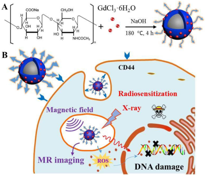
HA-Gd의 합성2 O3 NP
HA-Gd2 O3 NP는 다음과 같이 원 포트 열수 접근법을 사용하여 제조되었습니다. 먼저 0.1g의 HA를 주위 온도에서 밤새 격렬하게 교반하면서 20mL의 물에 용해했습니다. 이어서, 0.1 g의 GdCl3 ·6H2 O 및 0.5 mL의 NaOH(1 M)를 각각 첨가하였다. 그런 다음 혼합물을 5분 동안 더 교반하여 균질한 투명한 용액을 형성하고 50mL 오토클레이브에 옮기고 밀봉하고 120℃에서 6시간 동안 열수 처리하였다. 실온으로 자연 냉각시킨 후 투명한 현탁액을 0.22μm 멤브레인으로 여과하여 큰 덩어리를 제거하였다. 그런 다음 준비된 용액을 14-kDa 분자량 컷오프가 있는 투석 백에서 3 일 동안 물에 대해 투석했습니다. 투석액을 채취하여 진공동결건조기를 이용하여 동결건조하였다. HA-Gd2 O3 따라서 NP 분말을 얻고 추가 특성화를 위해 저장했습니다.
계측 및 특성화
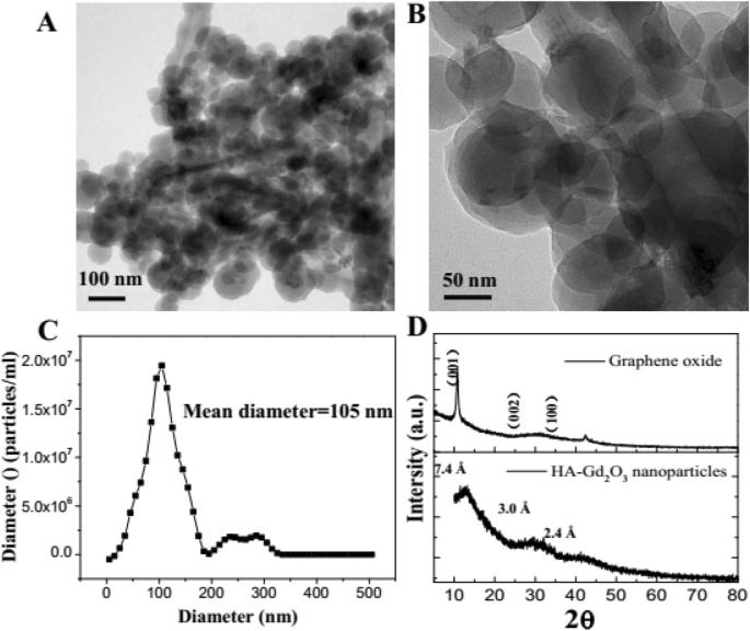
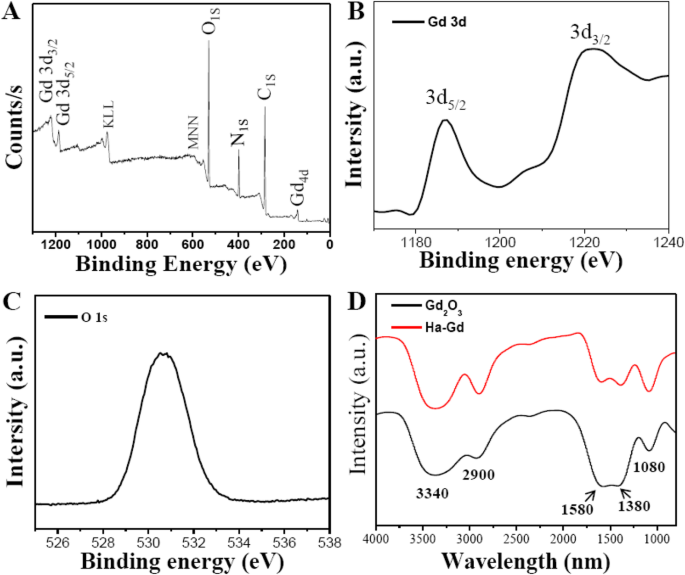
HA-Gd2의 형태 O3 NP는 200 kV의 가속 전압에서 JEM-2100 현미경(JEOL, Tokyo, Japan)에서 고해상도 투과 전자 현미경으로 검사되었습니다. HA-Gd2의 원소 구성 O3 NP는 X선 소스로 Al-Ka(1486.6 eV)와 푸리에 변환 적외선(FTIR) 분광계(Nicolet Nexus 470, GMI, Ramsey, MN, USA)를 사용하여 MK II 광전자 분광계에서 XPS 측정에 의해 결정되었습니다. HA-Gd2의 결정 구조 O3 NP는 Cu Kα(λ =0.15405 nm) 5 ~ 80° 범위에서 4°/분의 스캔 속도에서 방사선.
세포 생존 분석
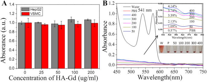
HA-Gd2의 영향 O3 세포 생존율에 대한 NP는 Cell Counting Kit CCK-8 분석(CCK-8 분석)을 통해 연구되었습니다. HepG2(인간 간암, ATCC 번호:HB-8065) 및 VSMC(혈관 평활근 세포)를 96웰 플레이트에 3 × 10
3
밀도로 시딩했습니다. 세포/웰 및 37 °C, 5% CO2에서 배양 다른 농도의 HA-Gd2를 함유한 DMEM을 사용하여 24시간 동안 배양 O3 성장 배지를 대체하는 NP(0 μg/mL, 25 μg/mL, 50 μg/mL, 100 μg/mL 및 200 μg/mL). 4 시간 더 배양한 후, 각 웰에 10μL CCK-8 용액을 첨가하고, 세포를 어두운 곳에서 4시간 동안 배양하였다. 흡광도는 Synergy HT Multi-Mode Microplate Reader(Bio Tek, Winooski, VT, USA)를 사용하여 490 nm에서 측정되었습니다. 처리되지 않은 세포(DMEM에서)는 대조군으로 사용되었으며 상대적인 세포 생존율(평균 SD, n =5) Abssample/Abscontrol × 100%로 표현하였다.
용혈 분석
간단히 말해서, 19-21 g, 6주령 암컷 BALB/c 생쥐는 중국 보건부의 동물 관리 규칙에 따라 친절하게 준비되었습니다. 헤파린 나트륨으로 안정화된 3밀리리터의 혈액은 안구를 제거하여 얻어졌습니다. 그런 다음 문헌에 따라 1200 rpm, 15 min으로 원심분리하여 상층액을 제거하였다. 그 후 침전물을 PBS로 5회 세척하여 마우스 적혈구(MRBCs)를 얻은 후 안구를 제거하여 혈액 약 3mL를 취하여 헤파린나트륨으로 안정화한 후 원심분리(1, 200 rpm, 15 min) 문헌[27]에 따라 상층액을 제거하기 위해 PBS로 5회 세척하여 마우스 적혈구(MRBC)를 얻었다. PBS로 10배 희석하여 0.1 mL MRBC를 HA-Gd2의 다양한 입자 농도(50–200 μg/mL)를 포함하는 0.9 mL PBS로 미리 채워진 1.5mL 튜브로 옮겼습니다. O3 NP, 각각 0.9 mL 물(양성 대조군) 및 0.9 mL PBS(음성 대조군). 혼합물을 부드럽게 흔든 후 실온에서 2시간 동안 인큐베이션한 다음, 12,000rpm에서 1분 동안 원심분리하였다. 마지막으로 모든 시료의 사진을 찍고 상층액(헤모글로빈)의 흡광도를 UV-2450 UV-Vis 분광광도계로 측정하였다. 541 nm에서 시료, 양성대조군, 음성대조군 간의 흡광도 차이를 나누어 서로 다른 시료의 용혈률을 계산했습니다.
체외 및 생체 내 MR 팬텀 연구
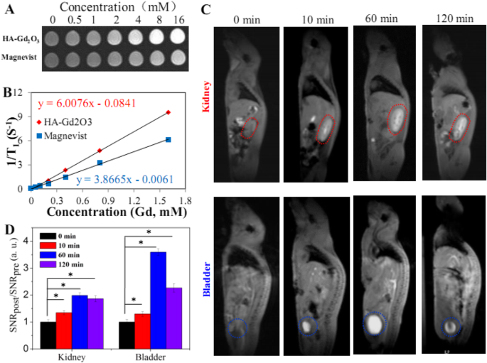
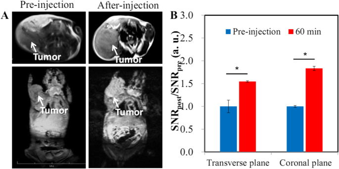
In vitro and in vivo MR images were acquired on 3 Tesla MRI scanner (Magnetom Trio Tim, Siemens, Germany). To study the T 1 phantom images in vitro, the solution of HA-Gd2 O3 NPs with different concentrations ranging from 0 to 16 mM was added to a 96-well culture plate, using the Magnevist (commercial MR contrast agent, Gd-DTPA) as control. For MR imaging in vivo, we chose the normal BALB/c mice as the model (n =4). Animal experiments were strictly conformed to the Animal Management Rules of the Ministry of Health of the People’s Republic of China. Ten milligrams per kilogram of HA-Gd2 O3 NPs filtering through sterilized membrane filters (pore size 0.22 μm) were intravenously injected into the animals, then immediately investigated with a MRI scanner. These samples for MR imaging in vitro and animals in vivo were imaged with the following parameters:TR/TE =300/10 ms, 256 × 256 matrices, slices =5, thickness =2 mm, averages =2, FOV =80 × 80.
Radiosensitization Effect In Vitro
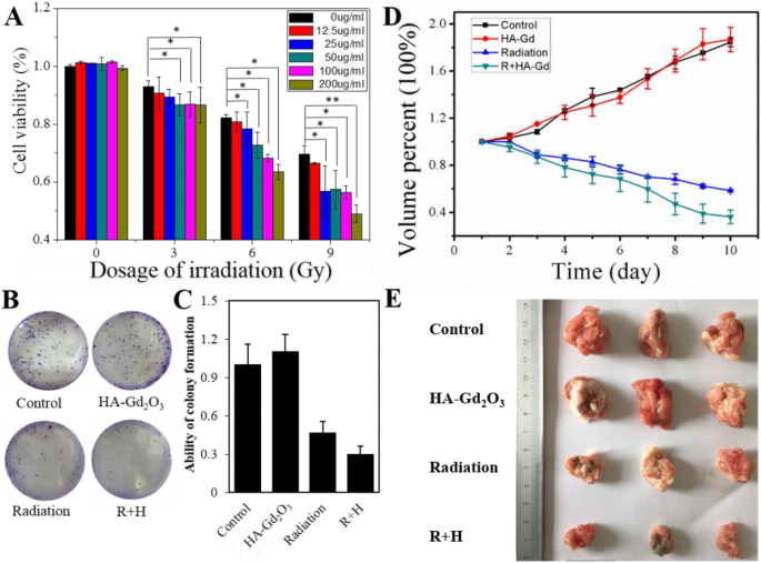
CCK-8 assay was used to evaluate radiosensitizing activity of HA-Gd2 O3 NPs in vitro. Cells were seeded in five 96-well plates at a density of 3 × 10
3
cells/well, and each plate was treated at the same condition:cultured at 37 °C in 5% CO2 incubator for 24 h, then using DMEM containing different concentrations of HA-Gd2 O3 NPs (0 μg/mL, 12.5 μg/mL, 25 μg/mL, 50 μg/mL, 100 μg/mL, and 200 μg/mL) replacing the growth medium. After incubation for 4 h, five plates were irradiated at different X-ray doses (0 Gy, 3 Gy, 6 Gy, and 9 Gy), 300 cGy/min, respectively. After radiation, all the plates were incubated for 4 h, then measuring the absorbance under the same parameters.
Clonogenic survival assay was also conducted to study the radiosensitization effect in vitro. HepG2 cells were seeded in six-well plates at 4.0 × 10
4
cells/well and allowed to grow for 16 h. The cells were incubated with HA-Gd2 O3 NPs diluted in cell culture medium for 6 h. The cells were then irradiated at 6 Gy using a clinical linear accelerator (Oncor, Siemens, Germany) with 6 MeV irradiation using a 10 cm × 10 cm radiation field at a source-to-skin distance (SSD) of 100 cm to cover the entire cells. Then, these irradiated cells were allowed to grow for 14 days, fixed with 4% paraformaldehyde at room temperature for 40 min, stained with a 1% crystal violet after washing the cells.
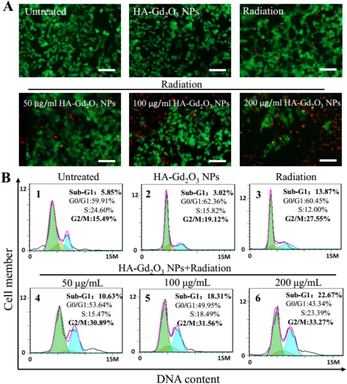
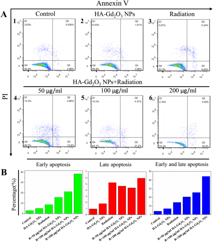
Live-Dead Staining Assay and Flow Cytometry
To study the radiosensitization effect of HA-Gd2 O3 NPs, HepG2 cells were seeded in six-well plates at a density of 4.0 × 10
4
cells/well and allowed to grow for 12 h and set six groups (control, HA-Gd2 O3 NPs, radiation, radiation + 50 μg/mL HA-Gd2 O3 NPs, radiation + 100 μg/mL HA-Gd2 O3 NPs and radiation + 200 μg/mL HA-Gd2 O3 NPs). When cells were grown to 80% in plates, the first group had no treatment, the second group was incubated with 100 μg/mL HA-Gd2 O3 NPs for 24 h, the third group was just irradiated, and the fourth group to the sixth group were irradiated and incubated with different concentrations of HA-Gd2 O3 NPs (50, 100, and 200 μg/mL) for 24 h, respectively. After that, FDA and PI working buffer were added for cell staining. The fluorescence of stained cells was observed under a fluorescence microscope (×20), and live cells showed green color and dead ones exhibited red color. Furthermore, cells by different treatments were washed three times with PBS, digested, collected, and centrifuged at a speed of 2000 rpm for 5 min, then fixed with 70% ethanol at − 20 °C overnight followed by PI staining. DNA fragmentation was quantified by the fluorescence intensity of PI on a flow cytometer (BD, Accuri, C6BD, Accuri, C6), analyzed by software (FLOWJO 7. 6. 2) to make clear the cell cycle distribution.
Radiosensitization Effect In Vivo
Female BALB/c mice with body weights of 19–21 g and ages of 6 weeks were obtained from the Yangzhou University Laboratory Animal Center under the standard conditions. Animal experiments were compliant with the Animal Management Rules of the Ministry of Health of the People’s Republic of China. A subcutaneous tumor model was established as the following procedures:First, 1 × 10
6
HepS cells were inoculated into mice intraperitoneal, and the ascites were collected after 5 days. Then, these ascites were injected into subcutaneous. When the tumor sizes reached approximately 100 mm
3
, subcutaneous tumors models were established and applied to the following experiments.
Eight mice bearing subcutaneous tumors per group were treated with radiation at 3 Gy per fraction to a total dose of 9 Gy within 7 days. The radiotherapy was conducted after 3 h intravenous injection of HA-Gd2 O3 NPs (10 mg/Kg), on a Siemens Primus clinical linear accelerator (6 MeV) using a self-made device to cover the entire tumor. These mice were anesthetized by 1% pentobarbital (50 mg/kg) to assure immobility during irradiation. The volume was measured and recorded every day, determined from caliper measurement, and calculated by the formulae:V =0.5 × a × b
2
, where V (mm
3
) is the volume of the tumor, and a (mm) and b (mm) are the tumor length and tumor width, respectively. Relative tumor volumes were normalized to their initial sizes. Each group was conducted on eight mice, wherein statistical analysis was performed using Student’s two-tailed t test (*p <0.05, **p <0.001).
데이터 및 자료의 가용성
The data sets supporting the results of this article are included within the article.